ADVERTIMENT. Lʼaccés als continguts dʼaquesta tesi queda ... · ology, Facu ogués Sanm r of the...
87
ADVERTIMENT. Lʼaccés als continguts dʼaquesta tesi queda condicionat a lʼacceptació de les condicions dʼús establertes per la següent llicència Creative Commons: http://cat.creativecommons.org/?page_id=184 ADVERTENCIA. El acceso a los contenidos de esta tesis queda condicionado a la aceptación de las condiciones de uso establecidas por la siguiente licencia Creative Commons: http://es.creativecommons.org/blog/licencias/ WARNING. The access to the contents of this doctoral thesis it is limited to the acceptance of the use conditions set by the following Creative Commons license: https://creativecommons.org/licenses/?lang=en
Transcript of ADVERTIMENT. Lʼaccés als continguts dʼaquesta tesi queda ... · ology, Facu ogués Sanm r of the...

ADVERTIMENT. Lʼaccés als continguts dʼaquesta tesi queda condicionat a lʼacceptació de les condicions dʼúsestablertes per la següent llicència Creative Commons: http://cat.creativecommons.org/?page_id=184
ADVERTENCIA. El acceso a los contenidos de esta tesis queda condicionado a la aceptación de las condiciones de usoestablecidas por la siguiente licencia Creative Commons: http://es.creativecommons.org/blog/licencias/
WARNING. The access to the contents of this doctoral thesis it is limited to the acceptance of the use conditions setby the following Creative Commons license: https://creativecommons.org/licenses/?lang=en

DOCTORAL THESIS
MOLECULAR STUDY OF IDIOPATHICNEPHROTIC SYNDROME
Gemma Bullich Vilanova
April 2016


M
Thes
Thes
Dr.Dire
olecula
sis realized ula
sis ascribed t
. Elisabet Arsector of the th
ar stud
Univ
nder the direaboratory of
to the DeparMed
s Criachhesis Dr.
y of idi
ThesiGemma
To fulfilversitat A
ection of Dr.Molecular B
rtment of Ceicine, Univer
PhD
RosDirect
GemmP
Barc
iopathi
is presenta Bullich V
the PhD dAutònoma
Elisabet ArsBiology of Fun
llular Biologyrsitat Autòno
in Cellular Bi
ser Torra Balctor of the the
ma Bullich ViPhD Candidatcelona, April
ic neph
ted byVilanova
degree atde Barce
s Criach andndació Puigv
y, Physiologyoma de Barc
iology
cellsesis Dr.
lanovate2016
hrotic s
lona
Dr. Roser Tovert, Barcelo
y and Immunelona
Carme NTuto
yndrom
orra Balcells ana
nology, Facu
Nogués Sanmor of the thes
me
at the
lty of
miquelsis


Als meus pares i al Tayo,

AGRAÏMENTS
Aquesta tesi és el resultat de la contribució de molta gent sense la qual no hagués estat possible arribarfins aquí. Tots els que citaré, i espero no deixar me ningú, heu posat el vostre granet de sorra (o unsorral sencer) en què aquesta tesis tirés endavant, moltes gràcies!
En primer lloc volia agrair de tot cor a les meves directores de tesi, l’Elisabet Ars i la Roser Torra, tot elque han fet per mi durant aquests anys. A l’Elisabet, moltíssimes gràcies per la teva enorme implicacióen tots els aspectes, per estar sempre disposada a dedicar me una estona, per ensenyar me a pensar i aprofunditzar, pel teu esperit crític, per tots els consells i per confiar en mi i en el meu criteri. De tu heaprès moltíssim a nivell científic però també a nivell personal, és un luxe treballar amb tu. A la Roser,moltíssimes gràcies per intentar trobar sempre un foradet en la teva atapeïda agenda, per resoldre totsels dubtes clínics, per aportar un punt d’optimisme als projectes, per implicar me en altres projectes,per valorar me tant, per haver me consolat en els moments més estressants, per aconseguir que tinguiun sou digne i per haver fet possible que d’aquí poquet me’n vagi a Holanda.
A la Carme Nogués, la tutora d’aquesta tesi, moltes gràcies per tota l’ajuda burocràtica, per facilitar meal màxim tots els tràmits i per resoldre els meus dubtes.
Moltíssimes gràcies a les companyes del laboratori de Biologia Molecular per pintar de colors els diesgrisos. A Patricia, por estar siempre dispuesta a ayudarme, por tus toques de humor que me encantan ypor enseñarme a ser una persona ordenada y organizada dentro del lab, estoy segurísima que dónde seaque vaya a trabajar echaré de menos una “lab manager” como tú. A la Laura, “la alegría de la huerta”,per les teves ganes d’aprendre el que calgui i per encomanar nos les teves rialles. A la Luz, l’altra“alegría de la huerta”, per ensenyar me que cal molt poc per ser feliç, per totes les xerrades d’ànims iper estar sempre disposada a ajudar me en l’àmbit científic i no científic. A Chiara, por escucharme enlos momentos bajos y filosofar sobre la vida y sobre si sirve para algo esto del doctorado, seguro quesí!!! Ah! y por resolverme las miles de dudas sobre el inglés! A l’Elena, per tota l’ajuda a tots els nivells,per preocupar te sempre per com estic, pels bons consells, i per ser tan optimista i positiva, ets un sol. Ala Bea, per haver me ajudat tantes vegades en mil temes: científics, de vestuari, de tesi, d’allotjament... iper ensenyar nos que si no hi ha risc físic no ens hem d’estressar, tan debò pogués fer ho! A la Irene,perquè si no fos per ella no faríem ni la meitat de sopars, ets la “lab manager” de la vida fora dellaboratori i el cotxe oficial que tants “apuros” ens ha solucionat, gràcies per estar sempre disposada aajudar! A la Judit, pels ànims en el moments tristos i per preocupar te per mi. A l’Iván, el profe debioinformàtica, per fer me perdre la por a la pantalla negra i per tota l’ajuda en la NGS i la no NGS, enlaborables i festius. A Dani, por su humor andaluz. I al Toni, la recent incorporació del laboratori, percuidar nos des de el primer dia amb croissants i braços de gitano, quin bon fitxatge!
Hi ha una sèrie de persones que ja no estan al laboratori però que sens dubte els he d’agrair la sevaajuda. A la Sheila, la meva “profe”, ha estat un autèntic plaer seguir els teus passos i tenir te com areferent en la SN, moltes gràcies per tot el que em vas ensenyar abans de marxar i per estar sempredisposada a resoldre’m tots els dubtes. A Deborah, mi gran compañera de atardeceres románticos en laPuigvert, ¡cómo te he echado de menos estos últimos años! Muchas gracias por todos tus consejos y porcompartir tantos momentos dulces y amargos. A l’Olga Sancho, la psicòloga, gràcies per ajudar me aencarar el doctorat (i la vida) d’una manera constructiva. A Paola, ¡suerte de tu optimismo durante elproyecto de los podocitos! Muchas gracias por todos los halagos y por valorarme tanto. A Ania por seruna buenaza y tener un gran corazón. A la Laia, la Xènia i la Lluïsa, sempre disposades a ajudar i a laMònica Vall, per animar me en els moments durs.
A part del laboratori de Biologia Molecular, la Fundació té grans persones a les quals he d’agrair la sevacontribució en aquesta tesi. A José Ballarín, muchas gracias por todo tu apoyo, por proporcionarme una

situación laboral digna, por valorar tanto nuestro trabajo y por estar siempre dispuesto a resolver misdudas. A la Sílvia Mateu, moltes gràcies per totes les gestions, pel “papeleo” i per tots els ànims. AMontse Díaz y Artur Oliver, por vuestra ayuda en el estudio de membranosa. A la Sílvia y la Charo, pelsànims i per fer me sentir que tot el daltabaix emocional que comporta escriure la tesi és normal. A lesdoctorandes nefròlogues, Nàdia, Iara iMónica per totes les paraules de suport i els ànims, en especial ala Nàdia, ha estat un plaer ajudar te en les nefropaties intersticials, només et queda escalar una micamés de muntanya fins arribar al cim, molts ànims! A l’Olga López pels elogis i la teva motivació. Al RicardPallejà, per tots els articles que m’has aconseguit amb una gran rapidesa. A la Núria Vinyolas per la sevadisposició a ajudar me en el què calgués. A tots els nois d’arxius, per tenir sempre apunt una paraulasimpàtica i un somriure. A l’Ignasi Gich, pel suport estadístic i a la Carolyn Newey per les correccions del’anglès.
Agrair també al Xavier Estivill per acollir me al seu laboratori del CRG durant la nostra col laboració. AlDani i al Justo per ensenyar me tots els trucs i secrets de la NGS.
Hi ha algunes persones que tot i no estar presents en el dia a dia han tingut un paper molt decisiu enl’inici d’aquesta tesi. Volia agrair al Pep per fer que una simple conversa es convertís en una granoportunitat. Al Josep Fargues per escoltar me, aconsellar me i ajudar me ja abans d’emprendre aquestcamí. Durant tots aquests anys me n’he recordat en molts moments de la teva frase: el doctorat és unacursa de resistència. Quina gran veritat! A la Naiara, moltes gràcies per tots els bons consells i perencomanar me la il lusió per la ciència.
A les bioquímiques, gràcies per no defallir en organitzar sopars! A la Georgina, moltíssimes gràcies perles trucades, per cuidar me i preocupar te per mi, pel punt d’optimisme i les ganes de tirar endavantque sempre em dónes, ets una gran amiga, hem compartit tot des de que vam entrar al món de laciència i no puc estar més orgullosa de l’amiga que he descobert, espero poder te ajudar en la teva tesitant com tu has fet amb la meva. A l’Àstrid, l’escriptora de tesi exprés (quina enveja!) i aventurera, pertransmetre aquesta motivació i ganes de tirar te a la piscina, espero que aviat organitzem un viatge aParís! A la Laura, la primera doctora del grup, gràcies per tots els consells i per compartir moltsmoments i dubtes sobre el futur durant el màster. A la Mireia perquè la seva motivació i ganes de fercoses alegren el dia a qualsevol i a la Hannah, quina sort coincidir al CRG i tornar nos a retrobar! I tambéa la resta de colla de la uni, Guillem, David, Raquel i Gemma per compartir penes dins i fora deldoctorat i perquè no em puc imaginar els anys d’universitat sense vosaltres.
Als “runners” moltes gràcies per transmetre superació km darrera km. A laMayte per aguantar totes lesvivències del doctorat i de la vida en general. Al Roger, René, Raúl, Lluís, Uri i a les 2 Patris, moltesgràcies per aguantar les angusties del doctorat i treure’m de casa de tant en tant. Al David i l’Òscar perajudar me a enderrocar tots els murs i per preocupar vos per mi, mai oblidaré els 100km!!!
Els amics te’ls vas trobant pel camí de la vida però sovint el destí els separa i no els pots veure tant comvoldries. Gemma, moltes gràcies per estar sempre disposada a ajudar me, per animar me en totmoment i per fer aquesta portada tant xula, ets genial!!! Martí, moltes gràcies per estar al meu costaten els moments difícils i per acceptar les meves decisions. Marta Cuenca, gràcies per no defallir enveure’ns encara que les respectives vides ho posin difícil i per estar sempre disposada a escoltar me,espero poder veure’t més sovint a partir d’ara. A les nenes de Ganàpies Eva, Anna Palou, Anneta iSantako, per tots els viatges que hem compartit, pels ànims que m’heu donat tots aquest anys i perentendre els “no puc he de fer tesi”. Als amics granollerins, Marta i Edmon, Elisenda i Joan, per aportarrialles i bons moments als caps de setmana de tesi, moltes gràcies!
A la família Castelluda, moltes gràcies per ser la millor segona família que podria tenir. A Pepe i Imma,per acollir me a la vostra família, per cuidar me i preocupar vos per mi i per valorar tant el que faig. A

Santi,Montse, José, Carol, Imma i Toni, gràcies per tot el suport que m’heu donat tots aquests anys, perinteressar vos per com m’anava el doctorat i per aguantar més d’una conversa sobre articles i cosesrares. A les xicotiues Júlia iMireia, per arrancar me sempre un somriure i fer me passar tots els mals.
A la Iaia, lluitadora i riallera, moltes gràcies per ensenyar me uns valors i una manera d’enfocar la vidaque ha sigut clau per fer el doctorat. A l’Avi moltes gràcies per ensenyar me la importància d’estudiar,de treure bones notes i per sentir te tant orgullós de mi. Sempre us portaré amb mi.
Als meus pares, Rosa i Francesc m’heu donat les ales per arribar fins aquí. Gràcies per creure sempre enmi, per valorar me tant, per estar al meu costat en els bons i mals moments, per acceptar les mevesdecisions, per fer me aixecar en els moments difícils. Gràcies per ensenyar me el valor de les coses, a serhumil, a lluitar pel què un vol. Sou el meu referent a la vida i si he arribat fins aquí és gràcies a vosaltres.
Al Tayo, per la seva ajuda infinita durant tots aquests anys. Ets una persona increïble i em sento moltafortunada de compartir la vida amb tu. Gràcies per estar al meu costat tots aquests anys, per creuretant en mi i en el què faig, per fer me enfocar els problemes de la manera més constructiva i per ajudarme a aixecar en tots els moments tristos. Ets el meu príncep blau, la parella que em cuida, l’amic queescolta, el pallasso que fa riure, l’aventurer amb qui viure experiències inoblidables, un mestre dins ifora de l’escola i una de les persones que més admiro del món.
Per últim voldria donar les gràcies a tots aquells pacients que ens han donat la seva mostra i han fetpossible aquesta tesi, així com a tots els clínics que han col laborat en l’estudi d’aquests pacients i enshan ajudat a seguir aprenent. Aquest estudi tampoc hauria estar possible sense el finançament per partdel Ministerio de Sanidad (FIS/FEDER 09/01506, FIS/FEDER 12/01523, FIS/FEDER PI13/01731 iFIS/FEDER PI15/01824) i la REDinREN.

TABLE OF CONTENTS
ABBREVIATIONS .................................................................................................................. 1
PRESENTATION ................................................................................................................... 5
INTRODUCTION ............................................................................................................................. 7
1. KIDNEY ANATOMY AND PHYSIOLOGY...................................................................................... 9
2. THE GLOMERULAR FITLRATION BARRIER .............................................................................. 11
2.1. FENESTRATED ENDOTHELIUM .................................................................................... 11
2.2. GLOMERULAR BASEMENT MEMBRANE...................................................................... 13
2.3. PODOCYTES ................................................................................................................. 16
3. NEPHROTIC SYNDROME......................................................................................................... 19
3.1. CLINICAL MANIFESTATIONS ........................................................................................ 20
3.1.1. Proteinuria....................................................................................................... 20
3.1.2. Hypoalbuminemia ........................................................................................... 21
3.1.3. Edema.............................................................................................................. 21
3.1.4. Hyperlipidemia ................................................................................................ 22
3.2. COMPLICATIONS ......................................................................................................... 23
3.2.1. Thrombosis ...................................................................................................... 23
3.2.2. Acute renal insufficiency ................................................................................. 23
3.2.3. Endocrin alterations ........................................................................................ 24
3.2.4. Infections......................................................................................................... 24
3.3. ETIOLOGY OF NEPHROTIC SYNDROME ....................................................................... 24
3.3.1. Congenital Nephrotic syndrome of Finnish Type............................................ 24
3.3.2. Diffuse mesangial sclerosis.............................................................................. 26
3.3.3. Minimal change disease .................................................................................. 26
3.3.4. Focal segmental glomerulosclerosis ............................................................... 27
3.3.5. Membranous nephropathy ............................................................................. 29
3.4. TREATMENT................................................................................................................. 30
3.4.1. General management...................................................................................... 30
3.4.2. Treatment in CNS ............................................................................................ 31
3.4.3. Treatment in children and FSGS/MCD adults ................................................. 31

3.4.3.1. Corticosteroid therapy ................................................................................ 31
3.4.3.2. Immunosuppressive therapy ...................................................................... 32
3.4.4. Treatment in MN............................................................................................. 32
4. PATHOGENESIS OF IDIOPATHIC NEPHROTIC SYNDROME ..................................................... 34
4.1. IMMUNE COMPLEXES DEPOSITION: MN .................................................................... 35
4.2. T CELL DEPENDENT CIRCULATING FACTOR: SSNS AND SDNS ..................................... 37
4.3. GENETIC CAUSE: SRNS................................................................................................. 38
5. GENETICS OF SRNS ................................................................................................................. 42
5.1. Autosomal recessive isolated SRNS and FSGS............................................................. 42
5.1.1. NPHS1 gene ..................................................................................................... 43
5.1.2. NPHS2 gene ..................................................................................................... 45
5.1.3. PLCE1 gene ...................................................................................................... 47
5.1.4. CD2AP gene ..................................................................................................... 49
5.1.5. PTPRO gene ..................................................................................................... 50
5.1.6. MYO1E gene .................................................................................................... 50
5.1.7. ARHGDIA gene................................................................................................. 51
5.1.8. ADCK4 gene ..................................................................................................... 51
5.1.9. TTC21B gene.................................................................................................... 52
5.1.10. CRB2 gene........................................................................................................ 54
5.1.11. NUP107 gene................................................................................................... 54
5.1.12. NUP93 gene..................................................................................................... 55
5.2. Genes implicated in autosomal dominant isolated SRNS and FSGS ........................... 56
5.2.1. WT1 gene ........................................................................................................ 56
5.2.2. ACTN4 gene ..................................................................................................... 58
5.2.3. TRPC6 gene...................................................................................................... 59
5.2.4. INF2 gene......................................................................................................... 61
5.2.5. LMX1B gene..................................................................................................... 63
5.2.6. ANLN gene....................................................................................................... 63
5.2.7. PAX2 gene........................................................................................................ 64
5.3. Genes implicated in syndromic forms of SRNS and FSGS ........................................... 65
6. CLINICAL UTILITY OF GENETIC TESTING ................................................................................. 67
AIMS ............................................................................................................................... ............. 71

RESULTS............................................................................................................................... ........ 75
STUDY I ............................................................................................................................... .... 77
HLA DQA1 and PLA2R1 Polymorphisms and Risk of Idiopathic Membranous Nephropathy 77
STUDY II ............................................................................................................................... ... 93
Targeted next generation sequencing in steroid resistant nephrotic syndrome: mutations in
multiple glomerular genes may influence disease severity................................................... 93
STUDY III ............................................................................................................................... 113
Contribution of TTC21B gene to glomerular and cystic kidney diseases............................. 113
STUDY IV............................................................................................................................... 123
Clinical utility of a kidney disease gene panel for genetic diagnosis of cystic and glomerular
inherited kidney diseases..................................................................................................... 123
DISCUSSION............................................................................................................................... 151
IDIOPATHIC NEPHROTIC SYNDROME........................................................................................ 152
IDIOPATHIC MEMBRANOUS NEPHROPATHY: THE IMPORTANCE OF GENETICS....................... 152
THE KIDNEY DISEASE GENE PANEL: THE INTEGRATIVE APPROACH.......................................... 155
SRNS/FSGS: FROM GENETICS TO GENOMICS............................................................................ 159
TTC21B GENE: CAUSATIVE AND MODIFYING ROLE................................................................... 162
CONCLUSIONS ........................................................................................................................... 165
BIBLIOGRAPHY........................................................................................................................... 169
ANNEXES....................................................................................................................... 195


1
ABBREVIATIONS
A, Ala Alanine
AIIRA Angiotensin II receptor antagonists
ACEi Angiotensin converting enzyme inhibitors
AD Autosomal dominant
ADAS Autosomal dominant alport syndrome
ADPKD Autosomal dominant polycystic kidney disease
ADTKD Autosomal dominant tubulointerstitial kidney disease
AFP Alpha fetoprotein
ANP Atrial Natriuretic Peptide
AR Autosomal recessive
ARAS Autosomal recessive alport syndrome
ARPKD Autosomal recessive polycystic kidney disease
AS Alport syndrome
Ca2+ Calcium ion
CAKUT Congenital anomalies of kidney and urinary tract
C, Cys Cysteine
CD2AP CD2 Associated Protein
CI Confidence interval
CKD Chronic kidney disease
CN Copy number
CNS Congenital nephrotic syndrome
CNF Congenital nephrotic syndrome of Finnish type
CNV Copy number variant
CoQ10 Coenzyme Q10
D, Asp Aspartic acid
DAD Diaphanous regulatory domain
DAG Diacylglycerol
Deletion/insertion Indel
DID Diaphanous inhibitory domain
DMS Diffuse mesangial sclerosis
DNA DeoxyriboNucleic Acid
DSC Doubling of serum creatinine
E, Glu Glutamic acid

2
ELISA Enzyme linked immunosorbent assay
ESRD End stage renal disease
F, Phe Phenylalanine
FCGR3 Fc gamma receptor III
FH1, FH2 Formin homology domains 1, 2
FNIII Fibronectin type III
fs Frameshift
FSGS Focal segmental glomerulosclerosis
g, mg Gram, milligram
G, Gly Glycine
GBM Glomerular basement membrane
GEC Glomerular endothelial cell
GFB Glomerular filtration barrier
GFR Glomerular filtration rate
h Hour
h Heterozygous
H Homozygous
H, His Histidine
HDL High density lipoprotein
HLA DQA1 HLA complex class II HLA DQ chain1
IFA Indirect fluorescent antibody technique
IFT139 Intraflagellar transport protein 139
Ig Immunoglobulin
IKD Inherited kidney diseases
IMN Idiopathic MN
INF2 Inverted formin 2
INS Idiopathic nephrotic syndrome
IP3 Inositol 1,4,5 triphosphate
JATD Jeune asphyxiating thoracic dystrophy
K, Lys Lysine
kDa Kilodalton
l, dl, ml Liter, deciliter, milliliter
L, Leu Leucine
LG Laminin globular
LMX1B LIM homeodomain containing transcription factor 1B
LM 521 Laminin 5 2 1

3
LN Laminin N terminal
M, Met Methionine
m2 Metre square
MAF Minor allele frequency
MCD Minimal change disease
mDias Mouse Diaphanous related formins
MG Mutation group
min Minutes
MLPA Multiplex ligation dependent probe amplification
Mmol Millimole
MN Membranous nephropathy
mo Months
MOI Mode of inheritance
MYO1E Myosin 1E
n Sample size
N, Asn Asparagine
NC1 Noncollagenous domain
ND No data
NEP Neutral endopeptidase
NGS Next generation sequencing
Nm Nanometre
NPHP Nephronophthisis
NPHP RC Nephronophthisis related ciliopathies
NS Nephrotic syndrome
NSR Non spontaneous remission
OR Odds ratio
P P Value
P, Pro Proline
PCR Polymerase chain reaction
PH Pleckstrin homology
PKD Polycystic kidney disease
PLA2R1 M type phospholipase A2 receptor
PLC 1 Phospholipase C epsilon 1
PLC_X, PLC_Y Phospholipase catalytic domains
Q, Gln Glutamine
R, Arg Arginine

4
RA1, RA2 RasGTP binding domain from guanine nucleotide exchange factors
RAAS Renin angiotensin aldosterone system
RasGEF_CDC25 Guanine nucleotide exchange factor for Ras like small GTPases
Ref Reference
rs RefSNP
S, Ser Serine
sd Syndrome
SD Standard deviation
SDNS Steroid Dependent Nephrotic Syndrome
SNP Single nucleotide polymorphism
SOD2 Superoxide dismutase 2
SRNS Steroid Resistant Nephrotic Syndrome
SSNS Steroid Sensitive Nephrotic Syndrome
suPAR Soluble urokinase plasminogen activator receptor
T, Thr Threonine
THSD7A Thrombospondin type 1 domain containing protein 7A
TM Transmembrane
TRP Tetratricopeptide repeat domains
TS Tuberous sclerosis
UCV Unclassified sequence variants
uPAR Urokinase plasminogen activator receptor
V, Val Valine
VEGF Vascular endothelial growth factor
vs Versus
VS Variant score
WAGR Wilms tumor, aniridia, genitourinary abnormalities, and mentalretardation
WT1 Wilms Tumor 1
XLAS X linked alport syndrome
Y, Tyr Tyrosine
y, yr Years
* Stop codon

5
PRESENTATION
This thesis is a contribution to the knowledge of the molecular bases of idiopathic nephrotic
syndrome. It has been performed in the Molecular Biology laboratory of Fundació Puigvert in
Barcelona. This laboratory performs genetic diagnostic of inherited renal diseases since 2002
as well as research on the molecular bases of these diseases.
The first part of this thesis presents the association of genetic polymorphisms with the risk to
develop idiopathic membranous nephropathy and with its clinical course. Our results showed
that risk alleles within HLA DQA1 and PLA2R1 genes are associated with a higher risk of
idiopathic membranous nephropathy. In addition, the combination of the risk alleles for both
genes results in an increased risk of idiopathic membranous nephropathy. For the first time,
we presented evidence of the contribution of these polymorphisms to predict response to
immunosuppressive therapy and decline in renal function.
The second part of these thesis focuses on the study of the molecular basis of steroid resistant
nephrotic syndrome. Our results contribute to enhance the genotypic spectrum associated
with this disease in several aspects: 1) mutations in an SRNS/FSGS gene together with COL4A3
have been detected in patients with increased disease severity, 2) the homozygous p.P209L
mutation in TTC21B gene is not the only causative mutation of FSGS, and 3) heterozygous
deleterious TTC21B variants may aggravate the phenotype of patients with glomerular and
cystic kidney inherited kidney diseases. In addition our results also improved the genetic
diagnosis of inherited kidney diseases. We developed a kidney disease gene panel that allowed
a more comprehensive characterization of patients enabling the differential diagnosis of cystic
and glomerular inherited kidney diseases and the identification of structural variants, complex
inheritance patterns and mosaic mutations. This approach is currently used in routine genetic
diagnostic of patients from all over Spain and other countries, providing a nice example of
translational research.
This thesis consisted of six parts. The introduction section describes the generalities of the
glomerular filtration barrier, as well as the clinical features and molecular bases of idiopathic
nephrotic syndrome. The aims section reflects the global and the particular aims of the present
thesis. The Results section contains the four studies performed in this project with a summary
of each one at the beginning. These results are discussed the Discussion section. Finally, the
conclusions derived from our results are presented in the Conclusion section.

6

7
INTRODUCTION

8

9
1. KIDNEY ANATOMY AND PHYSIOLOGY
The kidney’s main function is to filter waste materials out of the blood and pass them out
of the body as urine. The kidney’s microscopic functional units that filter blood to produce
urine are nephrons. An adult human kidney is known to contain an average of one million
nephrons. Based on their location, there are two types of nephrons: cortical nephrons (85%),
which are located deep in the renal cortex, and juxtamedullary nephrons (15%), which lie in
the renal cortex close to the renal medulla (Figure 1).
Nephrons are functionally divided into a filtration unit called the renal corpuscle and a
reabsorption unit named the renal tubule:
The renal corpuscle is composed of the glomerulus and Bowman’s capsule, separated
by the urinary space also named Bowman’s space.
o The glomerulus is a network of tangled capillaries where the actual filtering
takes place. It is composed of three cell types: glomerular endothelial cells that
form the intricately tortuous inner capillary tuft; podocytes or visceral
epithelial cells that tightly wrap around the exterior of glomerular capillaries;
and mesangial cells that together with the mesangial matrix provide a
structural reinforcement for the glomerular vasculature. Glomerular
endothelial cells and podocytes share a common extracellular cell matrix
known as the glomerular basement membrane (GBM). These three layers form
the glomerular filtration barrier (GFB).
o Bowman’s capsule is a cuplike enclosure composed of parietal epithelial cells
that surround the tuft of capillaries.
The renal tubule is a long convoluted structure that emerges from the glomerulus. Its
function is to concentrate urine and recover non waste solutes from the primary urine.
It is divided into three parts based on their function: the proximal convoluted tubule,
the loop of Henle and the distal convoluted tubule, which empties its filtrate into
collecting ducts.

Figure2013)
T
arter
glom
affere
mole
in the
are n
glom
medu
proxi
capill
T
the u
into
desce
corte
distal
neph
collec
e 1. Basic kid).
The renal ar
ioles that ca
eruli throug
ent arteriole
cular weight
e urinary spa
necessary fo
eruli throug
ulla and sepa
mal and dist
laries conver
The primary
urinary pole o
the primary
ends deep in
ex. In the loo
l convoluted
rons enters
cting duct ca
ney anatomy
rtery enters
arry the bloo
gh the affer
e immediate
t plasma was
ace, while th
r maintainin
gh the effer
arate into th
tal tubules, a
rge into vein
urine formed
of the renal c
y urine are
nto the med
op of Henle
d tubule an
the collectin
arries the co
(adapted fro
the kidney
od to the re
rent arteriol
ely branches
ste products
he passage o
ng normal ho
rent arteriol
he peritubula
as well as the
s that end in
d in the urin
corpuscle. T
reabsorbed
ulla of the k
water and
nd finally ur
ng duct. In th
oncentrated
om: “Kidney S
y through th
enal cortex.
e in the va
s into the el
s are filtered
of albumin a
omeostasis i
les. The eff
ar capillaries
e loop of He
n the renal ve
nary space fl
here, much o
d. Then, the
kidney, make
ions are rea
rine from th
his structure
urine throu
Stucture.” Bou
he renal hil
In the rena
ascular pole
laborated gl
across the G
and larger m
s restricted.
ferent arteri
s. The peritu
nle, where t
ein.
ows to the p
of the water
e filtrate en
es a hairpin t
absorbed. Ne
he distal co
s water and
ugh the rena
undless Biolog
ius and bra
al cortex, blo
. Inside the
omerular ca
GFB forming
acromolecu
. The filtrate
oles descen
bular capilla
they are know
proximal con
r and nutrien
ters the loo
turn, and ret
ext, the filtr
onvoluted tu
ions are stil
al medulla a
gy. and Kurts
anches into
ood enters t
e glomerulus
apillary tuft.
the primary
les (>15 kDa
ed blood exit
nd into the
aries surroun
wn as vasa.
nvoluted tub
nts initially fi
op of Henle
turns to the
ate passes t
ubules of se
ll reabsorbed
and into the
10
et al.,
small
to the
s, the
Low
y urine
a) that
ts the
renal
nd the
These
bule at
ltered
e that
renal
to the
everal
d. The
renal

pelvis
kidne
2. T
T
betw
selec
prima
T
highly
arter
oncot
hydro
T
podo
diaph
Figurepodocfenestdiaph
2
T
fenes
s. In the ren
eys and into t
THE GLOM
The GFB lies
ween the blo
tive filter th
ary urine.
The driving c
y controlled
iole via diffe
tic pressure
ostatic press
The GFB is c
ocytes, visce
hragm (Figur
e 2. A) Schecytes, mesangtrated endothragm (Leeuw
2.1.FENEST
The glomeru
strae and are
nal pelvis, u
the ureter.
MERULAR
s between t
oodstream a
at sieves on
component o
by the reg
erent mecha
due to the
ure, exerted
composed o
eral epithelia
re 2).
matic represegial cells andhelium, glomis et al., 2010
TRATED EN
ular capillari
e not surroun
rine from m
FITLRATIO
the vasculat
and the prim
n the basis o
of the glom
ulation of th
nisms. Other
e high conce
d by a fluid at
f three laye
al cells with
entation of athe endotheerular basem).
NDOTHELIU
ies are hea
nded by smo
many collecti
ON BARRI
ture and th
mary urine.
of molecular
erular filtrat
he vascular
r factors det
entration of
t rest (Leeuw
ers: the fene
h interdigita
a glomerularlium. B) Enlarent membran
UM
vily perfora
ooth muscle
ng ducts co
ER
he urinary s
This structu
size and ele
tion is the g
resistance o
termining the
f intracapilla
wis et al., 201
estrated end
ating foot p
capillary loorged view ofne and podoc
ated with tr
cells. These f
ombines and
space being
ure is a dyn
ectrical charg
glomerular p
of both affe
e glomerular
ary plasma p
10).
dothelium, t
processes th
p showing inthe glomerulacyte foot pro
ranscellular
fenestrae ar
flows out o
the only b
namic and
ge generatin
pressure, wh
rent and eff
r filtration ar
proteins, an
the GBM an
hat form th
nteraction bear filtration bcesses with t
pores know
e 60 100 nm
11
of the
barrier
highly
ng the
hich is
ferent
re the
d the
nd the
he slit
etweenbarrier:the slit
wn as
m wide

and c
efficie
woul
1987
indica
T
glom
(Rost
glyco
are h
absor
steric
drag
free c
T
plasm
(Hjalm
glom
lume
Figureglycoc
comprise ap
ent portals f
d expect tha
). However,
ating that th
The glycocaly
erular capilla
tgaard & Q
osaminoglyca
heparin sulfa
rbed within
c hindrance t
through the
circulating pr
The cell surfa
ma proteins
marsson et
erular filtrat
n (Figure 3).
e 3. Schematcalyx compon
pproximately
for the rapid
at albumin (3
the fenestr
e glomerula
yx is a negati
aries and the
Qvortrup 20
ans, glycopro
te and hyalu
the glycoca
to protein fi
e glycocalyx
roteins (Hara
ace anchore
absorbed w
al., 2004). T
tion barrier
tic representaents. ESL, end
y 20% of th
passage of h
3.5 nm of rad
rated endoth
r endotheliu
ve charged g
e fenestral s
002). It is
oteins and g
uronan and
alyx by bindi
ltration. This
which allow
aldsson et al
ed glycocalyx
within the
This layer fo
and ensures
ation of thedothelial surfa
e endothelia
high volumes
dius) could p
helium has a
um plays an a
gel like surfa
urfaces, and
composed
glycolipids. A
they act as
ing multivale
s barrier fun
s for dynam
l., 2008).
x and the b
glycocalyx c
orms the firs
s that the al
glomerular fiace layer (ada
al surface, m
s of fluid. Ba
ass through
a very low s
active role in
ace structure
acts as a siz
d of proteo
Around 90%
a molecular
ent to glyco
ction will be
mic equilibriu
broad coat o
constitute t
st barrier to
bumin is lar
iltration barrpted from Ha
making glom
sed on the f
the fenestra
sieving coeff
n renal filtrat
e that lines t
ze barrier to
oglycans w
of these gly
scaffold. Pla
osaminoglyca
e further mo
m with glyc
of >200 nm
he endothe
o albumin pa
rgely confine
ier with a dearaldsson et al
merular capi
fenestrae size
ae (Levick & S
ficient of alb
ion (Sarin 20
he luminal fa
albumin filt
ith their b
ycosaminogl
asma protein
ans, thus cre
dified by the
ocalyx boun
thick form
elial surface
assage acros
ed to the cap
etailed viewl., 2008)
12
llaries
e, one
Smaje
bumin
010).
ace of
ration
bound
lycans
ns are
eating
e fluid
d and
ed by
layer
ss the
pillary
of the

13
2.2.GLOMERULAR BASEMENTMEMBRANE
The GBM is a 250 to 400 nm thin meshwork of extracellular matrix proteins. Most of the
GBM is situated between the glomerular endothelial cells and the podocytes although it is also
found between mesangial cells and podocytes.
The GBM provides structural support for the glomerular capillaries and harbors ligands for
receptors of the adjacent endothelial cells, podocytes, and mesangial cells (Yurchenco &
Patton 2009; Miner 2005). In addition, it acts as a size and charge selective filtration barrier,
impeding the passage of molecules negatively charged and molecules larger than
approximately 5 nm (Deen et al., 2001).
The GBM is composed of laminin, type IV collagen, heparan sulfate proteoglycan and
nidogen (Figure 4). These components are synthesized by podocytes and glomerular
endothelial cells and secreted to the extracellular space (St John & Abrahamson 2001). During
glomerulogenesis the separate podocyte derived and endothelium derived basement
membranes fuse to form the immature GBM. After glomerular maduration both podocytes
and endothelial cells are equally important for maintaining its structure and function (Suh &
Miner 2013).
Laminins are large heterotrimeric glycoproteins composed of three different chains: ,
and . These chains assemble to form at least 15 distinct laminin trimers with
cruciform, Y shaped, or rod shaped structures. Each laminin trimer has the following
parts:
o A long arm formed by association of , and chains via coiled coil
interactions and disulfide bonding. At the distal end, there is a large laminin
globular (LG) domain formed exclusively by the C terminal of the chain that
link laminin trimers to the podocytes and endothelial cells via interactions with
integrins and dystroglycans receptors.
o Three short arms called laminin N terminal (LN) domains corresponding to the
N terminal globular domains of the three chains, which plays an essential role
in polymerization of trimers to form a network.
The major laminin trimer found in the mature GBM is laminin 5 2 1 (LM 521) (St
John & Abrahamson 2001). Once laminin is secreted to the extracellular space, laminin
trimers self polymerize by interactions among LN domains and form a laminin network

14
(Cheng et al., 1997). This polymerization step is a reversible and calcium dependent
process that requires a minimum concentration of laminin trimers to form an initiation
complex (Yurchenco & Cheng 1993). Therefore, laminin appears to be essential for the
initial formation of GBM. Then, interactions of laminin with other basement
membrane molecules (type IV collagen, nidogen, and sulfated proteoglycan) enable
the assembly of a GBM.
Type IV collagens represent half the total proteins of a mature GBM. There are 6
genetically distinct collagen IV chains ( 1 6) and they assemble to form three
different protomers, each composed of three chains: 1 1 2, 3 4 5 and 5 5 6.
Each collagen IV chain has three domains: the N terminal 7S domain, a central
collagenous domain and a noncollagenous domain (NC1) at the C terminus.
The central collagenous domain consists of many Gly X Y amino acid triplet repeats
(with X and Y usually being lysine or proline) that allow the assembling of the chains
into a triple helix. There are also multiple interruptions of the Gly X Y repeats scattered
throughout the collagenous domain that provide flexibility to the collagen IV
protomers and therefore, to the collagen IV network. Both the 7S and the NC1
domains are involved in linking trimers to each other to promote collagen IV network
formation. In addition, the NC1 domain is crucial for directing the composition of
collagen heterodimers.
Maturation of the GBM involves the transition of the 1 1 2 collagen to the 3 4 5
collagen protomers as the predominant collagen complex. This transition might be
required to accommodate the increased blood pressure after birth, since 3 4 5 type
IV collagen produces a more heavily cross linked and more protease resistant network
compared to the 1 1 2 type IV collagen network (Miner & Sanes 1994).
Nidogen 1 and nidogen 2 are two homologous glycoproteins containing three
globular like domains with two rod like domains that separate the globular like
domains. Nidogen 1 binds to both, laminin 1 chain short arm and type IV collagen.
Nidogens are supposed to provide extra stability to GBM under situations of unusual
stress, but they are not required for the initial formation of the GBM.
Heparan sulfate proteoglycans have a protein core with covalently linked sulphated
glycosaminoglycan side chains. The sulphated side chains give a negative charge to the
proteoglycan, however they do not seem to play critical roles in the GFB. The most

15
common heparan sulphate proteoglycan in mature GBM is agrin, although in
glomerulogenesis there appears to be co distribution of perlecan and agrin (Groffen et
al., 1998). The N terminal domain of agrin binds avidly to the LM 541 long arm, and its
C terminal contains domains that can bind to cell surface receptors such as
dystroglycan and integrins. These properties of agrin suggest that it could be
implicated in mediating charge selectivity within the GFB and in linking the GBM to the
adjacent cells. In addition, heparan sulphate side chains are important for binding and
sequestering growth factors in some contexts, such as the passage of vascular
endothelial growth factor (VEGF) from podocytes to fenestrated endothelium.
Figure 4: Components of the glomerular basement membrane and the podocyte glomerular basementmembrane interface (Lennon et al., 2014).
These components are distributed as a highly organized labyrinth of interconnected
polygonal fibrils of varying thickness ranging from 4 to 10 nm. The fibrils are more densely

packe
The
endo
(Scot
FigureFP, forecep
2
P
outer
barrie
count
produ
(Shan
M
body
in the
arise
foot
adjac
struct
ed within th
3 4 5 col
thelial side,
t & Quaggin
e 5. Moleculaoot processes;ptor (Scott & Q
2.3.PODOC
Podocytes ar
r aspect of
er to protei
teraction of
uction and
nkland 2006)
Mature podo
, major proc
e urinary sp
long major
processes. F
cent foot pr
ture of the
e core wher
lagen is con
whereas the
2015).
r organizationGBM, glome
Quaggin 2015)
CYTES
re highly spe
the GFB. Th
ns, a charge
the intraglo
secretion
).
ocytes consis
cesses, secon
pace and it c
processes t
Foot process
ocesses are
podocyte i
re they have
ncentrated
e laminin LM
n of the glomerular baseme).
ecialized, ter
hey perform
e barrier to
omerular pre
of VEGF re
st of morpho
ndary proces
contains the
that branche
es branch an
directly link
s achieved
e heterogene
at the core
M 521 and a
erular basement membrane
minally diffe
m highly spec
proteins, m
essure, synt
equired for
ologically an
sses and foo
nucleus and
ed into seco
nd interdigit
ked by the
by a uniqu
eous pores a
e with the m
grin are biom
ent membrane; GEC, glome
erentiated ep
cialized func
maintenance
hesis and m
r glomerula
nd functiona
ot processes.
d the cell m
ondary proce
tate with ne
glomerular
e cytoskelet
averaging 10
minor 1 1
modally dist
ne. ESL, endotrular endothe
pithelial cells
ctions, which
of the cap
maintenance
r endotheli
lly different
. The cell bo
machinery. Fr
esses, which
ighboring fo
slit diaphgra
tal architect
0 nm in diam
1 2 closer t
tributed (Figu
thelial surfaceelial cell; IR, in
s that enwra
h includes:
illary loop s
of the GBM
al cells int
component
ody essential
rom the cell
in turn spli
oot processes
am. This com
ture consisti
16
meter.
o the
ure 5)
e layer;ntegrin
ap the
a size
shape,
M, and
tegrity
s: cell
ly lies
body
it into
s, and
mplex
ing of

17
microtubule rich major and secondary processes, and actin based foot processes (Welsh &
Saleem 2012)(Figure 6).
Figure 6. A scanning electron micrograph of podocyte cells in a glomerulus shows the cell body (CB),major processes (MP), secondary processes (SP) and the finely interdigitating foot processes (DennisKunkel Microscopy, Inc. Welsh & Saleem 2012)
Foot processes contain three distinct membrane compartments: the basal side, the apical
side and the slit diaphgram. All three domains interact with the foot processes actin
cytoskeleton, influencing signaling pathways and motility of the podocyte. The basal side
anchors the podocyte to the GBM with several types of integrins and dystroglycans (Kreidberg
et al., 1996; Raats et al., 2000). The apical side is negatively charged due to the presence of the
anionic proteins podocalyxin, podoplanin, and podoendin (Kerjaschki et al., 1984; Matsui et al.,
1999). This negative charge contributes to limit the passage of albumin (also negatively
charged) and maintains the separation among adjacent podocytes.
The podocyte slit diaphgram is a unique cell cell contact with zipper like structure and a
constant width of 40 nm (Rodewald & Karnovsky 1974). The podocyte slit diaphgram
represents a separate class of specialized intercellular junction because it integrates structural
components of various cell junction types, including tight, adhesion, gap and neural junctions
(Reiser et al., 2000; Schnabel et al., 1990). Many of the molecular constituents of the slit
diaphgrams have been identified, although their topological assembly into a functional
complex is poorly understood. The ectodomains of several adhesion receptors in the slit
diaphgram likely organize the bridge linking juxtaposed foot processes via a combination of
homophilic and heterophilic receptor receptor interactions. By virtue of their large
ectodomains, nephrin and Fat1 are excellent candidates to associate in trans to connect
opposing foot processes (Inoue et al., 2001; Khoshnoodi et al., 2003) (Figure 7).

18
The slit diaphgram contains unique proteins that have to fulfill at least four different tasks:
they act as a macromolecular filter, anchor the filter to the GBM, connect the slit diaphgram to
the actin cytoskeleton via adaptor proteins, and are part of a signaling complex that integrates
and mediates extracellular and intracellular signals regulating the plasticity of food processes
(Grahammer et al., 2013).
The first unique slit diaphgram protein identified was nephrin, a type I transmembrane
protein with a cytoplasmatic C terminus, a transmembrane domain, a fibronectin type III
domain and eight extracellular C2 type immunoglobulins domains. Nephrin molecules interact
in trans configuration with each other, and probably overlap in the middle of the gap to form a
dense midline, giving the slit diaphragm its zipper like appearance. Therefore, nephrin is
considered the core biomechanical component of the molecular sieve (Tryggvason 1999;
Ruotsalainen et al., 1999).
The Neph 1, Neph 2 and Neph 3 proteins share certain homology with nephrin, but lack a
fibronectin domain and only have five extracellular C2 type immunoglobulin domains (Sellin et
al., 2003). These molecules are probably too short to participate in homophilic interactions in
trans configuration across the 40 nm gap of the slit diaphragm, but are likely to interact with
nephrin in both cis and trans configurations (Gerke et al., 2003; Barletta et al., 2003). All three
Neph proteins possess a THV motif near their C terminal end, which can bind to PDZ domains,
thereby enabling recruitment of scaffolding molecules, such as ZO 1 or the aPKC/PAR 3/PAR 6
polarity complex. Neph1 and nephrin seem to be involved in signaling events from the urinary
space to the podocyte cytosol (Hartleben et al., 2008).
The third unique slit diaphragm protein identified to date is podocin, a member of
stomatin family of proteins. Podocin is anchored to the membrane via its central part and its
N terminal and C terminal both face the cytoplasm. Podocin helps in the nephrin anchoring,
seems to recruit other slit diaphragm proteins to cholesterol rich membrane domains, and to
increase the signaling properties of the nephrin Neph1 complex (Huber, Simons, et al., 2003;
Garg et al., 2007).
The scaffold proteins ZO 1, CD2 associated protein and MAGI 2, as well as CASK and the
actin binding proteins IQGAP1 and actinin 4, anchor nephrin (and potentially other slit
diaphragm proteins) directly or indirectly to the actin cytoskeleton, which is crucial to both
foot process spacing and stability (Huber, Hartleben, et al., 2003; Palmen 2002). These
interactions probably strengthen the biomechanical properties of the slit diaphragm itself, and

19
might also enable it to relay signals to the actin cytoskeleton that, in turn, induce
morphological changes to primary and secondary foot processes (Grahammer et al., 2013).
Figure 7. The glomerular slit diaphragm. An ultrathin slit diaphragm spans the filtration slit between the
foot processes, slightly above the basement membrane (Tryggvason et al., 2006).
3. NEPHROTIC SYNDROME
Nephrotic syndrome (NS) is characterized by massive proteinuria, hypoalbuminemia, and
edema, although additional clinical features such as hyperlipidemia are also usually present. A
recently published definition of NS considers that nephrotic range proteinuria of glomerular
origin and a serum albumin concentration below the lower limit of normal are essential to NS
definition whereas edema, hyperlipidemia and lipiduria are commonly associated but not
essential (Glassock et al., 2015).
The central abnormality in all cases of NS is the development of massive proteinuria.
Physiologically, the liver tries to compensate for this excessive loss with increased protein and
lipoprotein synthesis. NS develops when the loss of protein in urine exceeds the rate of
albumin synthesis in the liver, resulting in hypoalbuminemia and edema.
NS represents one of the most common diagnoses in pediatric nephrology. The annual
incidence is estimated to be 2 7 per 100.000 children, with a prevalence of 16 per 100.000
children in Western countries (Giglio et al., 2015). There is a male preponderance among

20
young children, at a ratio of 2:1 to females, although this gender disparity disappears by
adolescence, making the incidence in adolescence and adults equal among males and females.
3.1.CLINICAL MANIFESTATIONS
3.1.1. Proteinuria
Proteinuria is a condition in which urine contains an abnormal amount of protein.
Nephrotic range proteinuria is defined as the proteinuria capable of producing NS and is
considered to be superior to 3.5 g/24h/1.73 m2 in adults or 40 mg/h/m2 in children. However,
the clinical manifestations of NS can appear with lower levels of proteinuria or be missing in
patients with higher levels of proteinuria. For this reason, it is preferred to consider nephrotic
range proteinuria, the proteinuria able to produce hypoalbuminemia.
The main proteins lost in NS have a molecular weight of 40 150 kDa, including albumin
(the most abundant protein in plasma), IgG, transferrin, ceruloplasmin and 1 acid
glycoprotein. There is also loss of small amounts of higher molecular weight proteins (200
kDa), like the small forms of high density lipoprotein (HDL). However, proteins higher than 200
kDa such as IgM, macroglobulins, fibrinogen, XIII factor, fibronectin and large lipoproteins are
not lost even in case of severe proteinuria.
The amount of proteinuria can be influenced by other factors different from the
glomerular damage such as the renin angiotensin system activity, the hepatic capacity for
albumin synthesis, the protein daily intake and the antihypertensive drug administration.
In healthy conditions, the GFB avoids the passage of molecules with a size superior to 70
kDa to the primary urine. Albumin has a molecular weight of 69 kDa and a net charge of 15
and is filtered through the GFB with a sieving coefficient of 0.0001. Interestingly, negatively
charged proteins have an increased clearance compared to positive charged ones due to the
negative charges in the GBM heparan sulfate proteoglycans.
Proteinuria in NS can be due to a structural alteration, disrupting the size selectivity of the
GFB and leading to massive nonselective proteinuria, or to an electrochemical alteration,
affecting the charge selectivity of the GFB and presenting with selective albuminuria.

21
3.1.2. Hypoalbuminemia
Hypoalbuminemia is a low level of albumin (inferior to 3 g/dl) in blood and it is a direct
consequence of proteinuria. Albumin is the most abundant plasma protein and represents 70
90% of the proteinuria detected in NS. This filtrated albumin is in part catabolized by the renal
tubule, which increases its catabolic rate. To compensate this loss of albumin in urine, the liver
increases the albumin synthesis even in a 300%. When the proteinuria and the renal albumin
catabolism exceed its hepatic synthesis, hypoalbuminemia appears. The severity of the
hypoalbuminemia is well correlated with the amount of proteinuria although other factors
such as the age, the nutritional state and the type of renal lesion also influence.
In addition to the hypoalbuminemia, there is a decrease in the serum immunoglobulins,
especially IgG, whereas high molecular weight immunoglobulins remain normal or even
elevated.
3.1.3. Edema
Edema is defined as an abnormal accumulation of fluid in the interstitial compartment.
Edemas are the main reason for diagnosis in children with NS. They are soft, pitting and mainly
localized in slope regions like foot or sacrum, and in regions with low tissue pressure such as
periorbital region. In advanced disease, patients may develop periorbital or genital edema,
ascites, or pleural effusion.
It is widely accepted that NS patients have an excess of total body sodium and water. Two
major pathophysiological mechanisms have been proposed to explain the development of
edema in NS based on the status of their intravascular volume: the underfilling hypothesis and
the overfilling hypothesis.
In the underfilling hypothesis (Figure 8A), the hypoalbuminemia causes a decrease in the
intravascular oncotic pressure. This in turn leads to fluid shift from intravascular to
extravascular compartment and edema formation. The homeostatic response to the
hypovolemia activates the renin angiotensin aldosterone system, the sympathetic nervous
system and the release of antidiuretic hormone. Gradually, the plasma volume normalizes as a
result of the increase of the extracellular space and the edema formation.
In the overfill hypothesis (Figure 8B), there is an intrinsic defect that enhances tubular
sodium and water reabsorption, independently of the hemodynamic situation. This causes

intrav
to ed
patie
show
reduc
circul
activa
Figureoverfi
T
that
intrav
and t
state
deve
the o
conti
H
frequ
vascular volu
dema format
nts with ove
w resistance t
ce the bloo
latory system
ation of the
e 8. Pathophyilling hypothe
The finding t
the overfill
vascular volu
the volume s
may be pr
lopment of
overfilled sta
nuing sodium
3.1.4. H
Hyperlipidem
uent lipid a
ume expansi
tion. Severa
erfilling of th
to Atrial Nat
od pressure
m. It has al
epithelial so
ysiological meesis (adapted f
hat most NS
ing hypothe
ume contrac
status may d
edominant
hypoalbumi
te may be p
m retention
Hyperlipide
mia is an abn
abnormality
ion and incr
l mechanism
he intravascu
triuretic Pep
by decreas
lso been su
dium channe
echanisms of efrom Hernand
S patients ha
esis might p
tion is found
depend on t
in the acute
nemia and a
predominant
due to persi
emia
normally hig
is hyperch
eased intrav
ms to explain
ular compart
ptide (ANP),
sing the am
ggested tha
el in the colle
edema formado Avendaño 2
ave normal o
prevail in m
d. Of note, bo
he stage of
e setting, in
an abrupt d
in the chron
stent low gr
gh concentra
olesterolem
vascular hydr
n this sodiu
tment have b
a polypeptid
mount of wa
at intrarenal
ecting duct.
tion. A) the u2008).
or elevated i
most cases.
oth hypothe
disease. It is
which mas
rop in plasm
nic phase du
ade hypoalb
ation of fats
ia appearin
rostatic pres
m and wate
been propos
de that is rel
ater, sodium
sodium ret
nderfilling hy
ntravascular
However, i
ses are not m
s possible th
sive protein
ma oncotic p
uring which p
buminemia.
or lipids in
ng in 85%
ssure, which
er retention
sed. Such pa
leased in ord
m and fat i
tention is d
ypothesis and
r volume sug
in some ch
mutually exc
hat the unde
uria causes
pressure, wh
patients may
blood. The
of patients
22
leads
in NS
atients
der to
n the
ue to
B) the
ggests
ildren
clusive
rfilled
rapid
hereas
y have
most
. The

23
hypertriglyceridemia is less frequent and only appears when the serum albumin is below 1 2
g/dl. Lipiduria, the excretion of fatty casts in urine, is also characteristic.
The pathogenesis of hyperlipidemia is multifactorial. Hypercholesterolemia seems to be
the consequence of the decrease in the intravascular oncotic pressure that results in an
increase in the hepatic synthesis of lipids and apolipoproteins. In addition, lipoproteins
clearance is decreased due to the low level of tissue low density lipoproteins receptors.
Hypertriglyceridemia is not due to the increase in triglycerides synthesis but a decrease in
triglycerides catabolism. It is correlated with the albumin clearance but not with the decrease
in the intravascular oncotic pressure.
The clinical significance of hyperlipidemia in NS is uncertain. Patients with NS and
hyperlipidemia have an increased cardiovascular mortality, although these patients also have
other cardiovascular risk factors. In animal models, hyperlipidemia accelerates the glomerular
damage, promoting the inflammatory infiltrate and renal fibrosis.
3.2.COMPLICATIONS
3.2.1. Thrombosis
Renal vein thrombosis and thromboembolic events in general constitute one of the most
severe complications in NS. Its incidence is estimated to be 5 60% of patients being more
frequent at the beginning of the disease. In addition, only 10% of patients with renal vein
thrombosis present with symptoms: flank pain, gross hematuria, increased renal size, and loss
of renal function. In children, thromboses are more severe and in half of cases affect the
arterial tree. In adults, arterial thrombosis is less common than venous thrombosis, but it is a
serious complication causing important morbidity.
Severe hypoalbuminemia (less than 25 g/l), high proteinuria (more than 10 g/24h), high
fibrinogen levels, low antithrombin III levels (less than 75% of normal), and hypovolemia are
significantly associated with an excessive risk of thrombotic complication.
3.2.2. Acute renal insufficiency
Acute renal insufficiency appears in patients treated with diuretics with severe
hypoalbuminemia. Its main cause is intravascular volume depletion.

24
3.2.3. Endocrin alterations
NS alters the regulation of several endocrine systems due to the urinary loss of hormones
or due to the change in their intravascular and extravascular distribution.
3.2.4. Infections
The risk of infections is increased in children. The most common and serious type of
infection is primary bacterial peritonitis, which is estimated to have an incidence of 5% in NS
children. Risk factors include low serum IgG levels due to urinary loss of IgG, abnormal T
lymphocyte function, and decreased levels of factors B and D, resulting in a decreased ability
to opsonize encapsulated bacteria. In addition, the administration of steroids and
immunosuppressive drugs during relapses further increases the risk of infection.
3.3.ETIOLOGY OF NEPHROTIC SYNDROME
NS may be caused by a variety of glomerular and systemic diseases. Approximately 90% of
the pediatric cases are primary or idiopathic but a small proportion of them are secondary to
infectious agents and other glomerular and systemic diseases. Separation of these two
categories is no always easy but it is important to decide the therapeutic approach. This thesis
is only focused on the idiopathic nephrotic syndrome (INS).
INS can be associated with a variety of distinct clinic and pathological conditions largely
dependent on the age at disease onset. The identification of the histological lesion may help to
elucidate the pathogenesis of the disease.
3.3.1. Congenital Nephrotic syndrome of Finnish Type
Congenital nephrotic syndrome (CNS) appears within the first 3 months of life. CNS of
Finnish type (CNF) represents the most severe form of NS. It was first described in Finland,
where it has an incidence of 1/8200 births but it has been reported worldwide.
CNF is an autosomal recessive (AR) inherited disorder characterized by massive proteinuria
even in utero, a large placenta, and characteristic microcystic dilatation of the proximal
tubules. Mutations in NPHS1 gene are the major cause of CNF, (explained in the next section)
(Kestilä et al., 1998).

25
A prenatal diagnosis of CNF is suspected as early as 16 to 18 weeks of gestation if elevated
fetoprotein levels are detected in the maternal serum or the amniotic fluid, reflecting fetal
proteinuria. The abnormally large placenta (placental/weight ratio over 0.25) constricts the
fetus likely causing premature delivery of the fetus with postural deformities. The child is
typically small for the gestational age, with widened cranial fontanelles, a small low bridged
nose, and edema in approximately 25% of cases. Proteinuria is detectable at birth and it is
extremely high ranging from 1 to 6 g/day. Initially, proteinuria is highly selective but it
becomes poorly selective as the disease progresses. Heavy and constant proteinuria inevitably
leads full blown NS soon after birth with life threatening edema, malnutrition, failure to thrive,
and secondary complications. Microhematuria or signs of tubular dysfunction (aminoaciduria
and glycosuria) may also be present. Renal function is usually normal at birth but it
progressively declines reaching end stage renal disease (ESRD) within the first 2 to 3 years of
life. There is a high mortality within the first year due to the complications of NS, particularly
infections and sepsis.
The histological lesions are detected more frequently after 3 months of age. The earliest
manifestation is focal cystic dilatation of proximal tubules (Figure 10), although it is not
present in all the cases, and/or an increase in mesangial cellularity and matrix, producing slight
glomerular enlargement. As disease progresses, the characteristic lesions of CNF appear
consisting of radial dilatations of the proximal tubules, mesangial proliferation and glomerular
lesions of focal segmental sclerosis. Immunofluorescence does not detect immune deposits
although as the disease progresses, IgM and complement C3 can be found. Effacement of
podocytes foot processes and disappearance of the slit diaphragm are seen in electronic
microscopy.
Figure 10. Histological lesions of congenital nephrotic syndrome of Finnish type at light microscopy.Hematoxylin eosin staining (Duicu et al., 2009).

26
3.3.2. Diffuse mesangial sclerosis
Diffuse mesangial sclerosis (DMS) accounts for approximately 10% of CNS cases and one
third of infantile NS cases (from 3 to 12 months) (Gbadegesin et al., 2008).
DMS is characterized by onset of NS in the first year of life and rapid progression to ESRD,
often within 1 to 3 months. This entity has been described as an isolated disorder or as part of
syndromes with renal and extrarenal abnormalities such as Denys Drash or Pierson Syndrome.
DMS is pathologically defined by mesangial expansion and sclerosis that evolves towards
obliteration of the capillary lumen and contraction of the glomerular tuft (Figure 11).
Immunofluorescence shows no glomerular immune deposits although nonspecific staining of
the glomerular mesangium for IgM, C3, and C1q may be identified in some glomeruli. The
major finding in electronic microscopy is extensive effacement of foot processes.
Figure 11. Histological lesions of diffuse mesangial sclerosis at light microscopy masson trichromestaining (from D’Agati et al., 2005).
3.3.3. Minimal change disease
Minimal change disease (MCD) is the major histological finding in INS children accounting
for more than 80%. The most frequent age at onset is 2 6 years (median 2.5) and is more
common in males than females (Bansal 2014). In adults, MCD is responsible for around 15 20%
of INS cases, there is a higher incidence in elderly than middle aged adults and the incidence is
equal between both genders (Cameron 1996; Korbet et al., 1988; Haas et al., 1997).
MCD virtually always manifests as NS at the onset, thus, the predominant findings are
heavy proteinuria, hypoalbuminemia, edema and elevated serum cholesterol. Proteinuria in
MCD is usually highly selective, consisting predominantly of albumin. Most children with MCD

27
do not have evidence of renal insufficiency, hematuria, or hypertension. This entity is highly
steroid responsive but in some cases may follow a remitting and relapsing course. Progression
to ESRD is not part of the natural history of MCD.
MCD is pathologically characterized by minimal or no glomerular alterations on light
microscopy (Figure 12). The “minimal” findings include podocyte swelling and mild mesangial
expansion. Immunofluorescence does not detect glomerular immune deposits although in a
minority of cases, there may be weak mesangial positivity for immunoglobulin IgM, with or
without C3. Electronic microscopy shows extensive effacement of foot processes in the
absence of other abnormalities of the peripheral capillary walls.
Figure 12. Histological lesions of minimal change disease at light microscopy. Masson trichrome staining(from kidney pathology website).
3.3.4. Focal segmental glomerulosclerosis
Focal segmental glomerulosclerosis (FSGS) is one of the most frequent patterns of injury
found in INS accounting for approximately 10% of children, 20 50% of adolescents and around
20 30% of adults (Hogg et al., 2007; Cameron 1996; Korbet et al., 1988; Haas et al., 1997).
FSGS lesions can occur as a result of many causes, including circulating factors, mutations in
podocyte genes or be secondary to drugs, infections or maladaptive responses to nephron loss
(Bose & Cattran 2014).
Ethnicity plays important roles in the incidence of FSGS being 3 7 times higher in young
black men as compared with whites (Freedman et al., 2009). The reported annual incidence
rates for FSGS is 5 cases per million population in whites, compared with 24 cases per million
population in African Americans. This increased incidence is partly explained by genetic risk
factors in 2 important podocyte function proteins: nonmuscle myosin heavy chain 9 (MYH9)
and apolipoprotein L1 (APOL1). Three polymorphisms in MYH9 gene were reported to confer

28
risk for idiopathic FSGS and hypertensive ESRD among blacks in AR model (Kopp et al., 2008).
Further investigation revealed two variants, called G1 and G2, in the last exon of the
neighboring gene APOL1 that had stronger association with FSGS and were under strong
selection only in Africa (Genovese et al., 2010).
Proteinuria is a defining feature of FSGS, typically accompanied by hypoalbuminemia,
hypercholesterolemia, and edema. Approximately 75 90% of children and 50 60% of adults
with FSGS have NS at presentation (D’Agati et al., 2011). FSGS is associated with a poor
response to corticosteroid treatment (only about 50% of patients respond to therapy). It is
usually a progressive disorder with <5% spontaneous remission and a 50% ESRD rate over a
period of 5 8 years from the time of the renal biopsy in patients that are either unresponsive
to treatment or not treated (Bose & Cattran 2014). Black race, increased degrees of
proteinuria, and renal insufficiency are associated with a worse outcome. Approximately 40%
of patients who undergo kidney transplantation develop recurrence of the disease in the
allograft (D’Agati et al., 2011).
FSGS is pathologically defined as glomerular sclerosis involving a subset of glomeruli (focal)
and a portion of the glomerular tuft (segmental) (Figure 13). Because juxtamedullary nephrons
are often affected first, adequate glomerular sampling is needed to identify the diagnostic
lesions. As the disease progresses, a more diffuse and global pattern of sclerosis evolves and
tubular atrophy and interstitial fibrosis develop. Immunofluorescence typically reveals coarse
segmental staining for IgM and C3 entrapped in areas of hyalinosis. On electronic microscopy,
the major finding is extensive effacement of the foot processes without other abnormalities in
the GBM. There are several FSGS variants based on the pathologic findings, from best to worst
prognosis: tip, FSGS not otherwise specified, cellular, collapsing, and perihilar, being the most
common FSGS not otherwise specified. Also, increased severity of interstitial fibrosis and
tubular atrophy in biopsy specimens are associated with worse outcome.

29
Figure 13: Histological lesions of focal segmental glomerulosclerosis at light microscopy. 400x, massontrichrome staining (from kidney pathology website).
3.3.5. Membranous nephropathy
Membranous nephropathy (MN) is one of the most common forms of NS in adults,
accounting for about 20% of cases(Glassock 2010). It comprises less than 2% of all idiopathic
NS cases in children under the age of 5 years, but its incidence increases progressively through
adolescence and into adulthood with a peak incidence in individuals aged 30 50 years. The
disease incidence is also influenced by sex being more common in males than females (sex
ratio 2:1). This condition is idiopathic in approximately 70 80% of cases, whereas the
remaining are secondary to a variety of autoimmune, infectious, neoplastic, and other
systemic diseases, or following exposure to certain medications (Glassock 2010).
The predominant clinical presentation of patients with MN is NS, occurring in 60 80% of
them. The remaining patients present with subnephrotic proteinuria, often asymptomatic that
may be detected as an incidental finding during a routine medical examination, and about 60%
of them will progress to full NS (Fervenza et al., 2008; Hladunewich et al., 2009). Microscopic
hematuria occurs in up to 50% of patients with MN, but red blood cells casts and macroscopic
hematuria are rare (Wasserstein 1997). About 80% of patients with MN have normal blood
pressure and glomerular filtration rate at presentation. Acute renal failure is uncommon
(Ronco & Debiec 2015).
IMN natural history follows three major clinical courses: spontaneous remission, stable but
persistent proteinuria, or progression to ESRD. Spontaneous remission occurs in approximately
one third of patients, usually within the first two years after presentation (Polanco et al., 2010;
Polanco et al., 2012). The other two thirds of patients can be divided equally into those with
persistent proteinuria who will maintain normal renal function long term and those patients
who will progress to renal failure despite immunosuppressive therapy (Glassock 2003). MN is
the second or third leading cause of ESRD in patients with primary glomerulonephritis and the
glomerulopathy that recurs most frequently after kidney transplantation (in about 40% of
cases) and threatens graft function (Ronco & Debiec 2015).
MN is pathologically defined as a capillary wall thickening, normal cellularity, positive IgG
and C3 staining along capillary walls on immunofluorescence, and accumulation of immune
deposits on the outer aspect of the GBM on electron microscopy called spikes (Figure 14).

Figuremicrostainibetweand cElectrglome
3
IN
but a
are c
the h
R
renal
the r
respo
P
both,
(AIIRA
angio
vasod
there
e 14: Histolooscopy: capillang at light meen the immucomplementron microscoerular baseme
3.4.TREATM
NS treatmen
lso the deriv
ommon in m
istological le
Renal biopsy
lesions bec
renal biopsy
ondent.
3.4.1. G
Proteinuria is
, angiotensin
A) as they
otensin II, a h
dilation of th
efore decreas
ogical lesionsary loops aremicroscopy: tune deposits. Cappear in apy: electronent membrane
MENT
nt requires a
ved systemic
most patients
esion.
is considere
ause the tre
is only perf
General ma
s treated wit
n converting
have a syn
hormone tha
he glomerula
ses protein l
of membrane thickened athere are chaC) Immunofludiffuse granudense imm
e (renal patho
global thera
c complicatio
s, whereas s
ed mandator
eatment app
formed whe
anagemen
h renin angi
enzyme inh
nergic effect
at has a vaso
ar efferent a
eakage.
nous nephropand prominearacteristic “uorescence miular patternune depositsology website
apeutic appr
ons. General
pecific treat
ry in adult pa
roach differs
n the NS is
t
otensin aldo
hibitors (ACE
t. These dru
oconstrictive
arteriole des
pathy. A) Hemnt but cellulaspikes” of glicrograph: subin the glomes are seen s).
roach to cou
measures to
ment depen
atients to di
s among the
steroid resis
osterone syst
Ei) and angio
ugs avoid t
e action and
creases the
matoxilin eosarity is not ilomerular babepithelial deerular basemscattered wit
unteract the
o treat prote
ds on the ag
stinguish am
e different en
stant as mo
tem (RAAS)
otensin II rec
he formatio
increases blo
intraglomer
sin staining aincreased. B)sement memposits of mainent membrathin the thic
glomerular
einuria and e
ge at onset a
mong the diff
ntities. In ch
st of patien
blockers incl
ceptor antag
on and acti
ood pressure
rular pressur
30
t lightSilver
mbranenly IgGne. D)ckened
lesion
edema
and/or
ferent
ildren
ts are
luding
onists
on of
e. The
re and

31
General measures to control edema include salt restriction, lying in supine position, and
use of diuretics drugs. Moderate exercise, a low cholesterol diet, hydroximetilglutaril CoA
reductase inhibitors, and weight lost in obese patients are recommended to reduce
hiperlipidemia. Hygienic, dietetic and pharmacological measures are needed to counteract the
high morbidity and mortality associated with NS and its complications.
3.4.2. Treatment in CNS
The goals of therapy during the first months are to control edema and possible uremia,
prevent and treat complications such as infections and thromboses, and provide optimal
nutrition so that the child grows and develops as normally as possible.
CNS patients are resistant to corticosteroids and immunosuppressive therapy, therefore
the only curative approach is renal transplantation. Bilateral nephrectomy and dialysis may be
necessary to avoid the complications of NS.
3.4.3. Treatment in children and FSGS/MCD adults
3.4.3.1. Corticosteroid therapy
Corticosteroids are the first line treatment for most INS. Children presenting with INS are
treated empirically with corticosteroids because there is a high incidence of MCD, which is
associated with a high rate of steroid responsive. A complete remission is considered if
proteinuria < 100mg/m2/day or < 1+ on urine dipstick for 3 consecutive days, and urine
protein/creatinine ratio < 20 mg/mmol. Partial remission consists of a proteinuria of 0.1 1
g/m2/day and absolute urine protein/creatinine ratio between 20 and 200 mg/mmol
(Trautmann et al., 2015).
Adults with FSGS or MCD as renal lesion are also initially treated with corticosteroids. A
complete remission is defined by proteinuria < 0.3 g/day and normalization of serum albumin
to at least 3.5g/dl. Partial remission is considered if proteinuria 0.3 3.5 g/day with a reduction
in proteinuria by at least 50% from the baseline.
Based on patients’ response to steroid therapy, INS can be divided into:

32
Steroid Sensitive Nephrotic Syndrome (SSNS): patients who achieve remission in
response to corticosteroid treatment alone. Approximately 80 90% of these patients
have one or more relapses, but they mostly continue to respond to corticosteroids
throughout their subsequent course and the long term prognosis, including the
maintenance of normal kidney function, is good. The most frequent renal histological
lesion of SSNS is MCD.
Steroid Dependent Nephrotic Syndrome (SDNS): patients that initially respond to
corticosteroid treatment by achieving complete remission but develop a relapse either
while still receiving steroids at lower doses or within 2 weeks of discontinuation of
treatment following a steroid taper. Such patients typically require continued low dose
treatment with steroids to prevent development of relapse.
Steroid Resistant Nephrotic Syndrome (SRNS): Patients who fail to achieve remission
after 8 weeks (children) or >4 months (adults) of corticosteroid treatment. The most
frequent renal histological lesions of SRNS are FSGS or DMS. Steroid resistant MCD
adults should be re evaluated for other causes of NS.
It is estimated that about 80% of children and 60% of adults with INS respond to steroid
therapy with complete resolution of proteinuria and edema. Among this steroid sensitive
group, the clinical course is variable, with up to 60% having frequent relapses or becoming
dependent on steroid therapy to maintain them in remission. The remaining 20% of children
and 40% of adults with INS do not achieve a sustained remission after standardized
corticosteroid therapy (Arbeitsgemeinschaft fur Padiatrische 1988; Antignac 2002; Troyanov et
al., 2005).
3.4.3.2. Immunosuppressive therapy
Immunosuppressive therapy is considered in patients with SRNS, frequent relapses or
steroid dependency. There are different immunosuppressive therapies available and different
therapeutic schemes depending on the age at onset and/or the histological lesion (reviewed in
KDIGO guidelines (Glomerulonephritis Work Group 2012)).
3.4.4. Treatment in MN
Spontaneous remission is a well known characteristic of MN occurring in approximately
one third of patients, but it may be delayed for as long as 18 24 months (Polanco et al., 2010).

33
Thus, the initial approach in MN patients consists of a 6 month observational period, in which
only antiproteinuric agents are given. Patients with persistent proteinuria and no decline after
6 months are treated with immunosuppressors accoding to KDIGO guidelines
(Glomerulonephritis Work Group 2012).
IMN patients presenting with non nephrotic proteinuria typically only receive ACEi and
AIIRA and they have a very favorable long term prognosis (Hladunewich et al., 2009; Ponticelli
& Glassock 2014). However, a delay in immunosuppressive treatment in patients presenting
with NS would expose them to the complications of NS (van den Brand et al., 2012). In these
patients, controversy exists regarding the proper timing of immunosuppression and the best
therapeutic regimen, due to the low therapeutic index of immunosuppressive drugs and their
side effects (Remuzzi et al., 2002; Perna et al., 2004; Glassock 2004; du Buf Vereijken et al.,
2005). A number of clinicians propose to restrict immunosuppressive therapy to patients with
sustained heavy proteinuria or in the face of worsening renal function (Cattran 2005).
Several markers of disease progression help clinicians to predict the clinical outcome of the
disease. Predictors of spontaneous remission are baseline proteinuria less than 8 g/day,
female sex, age younger than 50 years old and preserved renal function at presentation
(Cattran 2005). However, even in patients with large proteinuria, spontaneous remission can
occur in up to 20 25% of them (Polanco et al., 2010).
The Toronto Risk Score is calculated by a validated algorithm that correctly identifies
patients at risk of disease progression with 85 90% accuracy. This score is based on the level of
proteinuria during a 6 month period of maximum proteinuria, creatinine clearance at the start
of that period, and the change in creatinine clearance over the course of those 6 months.
According to the score obtained, patients are grouped into low , middle , and high risk for
progression and a different treatment approach is indicated (Figure 15) (Pei et al., 1992;
Cattran et al., 1997). The main disadvantage of this model is that an observational period of at
least 6 months (or more if the period of maximum proteinuria does not correspond with the
onset of the disease) is needed to calculate the score, which prolongs patient exposure to risks
associated with NS.

Figureconse
M
urine
(Bran
of th
and
speci
advan
al., 2
mark
4. P
IN
its in
invar
disea
abno
speci
on re
CNS a
defec
e 15. A treaervative and th
Measuremen
e samples w
nten et al., 20
e Toronto R
1 microglo
ficity with n
ntage that th
2012). The c
kers would be
PATHOGEN
NS is due to
nability to r
iable featur
ases, and it
rmalities (D
fic autoimm
enal biospy, a
and SRNS pa
ct is consider
atment algorhen immunos
nt of the uri
was also sho
005; Hofstra
Risk score w
bulin) and s
no significant
he prognosis
linical comp
e the best op
NESIS OF
the develop
restrict urina
e observed
can result
eegens et a
une disease
are considere
atients, the la
red to be res
rithm that csuppressive tr
nary excreti
own to acc
et al., 2008
with the urin
showed that
t differences
s can be esta
plexity of the
ption for pre
IDIOPATH
pment of str
ary loss of
not only in
from seve
al., 2008). B
. SSNS and S
ed to be cau
atter usually
sponsible for
ombines theeatment (Catt
on of 2 mi
urately pred
). Van den B
ary low mol
t both prog
s between th
ablished with
e disease su
ediction of cli
HIC NEPHR
uctural and
protein. Po
n NS patient
eral triggers
riefly, idiopa
SDNS patient
used by a T c
showing FSG
r the disease
e predictivetran 2005).
croglobulin
dict progres
rand et al. co
ecular weig
nostic meth
hem. Howev
h a single me
uggests that
inical outcom
ROTIC SYN
functional d
odocyte foot
ts but also
including i
athic MN (IM
ts, generally
ell dependen
GS or DMS a
.
factors and
and 1 micr
ssive loss of
ompared the
ht proteins
hods had hig
ver, urinary
easurement
a combinat
me.
NDROME
efects in the
t process e
in all prote
immune cau
MN) is cons
presenting w
nt circulating
as histologica
best evidenc
roglobulin in
f kidney fun
e prognostic
( 2 microglo
gh sensitivit
markers hav
(van den Bra
tion of prog
e GFB, result
effacement i
einuric glom
uses and ge
sidered an o
with MCD or
g factor. Fina
al lesion, a ge
34
ce for
n spot
nction
value
obulin
y and
ve the
and et
nostic
ting in
is the
erular
enetic
organ
r FSGS
ally, in
enetic

4
M
comp
depo
comp
altho
depo
1997
In
patho
Heym
targe
16) (
expre
(Herr
Figureantige
In
that
activa
C
neon
biolo
muta
alloim
pregn
4.1. IMMUN
MN is an im
plexes in the
sits consist
plex. IgG4 is
ough variable
sition of IgG
; Kuroki et a
n the past 10
omechanism
mann nephrit
et of circulat
(Heymann e
essed in hum
rmann et al.,
e 16. In situ foen (adapted fr
n humans, a
target pod
ation, functio
Circulating an
atal MN (D
gically activ
ations on MM
mmunized to
nancy and tr
NE COMPL
munological
e subepithel
of several c
s usually th
e amounts o
G1, IgG2 and
l., 2002; Oht
0 years, grea
ms of human
tis rats led t
ing antibodi
et al., 1959
man podocyt
2012).
ormation of imrom Ronco &
a number of
ocyte antige
onal impairm
ntibodies ag
Debiec et al
ve peptides
ME gene, wh
o the pater
ransplacenta
LEXES DEPO
ly mediated
lial space, w
components
he most pro
of IgG1 are
d IgG3 excee
tani et al., 20
at progress h
n MN inspir
o the conce
es leading to
9; Kerjaschk
tes nor detec
mmune deposDebiec 2015)
f podocyte a
ens accumu
ment of the G
ainst neutra
l., 2002). N
(Turner et
hich codifies
rnally inheri
ally transferr
OSITION: M
disease cha
which causes
, including I
ominent IgG
also associa
eds depositio
004; Segawa
has been ma
red by stud
pt that a po
o the in situ
i & Farquh
cted in the s
stis. Circulatin.
antigens hav
ulate as im
GFB and prot
l endopeptid
EP is a me
al., 2001).
for a memb
ted NEP an
red NEP anti
MN
aracterized b
s a thickenin
IgG, antigen
G subclass d
ated with im
on of IgG4 i
et al., 2010)
ade in the un
ies of expe
docyte antig
u formation o
ar 1982). H
subepithelial
ng antibodies
ve been iden
mune depo
teinuria (Ron
dase (NEP) w
embrane bou
The mothe
rane metallo
ntigen expre
ibodies to th
by the depo
ng of the G
s, and the m
deposited in
mmune depo
in secondary
).
nderstanding
rimental mo
gen, megalin
of immune c
However, m
deposits in
bind to an en
ntified (Figur
osits leading
nco & Debiec
were identifi
und enzyme
r was NEP
o endopeptid
essed in the
he child caus
osition of im
BM. The im
membrane a
n idiopathic
osits; by con
y MN (Imai
g of the mole
odels. Studi
n, could serv
complexes (F
megalin is n
patients wit
dogenous pod
re 16). Antib
g to comple
c 2012).
ied in a child
e that can
deficient d
dase. She be
e placenta d
sing MN. Th
35
mune
mune
attack
IMN,
ntrast,
et al.,
ecular
es on
e as a
Figure
either
th MN
docyte
bodies
ement
d with
digest
ue to
ecame
during
e fact

36
that rabbits injected with the IgG fraction from the mother also developed MN and proteinuria
was an additional proof that the disease was related to circulating anti NEP antibodies (Debiec
et al., 2002). In the five cases with alloimmune neonatal MN identified so far, the mothers
were NEP deficient due to homozygous truncating deletion in exon 7 or compound
heterozygote for this mutation and a second mutation in exon 15 of the MME gene (Debiec et
al., 2004; Vivarelli et al., 2015). This finding provided the proof of concept that a podocyte
antigen could be responsible for human MN, as is the case for megalin in rats, and laid the
foundation for the identification of podocyte autoantigens in adults with IMN.
The identification of antibodies to the M type phospholipase A2 receptor (PLA2R1) in
about 70% of patients with IMN indicated that PLA2R1 is the major target antigen in this
disease (Beck et al., 2009). Recently, Tomas et al. detected circulating antibodies to
thrombospondin type 1 domain containing protein 7A (THSD7A) in 8 14% of patients negative
for anti PLA2R1, indentifying THSD7A as the second major autoantigen in IMN (Tomas et al.,
2014). PLA2R1 and THSD7A proteins are detected in normal human podocytes, and both
antigens colocalize with IgG4 in subepithelial deposits. Both proteins have quite similar
structural and biochemical properties consisting of a large extracellular region with multiple
and repeated disulfide bonded and N glycosylated domains. IgG4 antibodies against these
antigens were present in sera from patients with IMN but not in the serum of healthy controls,
or patients with secondary MN. Furthermore, IgG eluted from biopsy samples reacted with
recombinant PLA2R or THSD7A. Autoantibodies to both proteins recognize their target
antigens only under non reducting conditions. Interestingly, patients with IMN have an
autoimmune response against either PLA2R or THSD7A, but not both. This finding suggests
PLA2R associated and THSD7A associated MN are two separate entities (Beck et al., 2009;
Tomas et al., 2014).
Antibodies against the cytosolic proteins superoxide dismutase 2 (SOD2), aldose reductase,
and enolase were identified in both, serum and glomeruli of patients with IMN (Prunotto et
al., 2010; Bruschi et al., 2011). These proteins are not present or minimally expressed on
normal podocyte membranes but are “neo expressed” in glomeruli of patients with IMN.
Mechanisms of translocation of these intracellular molecules have been proposed to explain
the development of these autoantiboides. In vitro studies have shown that SOD2 can be
induced by oxidative stress such as exposure to hydrogen peroxide. Anti enolase antibodies
are not specific for MN as they have been detected in a wide range of autoimmune diseases

37
without glomerular involvement and in healthy individuals. The pathogenic role of these
antibodies remains uncertain.
There are still 15 25% of IMN cases in which no target antigen has been identified
indicating that there may be as yet unidentified antigens or that these patients have been
wrongly classified as idiopathic owing to an undetected secondary cause of the disease (Tomas
et al., 2014).
It has long been known that IMN is associated with certain HLA class II immune response
genes (Klouda et al., 1979; Le Petit et al., 1982; Vaughan et al., 1989; Ogahara et al., 1992;
Chevrier et al., 1997). The single nucleotide polymorphism (SNP) rs35771982 in PLA2R1 gene
was associated with IMN in Korean and Taiwanese cohorts (Liu et al., 2010; Kim et al., 2011).
Genome wide association studies have reported highly significant association of an HLA DQA1
allele on chromosome 6p21 and a PLA2R1 allele on chromosome 2q24 with IMN in white
European patients. Interestingly, carrying the risk alleles of both genes had an additive effect,
thus, for a person who is homozygous for both risk alleles the odds ratio for developing IMN is
close to 80, compared to individuals homozygous for the protective alleles. These findings
suggest that genetic background plays a significant role in the predisposition of the primary
MN (Stanescu et al., 2011).
4.2.T CELL DEPENDENT CIRCULATING FACTOR: SSNS AND SDNS
An underlying immune defect is supposed to cause SSNS, SDNS and the subset of SRNS
that respond to immunosuppressive agents and/or relapse after kidney transplantation. These
patients generally present with MCD or FSGS on renal biopsy.
In 1954, Gentili et al. hypothesized that INS was caused by a circulating factor, based on
daring experiments in which they administered plasma from infants with INS to non nephrotic
children and observed a minimal increase in proteinuria (Gentili et al., 1954). In 1974,
Shalhoub suggested that INS was mediated by a T cell dependent circulating factor that would
affect glomerular permeability (Shalhoub 1974).
Clinical evidence for the existence of a circulating permeability factor in idiopathic MCD
was based on the responsiveness of most forms of primary MCD to corticosteroids, alkylating
agents, calcineurin inhibitors, and mycophenolate mofetil, all of which are known inhibitors of
T lymphocyte function (Gbadegesin & Smoyer 2008). Regarding FSGS, the best evidence for

38
the existence of circulating permeability factor came from clinical observations of proteinuria
and FSGS recurrence after kidney transplantation (Hoyer et al., 1972). Additional evidence
came from studies reporting remission of proteinuria by plasma exchange or
immunoadsorption, especially if instituted early in the course of recurrent disease (Dantal et
al., 1994; Artero et al., 1994; Deegens et al., 2004). Moreover, serum or plasma from patients
with recurrent FSGS induced proteinuria in rats and increased albumin permeability in isolated
glomeruli. In the past four decades, many investigators have searched the responsible factors,
thus far with little success (Maas et al., 2014).
In 2011, soluble urokinase plasminogen activator receptor (suPAR) was proposed to be the
permeability factor causing FSGS. suPAR is a 20 50 kD protein with three homologous domains
produced by cleavage and release of membrane bound urokinase plasminogen activator
receptor (uPAR) (Behrendt et al., 1990). In cultured podocytes and in animal models, increased
podocyte uPAR expression resulted in foot process effacement and actin cytoskeleton
rearrangement due to the activation of integrin 3, one of the main proteins anchoring
podocytes to the GBM (Wei et al., 2011).
In humans, a significantly increased serum suPAR levels was found in patients with FSGS
compared to healthy and disease controls but these relationships disappeared when adjusting
for renal function. Of note, the ELISA test used in these studies did not discern between full
length glycosylated suPAR from fragments. In addition, both short term and prolonged
administration of recombinant suPAR in mice did induce neither albuminuria, nor foot
processes effacement. Based on these findings, full length suPAR is not the cause of FSGS.
However, the specific pathologic type of suPAR could be unglycosylated and/or suPAR
fragments, likely explaining the negative results in ELISA experiments and with recombinant
suPAR (Deegens & Wetzels 2014; Reiser et al., 2014).
4.3.GENETIC CAUSE: SRNS
Inherited structural defects of GFB are responsible of a proportion of SRNS patients. These
patients are mostly resistant to immunosuppressive agents, almost invariably progress to ESRD
and do no relapse after renal transplantation (Machuca, Benoit, et al., 2009).
Mutations in more than 30 genes have been associated with SRNS to date. Most of these
mutations are in podocyte genes and disrupt podocyte function either through slit diaphragm
disassembly, damaging cell architecture or metabolism, disturbing cell matrix interactions,

39
and/or impeding signaling pathways. Syndromic forms of NS are caused by inherited defects in
structures nonspecific to the kidney, such as mitochondrial and lysosomal organelles that may
also lead to podocyte dysfunction. Mutations in a few genes encoding GBM proteins have also
been associated with NS (Figure 17) (Vivante & Hildebrandt 2016).
Figure 17. Genetic causes of SRNS. Most of the mutated proteins reside in the podocyte (MalagaDieguez & Susztak 2013).
Inherited genetic defects in all these genes only explained approximately 30% of SRNS
cases consisting of 70% of familial and 15 25% of sporadic SRNS cases (Santín, Bullich, et al.,
2011; McCarthy et al., 2013). The percentage of patients with mutations decreases as the age
at onset increases (Figure 18). Mutation screening of the largest cohort of SRNS patients
studied so far, consisting of 2016 patients (1783 families), identified a genetic disease cause in
29.5% of cases. Specifically, mutations were identified in 69.4% of congenital cases (onset from
0 3 months), 49.7% of infantile cases (from 4 to 12 months), 25.3% of young children cases
(from 13 months to 6 years), 17.8% of older children cases (from 7 to 12 years), 10.8% of
adolescents (from 13 18 years) and 21.4% of young adults (from 19 to 25 years). In this study
the most frequent mutated gene was NPHS2 accounting for 49.8% of patients, followed by
WT1 in 17.3% of patients and NPHS1 in 14.8% of patients. Most of the remaining genes
identified are rare mutations involving a few families (Sadowski et al., 2014).

Figureanalyz
M
the d
statu
testin
respo
popu
in th
patie
frequ
muta
spora
onset
NPHS
et al.,
M
in NP
and a
gene
years
ESRD
these
e 18. Percenzed in relation
Mutation scr
distribution
s, and the m
ng approach
onsible for m
lations. Mos
e NPHS1 ge
nt in whom
uent cause o
ations althou
adic patients
t patients th
S2 gene and
, 2011).
Most of the g
PHS1, NPHS2
autosomal do
rally charact
s) or adult (
D (Conlon et
e forms are u
tage of patien to age of on
eening of th
of the caus
mode of inh
h in children
most of the C
st patients in
ene, wherea
NS began m
f infantile an
ugh NPHS1 m
s with an ons
e p.R229Q v
INF2 mutat
genes involve
2, and PLCE1
ominant (AD
terized by a
>18 years) o
al., 1995; Ra
usually referr
ents with caunset of protein
he most freq
sative gene
eritance. Ba
n and adult
CNF patients
n whom the
as mutations
more than 1
nd childhood
mutations are
set ranging fr
variant in com
ions are the
ed in pediatr
among othe
D) pattern. T
milder dise
onset, varia
ana et al., 20
red to as AD
usative mutatnuria (Sadows
quently muta
depends on
ased on thes
ts with SRN
s but CNS is
disease beg
s in NPHS2
1 month afte
d onset SRNS
e also found
rom congeni
mpound hete
most frequ
ric SRNS follo
ers. The exce
he AD forms
ease course
ble degrees
003). Since m
FSGS (Boyer
tion detectedki et al., 2014
ated genes i
n the age at
se criteria, o
NS (Figure 1
genetically h
gan in the fir
were the m
er birth (Mac
S (from 13 m
d. WT1 relate
ital to childh
erozygosity w
ent causes o
ow an AR inh
eption is WT
s of SRNS (IN
with typical
of proteinu
most patient
r et al., 2015
d in one of t4).
n large coho
t onset, the
our group pr
9). Mutatio
heterogeneo
rst week of l
main genetic
chuca et al.,
months to 12
ed SRNS is m
ood. In adole
with a patho
of the diseas
heritance, in
T1 related SR
NF2, TRPC6 a
ly adolescen
uria, and slo
s do no exhi
5).
the 21 SRNS
orts revealed
familial/spo
roposed a ge
ns in NPHS
ous in non F
ife had muta
cause of C
, 2010). The
years) are N
mostly detec
escence and
ogenic mutat
se (Santín, B
cluding muta
RNS which fo
among other
nt (from 13
ow progressi
ibit full blow
40
genes
d that
oradic
enetic
S1 are
innish
ations
CNS in
most
NPHS2
ted in
d adult
tion in
ullich,
ations
ollows
rs) are
to 18
ion to
wn NS,

41
Figure 19. Genetic approach in children and adults with SRNS (Santín, Bullich, et al., 2011).
Of note, the renal histological lesion is not considered in our genetic testing algorithm
because in most children renal biopsy is not performed. In addition, the renal histological
lesion is not specific as most cases present with FSGS independently of the mutated gene. The
exception is DMS lesion, which is suggestive ofWT1 or PLCE1mutations.
The high genetic heterogeneity and phenotypic variability of SRNS hampers the genetic
diagnosis of the disease. The advent of next generation sequencing technologies has
revolutionized the genetics field facilitating the identification of new genes associated with
SRNS and providing the tools to deal with the high genetic heterogeneity of SRNS. However,
there are still a large proportion of SRNS patients with no underlying genetic defect found;
therefore, numerous further genes are expected to be identified as causative of SRNS.

42
5. GENETICS OF SRNS
5.1.Autosomal recessive isolated SRNS and FSGS
Table1. Genes causative of autosomal recessive isolated SRNS and FSGS.
Gene Reference MOITypical age atdiagnosis ofproteinuria/NS
Tyicalhistology
Age atESRD
Number of families(patients)published
NPHS1 Kestila M et al., 1998 AR 0 10 yearsCNF, MCD,FSGS
7 months –15 years
> 270 (>300)
NPHS2 Boute N et al., 2000 AR 0 40 years MCD, FSGS 2 50 years >600 (>850)
PLCE1 Hinkes B et al., 2006 AR 0 8 years DMS, FSGS5 months –12 years
50 (71)
CD2AP Lowik MM et al., 2007AR(AD)
10 months FSGS 3 years 1 (1)*
CUBN Ovunc et al., 2011 AR 4 5 years ND ND 1 (2)
PTPRO Ozaltin F et al., 2011 AR 5 14 years MCD, FSGS 18 years 2 (5)
MYO1E Sanna Cherchi S et al., 2011 AR 2 months 9 years FSGS6 >15years
10 (14)
CFH Sethi et al., 2012 AR childhood FSGS ND 1 (1)
ARHGDIA Gupta IR et al., 2013 AR 2 3 weeks DMS 3 months 3 (6)
ADCK4 Ashraf S et al., 2013 AR <1 21 years FSGS 7 23 years 20 (41)
TTC21B Cong EH et al., 2014 AR 9 30 years FSGS 12 35 years 7 (13)
EMP2 Gee et al., 2014 AR 3 years MCD ND 1(1)**
CRB2 Ebarasi L et al., 2015 AR 9 months 6 years FSGS NA 5 (4)
NUP107 Miyake et al., 2015 AR 2 11 years FSGS 4 12 years 5 (9)
NUP93 Braun et al., 2016 AR 1 6 years FSGS 1 11 years 6 (7)
Abbreviations: AD, autosomal dominant; AR, autosomal recessive, CNF, congenital nephrotic syndromeof Finnish type; DMS, diffuse mesangial sclerosis; ESRD, end stage renal disease; FSGS, focal segmentalglomerulosclerosis; MCD, minimal change disease; MOI, mode of inheritance; ND, no data; NS,nephrotic syndrome*only the AR patient is considered**Mutations in this gene in 3 families (4 patients) with steroid sensitive NS have been reported.

T
1994
trans
belon
and a
and a
heter
perm
Hartl
Figurediaph
M
(Kest
muta
pair d
(p.R1
prem
to th
popu
muta
5.1.1. N
The NPHS1 g
). NPHS1 e
smembrane
ngs to the im
a fibronectin
a cytosolic C
rodimers wit
meability and
eben et al., 2
e 20. Structurragm (adapte
Mutations in
ilä et al., 19
ations. The F
deletion in ex
1109*) accou
mature stop c
e absence o
lations, the
ations. The fr
NPHS1 gen
ene spans 2
ncodes nep
adhesion p
mmunoglobu
n type III rep
C terminal e
th NEPH1. N
d podocyte
2008).
re and locationed from Mach
NPHS1 are
998). In Finl
in major (p.
xon 2 that re
unts for 16%
codon leadin
f nephrin in
Fin major a
requency of
e
6 kb on chro
phrin, the m
rotein of 1
ulin superfam
peat in the e
nd. The extr
Nephrin NEP
cell polarity
n of the nephuca, Benoit, e
e the major
and, two fo
L41fs*91) ac
esults in a tru
of patients a
g to a protei
the podocy
nd Fin mino
NPHS1 muta
omosome 19
major struct
241 amino
mily. This pr
xtracellular
racellular do
H1 interactio
y (Figure 20
rin protein inet al., 2009).
cause of CN
ounder muta
ccount for 78
uncated prot
and is a nons
in of 1108 am
yte slit diaph
or mutations
ations in non
9q13 and con
tural protein
acids and
rotein conta
regions, a si
omain of nep
ons control
0) (Garg et
the slit diaph
NS including
ations accou
8% of patien
tein of only 9
sense mutati
minoacids. T
ragm (Kestil
are rare an
n Finnish pa
ntains 29 ex
n of the G
approximate
ins 8 C2 typ
ngle transm
phrin forms
nephrin sign
al., 2007;
hragm. FP, foo
g CNF, the m
unt for more
nts and cons
90 amino ac
ion in exon 2
hese two mu
ä et al., 199
nd most pati
tients is low
ons (Kestila
FB. Nephrin
ely 135 kDa
pe Ig like do
embrane do
homodimer
naling, glom
Liu et al.,
ot processes; S
most severe
e than 94%
ists of a two
ids. The Fin
26 that result
utations both
98). In non F
ents have p
wer than in F
43
et al.,
n is a
a that
mains
omain,
rs and
erular
2003;
SD, slit
form
of all
o base
minor
ts in a
h lead
innish
private
innish

44
patients, accounting for 39% to 58% of congenital NS cases (Hinkes et al., 2007; Heeringa et al.,
2008; Schoeb et al., 2010).
To date, more than 250 different mutations have been described, including nonsense,
missense, frameshift insertions/deletions, and splicing mutations spanning over the whole
gene. In vitro functional assays have shown that most NPHS1 missense mutations lead to
protein retention in the endoplasmic reticulum, possibly as a result of protein misfolding,
resulting in defective intracellular nephrin trafficking out to the cell surface (Liu et al., 2001).
Interestingly, in vitro treatment with chemical chaperone rescued several of the missense
mutants from the endoplasmic reticulum to the cell surface (Liu et al., 2004).
The spectrum of renal disease related to NPHS1 mutations has broadened with the
identification of patients with NPHS1 mutations and childhood and even adult onset SRNS
(Philippe et al., 2008; Santín, García Maset, et al., 2009). Philippe et al. found NPHS1mutations
in 10 of 160 patients presenting with SRNS at a mean age of 3 years (from 6 months to 8
years), MCD or FSGS on renal biopsy, and ESRD at a mean age of 13.6 years (from 6 to 25
years). Santín et al. detected NPHS1 mutations in a patient diagnosed at 27 years of age and
with unimpaired renal function after two years of follow up (Santín, García Maset, et al.,
2009). All these patients carried a severe mutation and one mild mutation in compound
heterozygous state. Mild mutations exhibited normal trafficking to the plasma membrane and
maintained the abilities to form nephrin homodimers and to heterodimerize with NEPH1,
suggesting that partial structural and signaling function was preserved (Philippe et al., 2008).
Patients carrying two mutations in the cytoplasmic domain were also associated with a mild
phenotype (Machuca et al., 2010). The homozygous p.R1160* mutation has been reported to
result in a mild CNF disease in about 50% of cases, mostly females. These patients have
histological findings consistent with CNF and either mild proteinuria or complete remission up
to 19 years of age (Koziell et al., 2002). A summary of the mild mutations reported to date is
shown in Figure 21.

FigureMachmutattype I
T
exons
383 a
the k
insert
topol
doma
show
these
2003
betw
T
befor
progr
trans
are a
also a
adult
M
2002
e 21. NPHS1uca et al., 20tions with resII domain; TM
5.1.2. N
The NPHS2 g
s (Boute et
amino acids t
kidney at the
ted in the
logy with in
ain that allo
wn to accum
e specialized
). By also i
ween the slit d
The classical
re 6 years of
ression to ES
splantation. H
lso found (B
are a freque
t onset SRNS
Mutations in
; Weber et a
mutations de010, and Schspect to exonM, transmemb
NPHS2 gen
gene spans
al., 2000). N
that belongs
e podocyte s
membrane
ntracellular N
ows podocin
ulate in olig
microdoma
nteracting w
diaphragm a
phenotype a
f age), resist
SRD (before t
Histological f
Boute et al.,
ent cause of
(16%) (Bouc
NPHS2 gene
al., 2004; Be
etected in thehoeb et al., 2ns and proteibrane domain
e
approximate
NPHS2 encod
s to the stom
lit diaphragm
through a s
N and C ter
binding to
omeric form
ins of the slit
with CD2AP
and the podo
associated w
tant to cortic
the end of th
findings sho
2000; Webe
CNS (15 39%
chireb et al.,
e account fo
rdeli et al., 2
e following st2010. Scheman functional d(adapted from
ely 25 kb on
des podocin
matin family.
m. It is predi
short hydro
rminal ends
cholesterol
m in lipid raft
t diaphragm
and TRPC6
ocyte cytoske
with NPHS2 m
costeroids a
he first deca
w FSGS in ap
er et al., 200
%), infantile
2014; Sadow
or approxima
2007; Hinkes
tudies: Liu etatic representdomains. Ig,m Philippe et
n chromosom
, a 42 kDa i
Podocin is a
icted to be a
phobic regio
. It also con
in plasma
t microdoma
plasma mem
6, podocin r
eleton.
mutations is
nd immunos
ade of life) w
pproximately
04; Hinkes et
SRNS (29 3
wski et al., 20
ately 30 40%
s et al., 2007
t al., 2001; Phtation of theIg like domaial., 2008).
me 1q25 q3
ntegral mem
lmost exclus
an integral m
on resulting
ntains a pro
membrane.
ains and to
mbrane (Hub
represents a
early childh
suppressive
with low recu
y 80% of cas
t al., 2008).
5.2%) and, t
014).
% of familial c
7; Hinkes et
hilippe et al.,localizationn; FNIII, fibro
31 and conta
mbrane prot
sively expres
membrane p
g in a hairpi
ohibitin hom
This protein
recruit neph
ber, Simons,
an importan
ood NS (nea
agents, and
urrence after
ses although
NPHS2 muta
to a lesser e
cases (Karle
al., 2008) an
45
2008;of theonectin
ains 8
ein of
sed in
rotein
in like
mology
n was
hrin in
et al.,
nt link
arly all
rapid
r renal
h MCD
ations
xtent,
et al.,
nd 10
46
30% of sporadic (Caridi et al., 2001; Caridi et al., 2003; Berdeli et al., 2007; Gbadegesin et al.,
2008; Megremis et al., 2009; Jungraithmayr et al., 2011).
To date, more than 110 NPHS2 pathogenic mutations have been reported (LOVD mutation
database). They are distributed throughout the entire gene and consist of all kinds of
alterations, incuding missense, nonsense, frameshift, and splicing mutations. Missense
mutations are the most frequent type of alteration accounting for 42% of the mutations
(Bouchireb et al., 2014).
NPHS2 mutations are frequently found in patients from Central Europe, North America,
Turkey, the Middle East, North Africa, and South America. However, they are not a major
cause of SRNS in African American and Asian patients. Several founder mutations have been
identified: p.R138Q in Europe, p.R138* in the Israeli Arab populations, p.V260E in the Comoros
Islands, and p.A284V in South America. The R138Q mutation is the most prevalent mutation in
European patients accounting for 32% and 44% of all affected NPHS2 alleles in two large
European series (Weber et al., 2004; Hinkes et al., 2008). However, in Spanish population it
was only found in 2 of 14 alleles, accounting for 14% of all NPHS2 affected alleles (Santín,
Tazón Vega, et al., 2011). The arginine residue at position 138 is highly conserved among the
stomatin like protein family members and is crucial for podocin function (Boute et al., 2000).
The protein resulting from the substitution of glutamine for arginine is retained in the
endoplasmic reticulum, impairing the intracellular trafficking of podocin to cell membrane and
losing its ability to recruit nephrin in lipid rafts (Huber, Simons, et al., 2003).
The nature of the mutations correlates with the age at onset of NS (Figure 22). Patients
with frameshift or nonsense mutations in homozygous or compound heterozygous state have
an earlier age at NS onset than those carrying two missense mutations (mean 1.75 years vs.
4.17 years) (Hinkes et al., 2008). Missense mutations leading to retention of the mutant
protein in the endoplasmic reticulum (such as the p.R138Q or the p.R168H) result in an earlier
onset of disease than mutations that do not disrupt the mutant protein traffic to the plasma
membrane (20.8 ± 4 vs. 128.7 ± 9 months, respectively) (Roselli et al., 2004). In congenital and
infantile SRNS, the 94.1% of patients with NPHS2 mutations were found to carry at least one
truncating mutation or homozygous p.R138Q mutations, (Hinkes et al., 2007). Missense
mutations like the p.G92C, p.V180M and p.R238S are associated with a milder phenotype, a
later age at onset of NS and ESRD. These mutations allow the targeting of the podocin mutant
to the cell membrane, likely resulting in a partial function of the podocin that may explain the
later age at onset of NS (around 10 years) (Weber et al., 2004) and the less severe disease.

Figurenephr(adap
P
signif
cases
(Tsuk
NPHS
was f
(Mac
patie
studi
sugge
demo
muta
certa
p.R22
carry
in Sp
heter
haplo
T
encod
the p
e 22. The natrotic syndrompted from Hild
Patients carr
ficantly later
s with NPHS
kaguchi et a
S2 variant in
found in SRN
huca, Humm
nts and in o
es demonst
esting that t
onstrated th
ation. The p
in specific C
29Q podocin
ing the p.R2
panish patien
rozygosity w
otype (Santín
5.1.3. P
The PLCE1 ge
des the phos
phospholipas
ture of NPHSme of Finnishdebrandt & He
ying the p.R
r onset of th
S2 mutation
al., 2002). T
Caucasians
NS, particula
mel, et al., 20
only 1.98% o
trated decr
his variant m
at the patho
.R229Q vari
C terminal N
n through a
229Q in hom
nts with late
with the p.A
n, Tazón Veg
PLCE1 gene
ene spans 33
spholipase C
se family of
S2 mutation dtype; FSGS,
eeringa 2009)
R229Q and a
he disease.
s and they
The p.R229Q
(Franceschi
arly in Europ
009). In Spa
of controls (S
eased bind
may be path
ogenicity of t
ant lead to
NPHS2 muta
altered dime
mozygous sta
e onset SRN
A284V path
ga, et al., 201
e
34.4 kb on c
C epsilon 1 (P
proteins and
determines thfocal segmen.
a pathogeni
These patie
develop ESR
Q is the mo
ni et al., 200
pean or Sout
nish populat
Santín, Tazó
ing of the
ogenic (Tsuk
the p.R229Q
disease phe
ations, whic
eritzation a
te are asym
NS, the p.R2
hogenic mut
11).
chromosome
PLC 1) prote
d catalyzes t
he age at onntal glomerulo
c mutation
ents represen
RD at a me
st frequentl
06). A highe
th American
tion, p.R229
n Vega, et a
p.R229Q m
kaguchi et a
Q variant dep
enotype onl
ch exert a d
nd mislocali
ptomatic (To
29Q was fre
tation, whic
e 10q23 and
ein, a cytopla
he hydrolysi
set of SRNS.osclerosis; mo
have been a
nt almost al
ean age of 2
ly reported
r frequency
populations
Q was found
al., 2011). In
mutant pro
l., 2002). Re
pends on the
ly when it i
dominant ne
ization. In c
ory et al., 20
equently fou
ch is found
contains 34
asmic enzym
s of polypho
CNS FT, congo, months; y,
associated w
ll the adult
26.1 ± 18.9
non synony
of p.R229Q
s, than in co
d in 5.3% of
addition, in
otein to ne
ecently, Tory
e trans assoc
s associated
egative effe
contrast, pa
014). Interes
und in comp
in a cons
4 exons. This
me that belon
osphoinositid
47
genital, years
with a
onset
years
ymous
allele
ntrols
SRNS
n vitro
phrin,
et al.
ciated
d with
ect on
atients
stingly
pound
served
s gene
ngs to
des to

48
generate the second messengers: inositol 1,4,5 triphosphate (IP3) and diacylglycerol (DAG).
These products initiate the cascade of intracellular pathways of cell growth and differentiation.
This protein has two isoforms: PLC 1 isoform A of 2302 amino acids (258 kDa), and PLC 1
isoform B of 1994 aminoacids (224 kDa). The PLC 1 protein contains the following putative
protein domains: RasGEF_CDC25 (guanine nucleotide exchange factor for Ras like small
GTPases domain), PH domain (Pleckstrin homology domain), EF hand, phospholipase catalytic
domains (PLC_X and PLC_Y), C2 motif (protein kinase C conserved region 2, subgroup 2) and
RA1 and RA2 domains (RasGTP binding domain from guanine nucleotide exchange factors)
(Hinkes et al., 2006). PLC 1 is expressed in the podocytes, neural tissue, and skeletal muscle of
mouse embryos, as well as the skin, skeletal muscle and heart of adult mice (Tadano et al.,
2005; Wu et al., 2003; Bai et al., 2004). PLC 1 has an important role during glomerular
development, and recruits nephrin to the plasma membrane via IQGAP1.
Mutations in PLCE1 gene were identified in patients with early onset NS and DMS or FSGS
on renal histology (Hinkes et al., 2006). Studies in large cohorts revealed that PLCE1 mutations
are the major cause of idiopathic DMS, accounting for approximately 28.6 33% of idiopathic
DMS patients (Gbadegesin et al., 2008). Mutations in this gene were also detected in about 8%
of FSGS cases without NPHS2 mutations. Patients with PLCE1 mutations have an early onset
NS, mean age of 22.8 ± 5.0 months, and lead to ESRD at a mean age of 39.1 ± 6.3 months
(Boyer et al., 2010).
To date, more than 40 different mutations have been described, predominantly truncating
mutations with no clustering around a specific gene region. In the initial cohort reported, all
the individuals with homozygous truncating mutations presented DMS as histological lesion
whereas two patients carrying homozygous non truncating mutations presented FSGS (Hinkes
et al., 2006). However, in a more extensive study of 139 patients with early onset SRNS,
truncating and missense mutations led to similar renal presentation and evolution and were
detected in patients with both, DMS and FSGS histological lesions. Recently, an earlier age of
onset has been reported for patients carrying splice site mutations compared with C terminal
truncating mutations (after amino acid residue 1000) or missense mutations (Sadowski et al.,
2014). Renal prognosis is worst in patients with DMS compared to those with FSGS: mean age
at onset of NS 15.3 ± 6.0 months vs. 30.5 ± 7.7 months, and mean age at ESRD 20.9 ± 5.8 vs.
57.2 ± 7.3 months, respectively (Boyer et al., 2010).
In individuals with PLCE1 mutations, the presence of DMS lesions with the appearance of
immature glomeruli, together with the reduced nephrin and podocin expression, indicates that

49
PLC 1 is necessary for proper progression of glomerular development at the capillary loop
stage. PLC 1 might interact with IQGAP1 in the vicinity of foot processes and might serve as an
assembly scaffold for the organization of a multimolecular complex involved in morphogenetic
processes of glomerular development at the capillary loop stage (Hinkes et al., 2006).
Four families with transiently affected or asymptomatic family members carrying
homozygous truncating mutations have been reported (Gilbert et al., 2009; Boyer et al., 2010).
This finding raised the possibility that modifier genes or environmental factors could play a
role in the renal phenotypic variability observed in individuals carrying PLCE1 mutations, but
additional variants in 19 candidate genes, including 16 phospholipases, were ruled out (Boyer
et al., 2010). The effect of modifiers genes could also explain the absence of an obvious renal
phenotype in the Plce1 knockout mouse model (Hinkes et al., 2006).
5.1.4. CD2AP gene
CD2AP spans approximately 170 kb on chromosome 6p12.3 and contains 18 exons. This
gene encodes the CD2 Associated Protein (CD2AP), a protein of 639 aminoacids and 80 kDa.
CD2AP protein was originally cloned as an interaction partner of CD2, a cell adhesion molecule
found on the surface of T lymphocytes and natural killer cells that is implicated in T cell
adhesion to antigen presenting cells. In the kidney CD2AP is expressed in proximal tubules,
collecting ducts and podocytes, where it interacts with nephrin (Shih et al., 1999), podocin
(Schwarz et al., 2001) and F actin (Lehtonen et al., 2002) suggesting its role in anchoring the
slit diaphragm to the actin cytoskeleton.
The Cd2ap null mouse model was found to present not only an impaired T cell function in
vitro but also CNS leading to ESRD by 6 7 weeks of age (Shih et al., 1999). In addition, mice
with Cd2ap haploinsufficiency developed glomerular changes at 9 months of age and had
increased susceptibility to glomerular injury by nephrotoxic antibodies or immune complexes
(Kim et al., 2003).
To date, only one homozygous CD2AP mutation in an infantile form of NS (Lowik et al.,
2007), two compound heterozygous CD2AP mutations in a congenital onset NS (Al Hamed et
al., 2013), and five heterozygous mutations in pediatric and adult FSGS cases have been
described (Kim et al., 2003; Lowik et al., 2008; Gigante et al., 2009). The homozygous patient
had failure to thrive secondary to nephrotic range proteinuria and microscopic hematuria at
the age of 10 months, FSGS on histology, and progressed to ESRD by the age of 3 years with no

50
recurrence after renal transplantation. Both parents were heterozygous and asymptomatic,
discarding a haploinsufficient phenotype in this family (Lowik et al., 2007). Studies in large
international cohorts did not identify any mutation in CD2AP, suggesting that the contribution
of this gene to SRNS is limited (Sadowski et al., 2014).
5.1.5. PTPRO gene
PTPRO gene encodes the protein tyrosine phosphatase receptor type O. This protein was
identified in rabbit as a podocyte specific transmembrane protein localized to the apical side
of foot processes. This receptor might play a role in maintaining foot process structure and/or
function by regulating tyrosine phosphorylation of podocyte proteins (Thomas et al., 1994).
PTPRO mutations were identified in two Turkish consanguineous families (five individuals)
by genome wide analysis followed by homozygosity mapping. Patients with PTPRO mutations
had an onset of SRNS between 5 and 14 years old with FSGS or MCD on histology and severe
foot process effacement. One patient reached ESRD at the age of 18 years whereas the others
had preserved renal function at 7, 11, 12 and 15 years old (Ozaltin et al., 2011).
5.1.6. MYO1E gene
MYO1E encodes myosin 1E (MYO1E), a member of the ubiquitously expressed class I
myosins. In podocytes MYO1E is associated with the plasma membrane and is implicated in
podocyte actin cytoskeleton organization and motility (Nabet et al., 2009; Mele et al., 2011).
Mutations in the MYO1E gene have been described in 5 families (7 individuals) with
isolated SRNS, accounting for approximately 6% of patients. However, all the families reported
to date had some degree of consanguinity. Thus, MYO1E mutations do not appear to be a
common cause of SRNS in familial or sporadic cases but to account for a number of
consanguineous families (Mele et al., 2011; Al Hamed et al., 2013).
All the families presented with isolated SRNS and, in some cases, microhematuria was also
described. They had an age at onset ranging from 4 months to 9 years and developed ESRD
between 10 and 13 years of age, although four patients preserved normal renal function at 11,
13 and 14 (n=2) years old. Renal biopsy showed FSGS in all the cases except one with MCD
(Mele et al., 2011; Sanna Cherchi et al., 2011; Al Hamed et al., 2013). Affected members were
resistant to treatment with corticosteroids but some of them responded to
immunosuppressive treatment with cyclosporine (Mele et al., 2011)(Mele C et al 2011).

51
Myo1e knockout mice have proteinuria, podocyte foot processes effacement, and chronic
renal disease, mimicking the phenotype of individuals with MYO1E mutations (19 Mele).
Functional studies in some of the mutations found revealed an abnormal subcellular
localization of the protein or an impaired function due to the loss of important MYO1E
domains (Mele).
5.1.7. ARHGDIA gene
The ARHGDIA gene encodes the Rho GDP dissociation inhibitor alpha (RhoGDI ), which
sequesters Rho GTPases in an inactive state in the cytosol. A homozygous mutation in frame
deletion in ARHGDIA was found in two siblings of consanguineous Pakistani parents by whole
exome sequencing. Both siblings presented CNS with generalized edema, severe proteinuria
and hypoalbuminemia. The renal biopsy performed in one of them revealed DMS. One sibling
reached ESRD at 3 months whereas the other died. The mutation in this family led to a loss of
function of RhoGDI likely disturbing the balance of RHO GTPases within podocytes, which
would cause dearrangement of the actin cytoskeleton, podocyte injury, and NS (Gupta et al.,
2013). Gee et al. identified two more families with homozygous missense mutations in
ARHGDIA gene by combining homozygosity mapping with whole exome sequencing. These
families presented with early onset SRNS (from 14 days to 2.4 years), DMS on renal biopsy
(when performed), and rapidly progressed to ESRD (from 6 weeks to 3 years). Extrarenal
features such as intellectual disability, sensorineural hearing loss, seizures or cortical blindness
were reported in some patients. The missense mutations in these families abrogate interaction
with RHO GTPases, increase active GTP bound RAC1 and CDC42, and result in a migratory
phenotypic change in podocytes (Gee et al., 2013).
5.1.8. ADCK4 gene
The ADCK4 gene encodes the AarF Domain Containing Kinase 4, a protein of 544 amino
acids and approximately 60 kDa. This protein localizes to the mitochondria and foot processes
in podocytes and is suggested to be involved in coenzyme Q10 (CoQ10) biosynthesis (Ashraf et
al., 2013).
Recessive mutations in ADCK4 have been identified in a total of 20 SRNS families (41
individuals), accounting for approximately 2% of SRNS screened. However, this prevalence
might be overestimated by the fact that most families were consanguineous (Ashraf et al.,
2013; Korkmaz et al., 2015).

52
The disease first manifested in adolescence, typically with mild to moderate proteinuria
and no or mild edema. Hematuria was present in 25% of patients at diagnosis. All patients
showed FSGS on biopsy. Advanced CKD was present in almost half of patients at time of
diagnosis and progression to ESRD occurred exclusively in the second decade of life (Korkmaz
et al., 2015).
Patients with ADCK4 mutations were found to have reduced cellular CoQ10 content (Ashraf
et al., 2013; Desbats et al., 2015). Hereditary defects of CoQ10 biosynthesis cause SRNS as a
part of multiorgan involvement but may also contribute to isolated SRNS. Patients with ADCK4
mutations showed largely renal limited phenotype, with only seven patients presenting signs
and symptoms compatible with neurologic dysfunction (Ashraf et al., 2013; Korkmaz et al.,
2015).
5.1.9. TTC21B gene
The TTC21B gene spans 96.37 Kb on chromosome 2q24 and contains 29 exons. This gene
encodes the retrograde intraflagellar transport protein 139 (IFT139), consisting of 1216 amino
acids and 151 kDa. It contains multiple tetratricopeptide repeat domains (TRP) that bind
specific peptide ligands and are thought to mediate protein–protein interactions (Brinker et
al., 2002). IFT139 is a component of the intraflagellar transport A complex that associates with
the motor protein dynein2 to regulate retrograde trafficking in the primary cilium (Taschner et
al., 2012).
The primary cilium is a microtubule based antenna like organelle present on the surface of
most cells. It is composed of the axoneme (a microtubule based core structure) surrounded by
a ciliary membrane that is continuous with the plasma membrane. The primary cilium plays an
important role in sensing flow changes and mediating signaling pathways involved in the
establishment of cell polarity during development (Ishikawa & Marshall 2011). The
intraflagellar transport involves movement of large protein complexes, including IFT particles
consisting of two distinct complexes: A and B. The anterograde movement (from cytoplasm to
ciliary tip) is powered by kinesin 2 while the retrograde movement (from the ciliary tip to
cytoplasm) is powered by dynein2 (Figure 24).
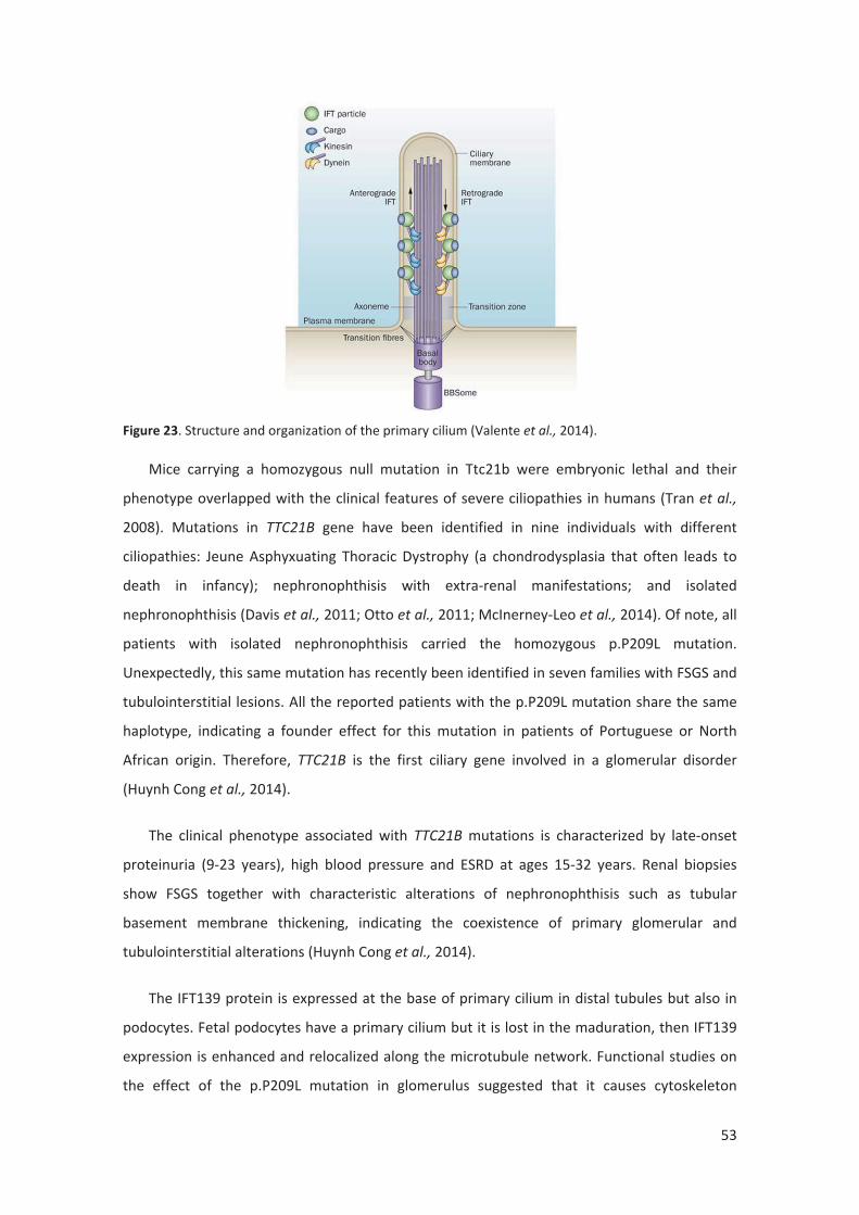
53
Figure 23. Structure and organization of the primary cilium (Valente et al., 2014).
Mice carrying a homozygous null mutation in Ttc21b were embryonic lethal and their
phenotype overlapped with the clinical features of severe ciliopathies in humans (Tran et al.,
2008). Mutations in TTC21B gene have been identified in nine individuals with different
ciliopathies: Jeune Asphyxuating Thoracic Dystrophy (a chondrodysplasia that often leads to
death in infancy); nephronophthisis with extra renal manifestations; and isolated
nephronophthisis (Davis et al., 2011; Otto et al., 2011; McInerney Leo et al., 2014). Of note, all
patients with isolated nephronophthisis carried the homozygous p.P209L mutation.
Unexpectedly, this same mutation has recently been identified in seven families with FSGS and
tubulointerstitial lesions. All the reported patients with the p.P209L mutation share the same
haplotype, indicating a founder effect for this mutation in patients of Portuguese or North
African origin. Therefore, TTC21B is the first ciliary gene involved in a glomerular disorder
(Huynh Cong et al., 2014).
The clinical phenotype associated with TTC21B mutations is characterized by late onset
proteinuria (9 23 years), high blood pressure and ESRD at ages 15 32 years. Renal biopsies
show FSGS together with characteristic alterations of nephronophthisis such as tubular
basement membrane thickening, indicating the coexistence of primary glomerular and
tubulointerstitial alterations (Huynh Cong et al., 2014).
The IFT139 protein is expressed at the base of primary cilium in distal tubules but also in
podocytes. Fetal podocytes have a primary cilium but it is lost in the maduration, then IFT139
expression is enhanced and relocalized along the microtubule network. Functional studies on
the effect of the p.P209L mutation in glomerulus suggested that it causes cytoskeleton
54
alterations that may destabilize podocyte architecture leading to FSGS (Huynh Cong et al.,
2014).
5.1.10. CRB2 gene
The CRB2 gene encodes the putative transmembrane Crumbs homolog 2 protein, a protein
required for podocyte foot process arboritzation, slit diaphragm formation, and proper
nephrin trafficking (Ebarasi et al., 2015).
Homozygous or compound heterozygous mutations in CRB2 gene have been identified by
homozygosity mapping and whole exome sequencing in four families with childhood onset (9
months 6 years) isolated SRNS and FSGS as histological lesions (Ebarasi et al., 2015). All
affected children carried a missense mutation in at least one allele, in accordance with the
embryonic lethality of Crb2 knockout mice (Xiao et al., 2011). Of note, CRB2 mutations were
also detected in five fetuses and one infant with cerebral ventriculomegaly and echogenic
kidneys with histopathological findings of congenital nephrosis (Slavotinek et al., 2015).
5.1.11. NUP107 gene
The NUP107 gene encodes the essential scaffold protein of the nuclear pore complex
NUP107. This protein facilitates the efficient transfer of macromolecules between the nucleus
and cytoplasm in a highly selective manner and plays pivotal roles in the nuclear framework
and gene expression (Strambio De Castillia et al., 2010).
Recessive mutations in NUP107 gene have been identified by whole exome sequencing in
five SRNS families (nine individuals). In four of the five families, all the individuals showed
early onset SRNS manifesting at 2 3 years of age and reached ESRD before 10 years of age. In
the remaining family, the two affected individuals had a disease onset after 10 years old, one
of them reached ESRD at 12 years old whereas the other has preserved renal function at the
current 34 years of age. In all the affected individuals renal biopsies revealed FSGS and they
were resistant to corticosteroid therapy but partially respondent to immunosuppressive drugs.
None of the five patients that underwent renal transplantation experienced recurrence of
SRNS (Miyake et al., 2015).
A total of four different mutations were found in the five families. Interestingly, the
heterozygous p.D831A mutation was common in all the five families. This mutation is likely to
be specific to East Asian because it has only been reported in Japanese population.

55
Furthermore, haplotype analysis confirmed that all the families shared the same haplotype
confirming a common ancestor for this mutation. Mutations in NUP107 are likely to cause
immature and/or hypoplastic podocytes, or at least impaired podocytes that are progressively
destroyed by increased filtration pressure after birth (Miyake et al., 2015).
5.1.12. NUP93 gene
Mutations in NUP93, encoding nuclear pore protein 93 likely involved in nuclear pore
complex assembly, have been recently identified in six families with SRNS (Braun et al., 2016).
Phenotypically, affected members had SRNS that manifested between 1 and 6 years of age
and caused ESRD between the ages of 1 and 11 years. Renal biopsy showed FSGS or DMS. In
addition, there was a renal tubular phenotype involving tubular dilation with protein casts and
interstitial cell infiltration. Electron microscopy showed partial podocyte foot process
effacement. One patient showed partial response to corticosteroids, and two patients
responded to immunosuppressive therapy with cyclosporine A (Braun et al., 2016).
Five different mutations were detected in homozygous or compound heterozygous state
including three missense mutations, one small deletion and one splicing mutation. The
mutations p.G591V and p.Y629C apparently represent European and Turkish founder alleles,
respectively. Functional studies revealed that NUP93 mutations caused disrupted nuclear pore
complex assembly and abrogated interaction with SMAD4, thus identifying aberrant SMAD
signaling as a new disease mechanism of SRNS (Braun et al., 2016).

56
5.2.Genes implicated in autosomal dominant isolated SRNS and FSGS
Table2. Genes causative of autosomal dominant isolated SRNS and FSGS.
Gene Reference MOITypical age at diagnosisof proteinuria/NS
Tyicalhistology
Age atESRD
Number of families(patients) published
WT1 Coppes et al., 1992 AD 3 59 years FSGS 10 69 years 7 (31)
ACTN4 Kaplan et al., 2000 AD 3 54 years FSGS 6 59 years 12 (>75)
TRPC6 Reiser et al., 2005 AD 2 75 years FSGS0 20 yearsafter onset
>28 (>62)
INF2 Brown et al., 2010 AD 5 72 years FSGS 13 70 years >60 (>221)
LMX1B Boyer et al., 2013 AD 5 70 years FSGS 28 70 years 8 (>28)
ARHGAP24 Akilesh et al., 2011 AD ND FSGS 12 29 years 1 (3)
ANLNGbadegesin et al.,2014
AD 9 69 years FSGS 35 75 years 2 (12)
PAX2 Barua et al., 2014 AD 7 68 years FSGS 30 58 years 7 (24)
Abbreviations: AD, autosomal dominant; ESRD, end stage renal disease; FSGS, focal segmentalglomerulosclerosis; MOI, mode of inheritance; ND, no data; NS, nephrotic syndrome.
5.2.1. WT1 gene
The WT1 gene spans 47.86 kb on chromosome 11p13 and contains 10 exons. This gene
encodes Wilms Tumor protein 1 (WT1), a zinc finger transcription factor of 449 amino acids
and 49 kDa that contains an N terminal transactivation domain (exon 1) and four zinc fingers
at the C terminal (exons 7 10). WT1 encodes numerous isoforms, which are products of
alternative translation start sites, alternative splicing, and RNA editing. Of particular interest
are WT1(+KTS) and WT1( KTS) variants, which differ by the presence of three amino acids (KTS)
between the zinc fingers three and four. The presence of this insert influences the molecular
biochemical properties of the resulting protein. While WT1( KTS) bind DNA efficiently and acts
as a transcriptional activator, WT1(+KTS) seem to have higher affinity to RNA and splicing
factors (Hammes et al., 2001).
WT1 protein plays a crucial role in kidney and genital tract development (Pritchard Jones
et al., 1990; Rivera & Haber 2005). In the fetal kidney, WT1 is abundantly expressed in areas of

57
active glomerulogenesis whereas in the adult kidney, WT1 expression persists in podocytes
and its integrity is required for proper GFB function (Morrison et al., 2008).
Mutations in WT1 gene are associated with a wide spectrum of AD syndromes in children
including Wilms tumor (Gessler et al., 1990), syndromic forms of glomerular disease and
genitourinary abnormalities: Denys Drash (Pelletier et al., 1991), Fraiser Syndromes (Klamt et
al., 1998) and WAGR syndrome (Wilms tumor, Aniridia, Genitourinary abnormalities, and
mental Retardation)(Miller et al., 1964), and isolated SRNS (Jeanpierre et al., 1998;
Schumacher et al., 1998; Ito et al., 1999; Denamur et al., 2000; Dharnidharka et al., 2001; Ito et
al., 2001). Interestingly, WT1 associated disorders may include SRNS either as an initial
symptom or as a later development in a patient diagnosed on the basis of extrarenal features.
The type and localization of the mutations has been clearly associated with the renal and
extrarenal phenotypes. Germline alterations, usually truncating mutations, located throughout
the entire coding sequence predispose to Wilms tumor. Heterozygous missense or truncating
mutations in exons 8 and 9, mostly de novo, cause Denys Drash syndrome or isolated SRNS
(Mucha et al., 2006). These mutations affect the DNA and RNA binding affinity of the second
and third zinc finger domains. Heterozygous mutations in the donor splice site of intron 9
result in Fraiser Syndrome. These mutations lead to a reduction of the +KTS isoform causing
the alteration of the normal ratio of +KTS/ KTS isoforms in the cell (Barbaux et al., 1997).
Finally, large genomic rearrangement affecting chromosome 11p13 may disrupt WT1 among
several genes, resulting in WAGR syndrome.
The renal phenotype associated with WT1 mutations is characterized by an early onset
SRNS and rapid progression to ESRD. In patients with mutations in exons 8 and 9 affecting the
DNA binding capacity (classically associated with Denys Drash Syndrome) NS presents in the
first months of life, may be preceded by isolated proteinuria, and is always resistant to
corticosteroid therapy. Progression to ESRD occurs most often before 4 years of age, and no
recurrence is observed after renal transplantation (Habib et al., 1985). Histological findings
show DMS in most patients. In contrast, patients with KTS mutations, causing Fraiser
syndrome, have a childhood onset proteinuria usually between 2 and 6 years of age, almost
invariably FSGS on biopsy, and slow progression leading to ESRD in adolescence or early
adulthood.
The renal phenotype of patients carrying other exonic mutations is determined by the type
of mutation. Most subjects with truncating mutations develop Wilms tumor early in life

58
whereas SRNS occurs later, typically in the second decade after nephrectomy. Missense
mutations affecting amino acids in non DNA binding positions are characterized by an
intermediate renal phenotype manifesting within the first 5 years of life, usually presenting
with DMS on biopsy, and progressing to ESRD at approximately 10 years old.
Female patients (46, XX) carrying either missense or splice site WT1 mutations commonly
do not exhibit genital anomalies, have normal puberty, and therefore present with isolated
sporadic SRNS (Ruf, Schultheiss, et al., 2004; Mucha et al., 2006). Missense mutations in exon 8
or 9 (p.R458Q, S461F, or R471K) affecting the second and third zinc fingers have been
identified in families with isolated AD FSGS, including males without genital abnormalities or
developmental tumors who transmitted the mutation (Coppes et al., 1992; Benetti et al., 2010;
Guaragna et al., 2013; Zhu et al., 2013; Hall et al., 2015).
Recently, a detailed study compared 61 patients with WT1 related SRNS with 700 SRNS
patients negative for WT1 mutations and revealed several genotype phenotype correlations.
WT1 mutated patients presented more frequently with chronic kidney disease and
hypertension at diagnosis and exhibit more rapid disease progression compared to WT1
negative patients. FSGS is the most prevalent histopathological finding in WT1 nephropathy
but DMS is largely specific for WT1 disease and is present in 34% of DMS cases (Lipska et al.,
2014). This finding goes against the results obtained by Gbadegesin et al. who identified
mutations in PLCE1 in 28.6% of families with DMS whereas mutations in WT1 were only found
in 8.5% of these families (Gbadegesin et al., 2008). Extrarenal phenotypes are almost exclusive
toWT1 and mostly comprise: sex reversal and/or urogenital abnormalities (52%), Wilms tumor
(38%), and gonadoblastoma (5%) (Lipska et al., 2014).
5.2.2. ACTN4 gene
A genome wide scan performed in a 100 member kindred allowed Mathis et al. to map the
first locus of AD FSGS on chromosome 19q13 (Mathis et al., 1992; Mathis et al., 1998). Linkage
analysis including additional families helped to reduce the size of the region and led to the
identification of three nonconservative missense mutations in the ACTN4 gene (Kaplan et al.,
2000). ACTN4 gene comprises 21 exons and encodes the actinin 4 protein, a protein of 911
amino acids and about 105 kDa that belongs to the spectrin gene superfamily. actinin 4 is an
ubiquitously expressed actin binding protein, that interconnects actin filaments in podocyte
foot processes (Ichimura et al., 2003). Its structure is a head to tail antiparallel homodimer
with the following parts: two calponin homologous domains (CH1, CH2) with actin binding sites

59
in the N terminal, followed by four spectrin repeats and two EF hand domains that can bind
Ca2+ ions in the C terminal (Baron et al., 1987; Imamura et al., 1988; Leinweber et al., 1999).
Affected cases typically present with proteinuria starting in the adolescence or later, with
FSGS lesions on kidney histology, and slowly progress to ESRD in the fifth decade of life. Also,
heterozygous ACTN4 mutations have seldom been reported in childhood onset NS (Kaplan et
al., 2000; Weins 2005; Pollak et al., 2007; Choi et al., 2008). Disease incomplete penetrance
and germline mosaicism has been described (Weins 2005; Choi et al., 2008).
Mutations in the ACTN4 gene were reported to account for approximately 2% of AD FSGS
(Barua et al., 2013). However, recent studies in large cohorts did not identify any patient
carrying ACTN4 mutations, suggesting an even lower contribution of mutations in this gene to
AD FSGS (Sadowski et al., 2014).
To date, 16 mutations have been reported. Most of them are missense mutations involving
residues within or next to the actin binding site of actinin 4 (Kaplan et al., 2000; Ichimura et
al., 2003; Weins 2005; Barua et al., 2013; Giglio et al., 2015). Some of these mutations have
been shown to increase the affinity of actinin 4 to F actin and/or induce actinin 4
mislocalization and the formation of actinin 4 and F actin aggregates around the nucleus
with subsequent impairment of podocyte spreading and motility (Kaplan et al., 2000; Weins
2005; Yao et al., 2004; Michaud 2003; Michaud et al., 2006). Knock in and knockout mouse
models and in vitro data suggest both gain of function and loss of function mechanisms (Yao
et al., 2004).
5.2.3. TRPC6 gene
TRPC6 spans approximately 421 kb on chromosome 11q22 and contains 13 exons. This
gene encodes the short transient receptor potential canonical 6 (TRPC6), a protein of 931
amino acids and around 106 KDa. TRPC channels are tetrameric proteins composed of six
transmembrane spanning domains and N and C terminal facing the cytosol (Venkatachalam &
Montell 2007). The fifth and sixth transmembrane domains line the pore of the ion channel
(Hofmann et al., 2002) (Figure 26). TRPC6 is a receptor operated non selective cation channel,
which allows intracellular Ca2+ to flow into the cell in response to activation by DAG secondary
to phospholipase C mediated signal coming from stimulation of specific cell surface receptors
(Hofmann et al., 1999; Montell 2005). In the kidney, TRPC6 is expressed in renal tubules and in
glomeruli, with predominance in podocytes. TRPC6 co localizes with podocin and nephrin and

immu
indica
There
et al.
the a
Figurediaphfrom
L
11q2
genes
muta
on th
Schill
p.P11
with
al. w
incom
B
AD FS
signif
Child
Ars, e
2012
Liako
unoprecipita
ating that th
efore, TRPC6
, 2005). In a
ctin cytoske
e 24. Structurragm. Ank, aMachuca et a
Linkage analy
1 q22 that li
s as well as
ations by dire
he basis of re
ing 1997). S
12Q in exon
an autosom
ho identified
mplete penet
Based on the
SGS and vari
ficant numb
hood onset
et al., 2009;
). Renal biop
opoulos et a
tion studies
he channel
6 is critical in
addition, Mo
leton in the
re and locatioankyrin repeaal., 2009; Drye
ysis of a 39
inked to the
multiple no
ect sequenci
eports of det
Sequencing
2 that segre
al dominant
d five familie
trance was o
ese two stud
iable rate of
ber of spor
SRNS has b
Heeringa e
psy typically
al., 2011) an
showed phy
is integrate
n adjusting th
oller et al. de
podocyte (M
n of the TRPCat; cc, coileder & Reiser 20
9 member f
disease (Wi
vel and pred
ng. After exa
tection of TR
of the 13
egated with t
t pattern (W
es with mut
observed (Re
ies the phen
progression
radic and c
een reporte
t al., 2009;
showed FSG
d MCD hav
ysical intera
d into the s
he cationic i
emonstrated
Möller et al.,
C6 protein incoil; CIRB, ca10).
family with
inn et al., 19
dicted genes
amination of
RPC6 mRNA i
exons of th
the disease i
Winn 2005). A
ations in TR
eiser et al., 2
notype assoc
n to ESRD (W
childhood on
ed in nine pa
Buscher et a
GS but collaps
e been repo
ction betwe
signaling co
on flux close
d that TRPC6
2007).
the slit diaphalmodulin and
FSGS identif
999). This reg
s, which wer
f 42 genes, T
in the kidney
his gene ide
in all availab
Additional wo
RPC6 that seg
005).
ciated with T
Winn 2005; Re
nset FSGS c
atients carry
al., 2012; Gi
sing glomeru
orted (Gigan
en TRPC6 an
mplex at th
e to the slit d
6 is function
ragm. FP, food IP3 recepto
fied a locus
gion contain
re systematic
TRPC6 emerg
y (Riccio et a
entified a m
ble family me
ork was repo
gregated wit
TRPC6 mutat
eiser et al., 2
cases have
ying TRPC6 m
gante et al.,
ulopathy (Gig
nte et al., 20
nd these pro
he slit diaph
diaphragm (R
nally connect
ot processes; Sor binding (ad
s on chromo
ned several k
cally screene
ged as a cand
al., 2002; Ga
missense mut
embers cons
orted by Rei
th FSGS, alth
tions is late
2005). Howe
been iden
mutations (S
, 2011; Mir
gante et al.,
011; Mottl e
60
oteins,
ragm.
Reiser
ted to
SD, slitdapted
osome
known
ed for
didate
rcia &
tation
sistent
ser et
hough
onset
ever, a
tified.
Santín,
et al.,
2011;
et al.,

61
2013). This high phenotypic variability can also occur within members of the same family even
with mutation carriers showing either no signs or mild signs of the disease, indicating that the
disease is not fully penetrant (Reiser et al., 2005; Santín, Ars, et al., 2009). Studies in large
cohorts revealed that TRPC6 mutations account for approximately 8% of AD FSGS patients
(Sadowski et al., 2014).
To date, 19 different mutations have been reported in 22 familial/sporadic patients from
different ethnic background. Three mutations are located in the ANK1, two in the ANK2, one in
ANK3, one in ANK4, and five additional N terminal mutations are located outside of the
predicted ANK domains (Reiser et al., 2005; Winn 2005; Santín, Ars, et al., 2009; Gigante et al.,
2011; Heeringa et al., 2009; Buscher et al., 2012; Hofstra et al., 2013; Mir et al., 2012). Seven
other TRPC6 mutations were located to the C terminal cytoplasmic tail (Reiser et al., 2005;
Santín, Ars, et al., 2009; Gigante et al., 2011; Zhu et al., 2009; Mottl et al., 2013), four of them
mapping to a predicted coiled coil domain at the C terminal (Gudermann et al., 2004). No
mutations have been identified in the transmembrane, extracellular or pore forming regions of
the channel.
Several of these mutations are gain of function mutations leading to increased channel
activity by increasing calcium current amplitudes or by delaying channel inactivation. The
p.N143S, p.S270T and p.K874* mutations did not modify the calcium influx during the
functional assays, suggesting that an abnormality other than increased current amplitude is
the cause of disease in individuals with these mutations. The several possibilities include
altered channel regulation (despite normal amplitude), altered interaction with other slit
diaphragm proteins and altered protein turnover (Reiser et al., 2005).
5.2.4. INF2 gene
The INF2 gene spans approximately 33 kb on chromosome 14q32 and contains 23 exons.
This gene encodes the inverted formin 2 (INF2), a protein of 1249 amino acids and around 136
KDa that belongs to formin family. Formins are widely expressed proteins governing several
dynamic events that require remodeling of the actin cytoskeleton such as cell polarity, cell and
tissue morphogenesis, and cytokinesis (Boyer, Benoit, et al., 2011). The INF2 protein has the
unique ability to accelerate both polymerization and depolymerization of actin (Chhabra et al.,
2009). This protein contains the following domains, from N terminal to C terminal: the
diaphanous inhibitory domain (DID), the formin homology domains FH1 and FH2, and the
diaphanous regulatory domain (DAD). The FH2 domain directly mediates actin assembly, and

62
the FH1 domains accelerate filament elongation. Interaction between DID and DAD blocks the
ability of FH2 to interact with actin, inhibiting actin depolymerization but not actin
polymerization. This step is inhibited by the binding of Rho GTPases to DID impeding its
interaction with DAD (Boyer, Benoit, et al., 2011; Gbadegesin et al., 2012).
Mutations in INF2 gene were identified as a cause of AD FSGS by Brown et al. (Brown et al.,
2010). Studies in large cohorts described that INF2 mutations account for approximately 9%
17% of familial AD FSGS and <1% of sporadic cases (Boyer, Benoit, et al., 2011; Gbadegesin et
al., 2012; Barua et al., 2013). Mutations in this gene are also the main cause of Charcot Marie
Tooth disease with FSGS (Boyer, Nevo, et al., 2011).
The clinical phenotype of individuals with INF2 mutations is characterized by moderate
proteinuria, without overt NS, manifesting at early adolescence or adulthood that is
progressive and often led to ESRD. However, nephrotic range proteinuria and full blown NS at
onset have been described. Microscopic hematuria and hypertension at onset have been
noted in some individuals. Most individuals have an onset of the disease in the second to
fourth decade of life with onset of renal failure in the third decade (Barua et al., 2013) but
significant variability has been reported. Considering all the patients published to date, age at
proteinuria onset ranges from 5 years to 72 years and age at ESRD onset from 13 to 70 (Brown
et al., 2010; Boyer, Benoit, et al., 2011). Intra familial variability in the clinical expression of the
disease is also wide with clinically unaffected members carrying the same mutation that their
affected son or daughter, consistent with incomplete penetrance that is often seen in AD
diseases (Lee et al., 2011; Boyer, Benoit, et al., 2011; Gbadegesin et al., 2012).
All the mutations described to date (more than 40) are highly conserved residues within
the DID domain. Most of them cluster in exon 4 and 2, accounting for approximately 80% and
10% of mutations, respectively. Mutations in exons 3 and 6 have been reported in two and one
families, respectively (Barua et al., 2013; Sanchez Ares et al., 2013).
Many of the mutations reported are recurrent with certain codons appearing to be
hotspots, such as Arg218 or Arg214. Mutations in Arg218 have been described in eight
unrelated families, six of them carry the p.R218Q mutation (Brown et al., 2010; Boyer, Benoit,
et al., 2011; Gbadegesin et al., 2012; Barua et al., 2013) whereas the remaining 2 families carry
the p.R218W mutation (Barua et al., 2013). Mutations in Arg214 have been described in eight
families four of them carry the p.R214H (Brown et al., 2010; Gbadegesin et al., 2012) mutation
while the remaining four families carry the p.R214C mutation (Boyer, Benoit, et al., 2011;

63
Gbadegesin et al., 2012; Barua et al., 2013). A possible founder effect of these mutations was
studied by haplotype analysis but no common ancestral disease associated haplotype was
found (Boyer, Benoit, et al., 2011).
INF2 mutations have been suggested to cause defects in actin mediated podocyte
structural maintenance and repair (Brown et al., 2010). Most of the mutations reported have
been shown to cause mislocalization of INF2 in podocyte culture and some of them also inhibit
the interaction between INF2 DID and mDias DAD (mouse Diaphanous related formins),
suggesting that changes are functionally important for regulation of actin cytoskeleton
(Gbadegesin et al., 2012).
5.2.5. LMX1B gene
The LMX1B gene encodes the LIM homeodomain containing transcription factor 1B
(LMX1B) that is essential during development. Mutations in LMX1B cause Nail Patella
syndrome associating dysplasia of the patellae, nails and elbows, iliac horns, glaucoma and, in
some cases, FSGS with specific lesions characterized by the presence of type III collagen fibrils
in the GBM by electron microscopy (Dreyer et al., 1998). However, LMX1B mutations have
been identified in four families (14 individuals) with AD FSGS but no glomerular membrane
anomaly suggestive of nail patella like renal disease by electron microscopy and no extrarenal
features (Boyer et al., 2013; Edwards et al., 2015). These patients presented significant
albuminuria between 5 and 70 years of age, and some of them reached ESRD between 26 and
70 years of age. The three different mutations detected (p.R246Q, p.R246P and p.R249Q) are
located in the LMX1B homeodomain. The affected residues are likely to play an important role
in strengthening the interaction between the homeobox domain of LMX1B and DNA, by
holding interactions with the DNA (Boyer et al., 2013).
5.2.6. ANLN gene
The ANLN gene encodes for anillin, an F actin binding protein that is enriched in the
cytoskeleton. Heterozygous missense mutations in ANLN have recently been described in two
families with AD FSGS by genome wide linkage study and whole exome sequencing. The age at
proteinuria onset varied from 9 to 69 years and ESRD occurred between 35 and 75 years
(Gbadegesin et al., 2014).

64
5.2.7. PAX2 gene
PAX2 is a transcription factor expressed from the fourth week of gestation in the kidney
and play a critical role in kidney development (Nakanishi & Yoshikawa 2003). Mutations in this
gene have typically been associated with congenital abnormalities of the kidney and urinary
tract (CAKUT) as part of a syndrome known as renal coloboma or papillorenal syndrome
(Sanyanusin et al., 1995). Barua et al. identified a PAX2 mutation in a family with AD FSGS by
exome sequencing. Sequencing of the PAX2 gene in FSGS families revealed a PAX2 mutation in
7 of the 175 families analyzed (24 patients), suggesting that mutations in PAX2 might account
for approximately 4% of families with FSGS (Barua et al., 2014).
The affected patients had various degrees of proteinuria diagnosed between 7 and 68
years old (mostly second to fourth decades) and FSGS on biopsy. ESRD occurred in nine
patients between 30 and 58 years old. Kidney ultrasound in patients with PAX2 related FSGS
either was normal or could reveal echoic or small kidneys, dilated renal pelvis, or calyceal
diverticulum. Most patients did no exhibit any extrarenal symptom, but renal coloboma was
diagnosed retrospectively in one family (Barua et al., 2014).
Interestingly, a patient with a PAX2 mutation together with the homozygous p.R229Q in
the NPHS2 gene has been reported. This patient presented with sub nephrotic proteinuria at 7
months. At 20 years old he presented nephrotic range proteinura, FSGS on renal biopsy and
his renal function progressively deteriorated reaching ESRD at 33 years of age. No recurrence
of the disease was observed after one year of renal transplantation. Additionally, he presented
ocular abnormalities in accordance with the ocular abnormalities seen in renal coloboma (Kerti
et al 2013).

65
5.3.Genes implicated in syndromic forms of SRNS and FSGS
Gene Reference MOITypical age at diagnosis
of proteinuria/NSTyical histology Age at ESRD Syndrome: recurrent extrarenal involvement
Nº families (patients)
published
SRNS with genitourinary features
WT1 Pelletier et al., 1991 AD 0 – 10 years DMS 0 – 15 years Dennys Drash sd: Wilms tumor in both genders, genital anomaliesin 46,XY patients
>170
WT1 Klamt et al., 1998 AD 7 months to 34 years FSGS 5 to 34 years Fraiser sd: Pseudohermaphroditism, gonadoblastoma in 46,XYpatients
>60
SRNS with osseous dysplasia
LMX1B Dreyer et al., 1998 AD 9 – 76 yearsFSGS, NPS
nephropathy22 52 Nail Patella sd: hypoplastic nails/patellae, iliac horns >35 (>54)
SMARCAL1 Boerkoel et al., 2002 AR 2 – 12 years FSGS 3.8 – 22 yearsSchimke Immuno osseous dysplasia: spondyloepiphyseal dysplasia,T cell deficiency, cerebral ischemia, broad nasal tip,hyperpigmented macules
>90
Mitochondriopathies with SRNS
MTTL1 Goto et al., 1990Matern
al5 – 47 years FSGS 13 – 51 years MELAS: myopathy, encephalopathy, lactic acidosis, and stroke like
episode, diabetes, and deafness>30 (>45)
COQ2 Quinzii et al., 2006 AR 0 – 30 months FSGS 0 to 5 years COQ10 deficiency: encephalomyopathy, hypotonia, seizures,lactate acidosis
10 (13)
PDSS2 Lopez et al., 2006 AR 0 23 months ND No data COQ10 deficiency: encephalomyopathy, hypotonia, seizures,lactate acidosis
3 (3)
COQ6 Heeringa et al., 2011 AR 2 months to 6.4 years FSGS5 months to
9.3 yearsCOQ10 deficiency: sensorineural deafness, seizure 8 (16)
SCARB2 Balreira et al., 2008 AR 9 to > 59 years FSGS 9 to > 59 years Progressive myoclonic epilepsy, cerebral and cerebellar atrophy,peripheral neuropathy, hearing impairment
18 (24)
SRNS with cutaneous features

66
Gene Reference MOITypical age at diagnosis of
proteinuria/NSTyical histology Age at ESRD Syndrome: recurrent extrarenal involvement
Nº families (patients)
published
ITGB4 Kambham et al., 2000 AR 2 months to 10 years FSGS NA Junctional epidermolysis bullosa, nail dystrophy, recurrenthemorrhagic cystitis, desquamation of laryngeal mucosa
2 (2)
LAMB3 Hata et al., 2005 AR 4 months DMSNA (5
months*)Epidermolysis bullosa, nail dystrophy 1 (1)
ITGA3 Nicolaou et al., 2012 AR 0 4 years FSGS2 weeks to 4
yearsInterstitial lung disease, epidermolysis bullosa 6 (6)
CD151 Karamatic et al., 2004 AR ND NDThickening of
GBMPretibial epidermolysis bullosa, sensorineural deafness, lacrimalduct stenosis, nail dystrophy
2 (2)
SRNS with intellectual disability
ARHGDIA Gupta et al., 2013 AR 0 – 2.4 years DMS6 weeks to 3
yearsIntellectual disability 3 (6)
KANK4 Gee et al., 2015 AR 2 months FSGS NDIntellectual disability, short stature, facial dysmophism, atrial septal
defect, dilative cardiomyopathy, leukocytosis2 (2)
XPO5 Braun et al,, 2016 AR 2 years MCD ND Speech development delay 1 (1)
SRNS with other features
LAMB2Hasselbacher K et al.,
2006AR 0 – 6 years DMS 0 21 years Pierson sd: microcoria, abnormal lens, VI, hypotonia, motor delay >60 (>70)
WDR73 Colin et al., 2014 AR 5 to > 13 years FSGS 5 yearsGalloway Mowat sd: secondary microcephaly, intellectualdeficiency, cortical and cerebellar atrophy, facial dysmorphy, opticatrophy
2 (3)
INF2 Boyer et al., 2011 AD 10 – 46 years FSGS 12 – 47 years Charcot–Marie–Tooth neuropathy, deafness 27 (44)
NUP205 Braun et al., 2016 AR 2 3 years FSGS 7 years Bicuspid aortic valve, aortic insufficiency, aortic root enlargement 1 (2)
Abbreviations: AD, autosomal dominant; AR, autosomal recessive, diffuse mesangial sclerosis; ESRD, end stage renal disease; FSGS, focal segmental glomerulosclerosis;MCD, minimal change disease; MOI, mode of inheritance; ND, no data; NPS, Nail patella syndrome; NS, nephrotic syndrome; sd, syndrome.

67
6. CLINICAL UTILITY OF GENETIC TESTING
The identification of the genetic cause of SRNS has several clinical implications:
Provides an unequivocal molecular diagnosis for the patients and their family
members and may diagnose previously unrecognized affected family members.
Predicts resistance to corticosteroid and/or immunosuppressive therapy. Therefore,
the unnecessary extension of corticosteroid treatment as well as the potential risk and
side effects associated with these treatments can be avoided (Weber et al., 2004; Ruf,
Lichtenberger, et al., 2004; Buscher et al., 2010; Giglio et al., 2015).
A low risk of relapse after renal transplantation, hence, living donor renal
transplantation is encouraged.
Genetic counseling (prenatal or preimplantation genetic diagnosis) for family planning
can be offered to couples with known risk for severe NS, in whom the causative
mutations have been identified in a first affected child.
Prognosis information depending on which gene is mutated and the type of mutations
considering the genotype phenotype correlations explained in the previous section.
Although these clinical implications are generally applicable to all patients with a genetic
cause of SRNS identified, specific particularities and/or exceptions have been described
depending on the mutated gene.
Most patients with NPHS1 mutations are resistant to corticosteroids and
immunosuppressive treatment. However, five patients (three families) with compound
heterozygous or homozygous missense mutations and partial remission have been reported
(Kitamura et al., 2007; Heeringa et al., 2008; Schoeb et al., 2010). Two siblings with frequent
relapsing NS that carried compound heterozygous p.C265R and p.V822M mutations in NPHS1
gene achieved remission of proteinuria without immunosuppressive treatment. Functional
studies showed that the p.C265R mutation was trapped in the endoplasmic reticulum whereas
the p.V822M mutation partially reached the plasma membrane, likely explaining their milder
disease (Kitamura et al., 2007). In the remaining three patients (two families) partial remission
due to steroid treatment was achieved (Heeringa et al., 2008; Schoeb et al., 2010).
Post transplantation recurrence in NPHS1 mutated patients is very low. However, in a
study of patients with Fin Major or Fin minor mutations (that lead to the absence of nephrin)
recurrence occurred in 25% of patients at a mean time of 12 months post transplantation. All

68
these patients were homozygous for the Fin major mutation, which leads to an early stop
codon and total absence of nephrin in the native kidney. In almost half of them antibodies
against nephrin were detected (Patrakka et al., 2002). Similarly, Kuusniemi et al reported post
transplant recurrence rate in 30% of patients homozygous for the Fin major mutation and 70%
of them showed anti nephrin antibodies (Kuusniemi 2007). Two patients with mutations
different from the Fin major and post transplant recurrence have also been reported
(Srivastava et al., 2006; Chaudhuri et al., 2012). In one of them anti nephrin antibodies were
detected (Chaudhuri et al., 2012). Thus, recurrence in these patients was considered an
immune process against the nephrin present in the graft and treatment with
immunosuppressive agents led to remission in some cases (Flynn et al., 1992).
During pregnancy, elevated AFP in the maternal serum and amniotic fluid is indicative of
CNS. However, elevated AFP is not a specific marker of CNS as it has also been observed in
NPHS1 heterozygous fetal carriers and in a fetus with Dennys Drash syndrome (Devriendt et
al., 1996; Patrakka et al., 2002). Therefore, prenatal genetic diagnosis is required to confirm
CNS before considering medical termination of pregnancy.
NPHS2mutated patients do not generally respond to corticosteroid or immunosuppressive
therapy, but a partial reduction of proteinuria has been reported with cyclosporine A in eight
patients (Ruf, Lichtenberger, et al., 2004; Malina et al., 2009; Machuca, Hummel, et al., 2009).
However, complete remission in SRNS caused by NPHS2 mutations have not been reported
(Ruf, Lichtenberger, et al., 2004; Buscher et al., 2010). Post transplant disease recurrence is
significantly low in patients with NPHS2 mutations than in patients with without mutations,
4.5% vs. 28.5%, respectively according to a recent study in a large international cohort
(Trautmann et al., 2015). However, post transplant disease recurrence has been described in
12 patients (Carraro et al., 2002; Bertelli et al., 2003; Billing et al., 2004; Weber et al., 2004;
Hocker et al., 2006; Becker Cohen et al., 2007; Trautmann et al., 2015). The most common
mutations in these patients was the p.R138* and the p.R138Q. There is no evidence of podocin
antibodies in these patients, thus the pathomechanism of disease recurrence in NPHS2
mutated patients remains unknown and is probably multifactorial (Holmberg & Jalanko 2014).
The identification of PLCE1 represents the first molecular cause of NS that might response
to therapy in some individuals. Two children responded to treatment with corticosteroids or
cyclosporine A achieving a complete remission and remained free of proteinuria after several
years of follow up (Hinkes et al., 2006). Based on these findings, it has been speculated that
the arrest of glomerular development through PLCE1 mutations may be reversible by

69
treatment with corticoids or cyclosporine A via induction of a redundant mechanism such as
the activity of another phospholipase C (Hinkes et al., 2006). Thus, corticosteroid and
immunosuppressive treatment is advised in patients with PLCE1 mutations. No post transplant
recurrence has been described to date.
Patients with WT1 mutations were described to be unresponsive to therapy. However, a
partial response to immunosuppressive agents has been described in some patients
(Gellermann et al., 2010; Buscher et al., 2010). Gellermann et al. reported a favorable
response to a therapy consisting of cyclosporine A in combination with corticosteroids in 3
patients with FSGS associated with WT1 mutations (Gellermann et al., 2010). Büscher et al.
reported 2 patients who achieved a partial response to cyclosporine A: one phenotypically
female Fraiser patient (karyotype XY) affected by aWT1 splice site mutations and one patients
(karyotype XX) with isolated FSGS affected by aWT1missense mutation (Buscher et al., 2010).
In patients with WT1 mutations, the karyotype of females should be investigated in order
to exclude the 46 XY karyotype with pseudohermaphroditism. These patients should also be
screened for the development of for the development of Wilms Tumor or gonadoblastoma
(Rood et al., 2012). Genetic counseling is essential in both female and males, as they have a
50% risk to transmit the mutation. Although most WT1 mutations are de novo, parents of a
child with a WT1 mutation should be advised of a low probability of recurrence due to the
possibility of germinal mosaicism, which has an unknown likelihood.
Potential therapy is available for some rare causes of SRNS. For instance, identification of
mutations in a gene encoding enzymes of the CoQ10 biosynthesis (COQ2, COQ6, ADCK4 or
PDSS2) may warrantee experimental treatment with CoQ10 (Heeringa et al., 2011; Montini et
al., 2008) as partial response to treatment with coenzyme Q10 has been described in
individuals with SRNS and mutations in COQ2 (Montini et al., 2008), COQ6 (Heeringa et al.,
2011) and ADCK4 (Ashraf et al., 2013). Similarly, individuals with TRPC6 mutations may
potentially be amenable to treatment with calcineurin inhibitors (Schlondorff et al., 2009),
patients carrying CUBN mutations may be amenable to treatment with vitamin B12, and
individuals with ARHGDIA mutations may theoretically be responsive to the eplerenone
treatment (Gee et al., 2013).
Genetic testing in SRNS should be strongly advised in those patients with a likely a genetic
cause of the disease: 1) in CNS patients, 2) in childhood onset SRNS patients, both familial and
sporadic, 3) in adult onset patients with a family history of the disease, and 4) in patients from

70
consanguineous marriages. This advice is based on the relatively high prevalence of genetic
causes in these groups.

71
AIMS

72
73
The global aim of the present thesis was to increase our knowledge on the contribution of
genetics to the pathogenesis and clinical outcome of idiopathic membranous nephropathy
(IMN) and SRNS/FSGS.
The first part of this thesis was focused on the association of genetic polymorphisms: SNPS
and Copy number variants (CNVs), with the risk to develop IMN and its clinical course. Specific
aims of this part were:
To validate the association of HLA DQA1 and PLA2R1 genes with the risk to develop
IMN in Spanish population.
To test the putative association of CNVs within FCGR3A and FCGR3B genes with
susceptibility to IMN in Spanish population.
To assess the contribution of these genetic factors to predict the clinical outcome and
renal function decline.
The second part of this thesis aimed to analyze the genetic causes of SRNS/FSGS as well as
to identify genetic modifiers of disease severity. Particular aims of this part were:
To improve the genetic testing of SRNS/FSGS.
To assess the putative influence of mutations in multiple glomerular genes on
SRNS/FSGS phenotype variability.
To study the causative and modifying role of TTC21B gene
The final aim of the present thesis was to develop an efficient and comprehensive genetic
diagnosis including all known genes causative of glomerular and cystic inherited kidney
diseases.

74