Nutritional changes in piglets and morphophysiologic ...
19
155 Vet. Méx., 43 (2) 2012 Cambios nutrimentales en el lechón y desarrollo morfofisiológico de su aparato digestivo Nutritional changes in piglets and morphophysiologic development of their digestive tract Recibido el 22 de mayo de 2011 y aceptado el 28 de noviembre de 2011. *Facultad de Ciencias Naturales, Universidad Autónoma de Querétaro, Av. de las Ciencias s/n, Juriquilla, Querétaro, 76230, México. **CENID Fisiología INIFAP, km 1, carretera a Colón, Ajuchitlán, Colón, 76280, Querétaro. ***Alumnos del Doctorado en Ciencias Biológicas, Facultad de Ciencias Naturales, Universidad Autónoma de Querétaro, Av. de las Ciencias s/n, Juriquilla, Querétaro, 76230, México. †Alumna de la Maestría en Recursos Bióticos Facultad de Ciencias Naturales, Universidad Autónoma de Querétaro, Av. de las Ciencias s/n, Juriquilla, Querétaro, 76230, México. Responsable de correspondencia: Tércia Cesária Reis de Souza, Tel.: (442) 1921200, ext. 5391, Fax: (442) 2342958, correo electrónico: [email protected] Abstract This review shows the development of the pig gastrointestinal tract (GIT) since intrauterine life. Immediately after birth, the GIT begins another important stage in its ontogenesis, which ensures for the piglets to have access to the protective substances in colostrum and milk nutrients. Another important stage in the development of the GIT begins after weaning. In general, life changes in the days around weaning leading to a decrease in feed intake, which, in turn, leads to nutritional stress that results in the atrophy of intestinal villi, diminishing the piglets ability to digest and absorb nutrients necessary for growth. The small intestine loses weight due to a net loss of intestinal mucosa. The decrease in voluntary feed intake also produces a reduction in both pancreatic enzyme activity, due to lack of substrate, and brush border enzyme activity, due to physical loss of enterocytes in the small intestine and to lack of substrate. Once the animals begin to eat solid food the organism goes through an adaptation process to all the adverse factors previously mentioned and GIT growth and digestive function are restored. During this process, the amount of feed provided to the newly weaned piglet and the ingredients used play an important role in GIT maturation. Key words: PIGLET NUTRITION, WEANING, DIGESTIVE PHYSIOLOGY, GASTROINTESTINAL TRACT DEVELOPMENT. Resumen En esta revisión se muestra el desarrollo que tiene el tracto gastrointestinal (TGI) desde la vida intrauterina. Inmediatamente después del nacimiento el TGI inicia otra fase importante de su ontogénesis, la cual garantiza el acceso de los lechones a sustancias protectoras del calostro y a los nutrimentos de la leche. Otra etapa de importancia en el desarrollo del TGI se da después del destete, en general, en los días alrededor del mismo se presentan cambios en la vida del lechón los cuales conducen a una disminución en el consumo de alimento por la confluencia de una serie de factores que llevan a un estrés nutricional, y tiene como consecuencia la atrofia en las vellosidades intestinales, disminuyendo la capacidad del lechón para digerir y absorber los nutrimentos necesarios para su buen crecimiento. El peso del intestino delgado se ve afectado debido a una pérdida neta de mucosa intestinal. La disminución en el consumo voluntario también produce una reducción en la ac- tividad de las enzimas pancreáticas, por la falta del sustrato necesario para su acción, y de las enzimas de borde en cepillo, en primer lugar por una pérdida física de enterocitos a nivel del intestino delgado y, en segundo lugar, por una ausencia de sustrato. Una vez que los animales empiezan a consumir alimento sólido, el organismo pasa por un proceso de adaptación a todos los factores adversos antes mencionados y los animales comienzan a recuperar el crecimiento del TGI y con ello sus funciones digestivas. En este proceso, la cantidad de alimento que se proporciona, así como las materias primas que lo componen, tienen un papel importante en la maduración del TGI de los lechones recién destetados. Palabras clave: NUTRICIÓN DE LECHONES, DESTETE, FISIOLOGÍA DIGESTIVA, DESARROLLO DEL TRACTO GASTROINTESTINAL. Tércia Cesária Reis de Souza* Gerardo Mariscal Landín** Konisgmar Escobar García*** Araceli Aguilera Barreyro*** Aline Magné Barrón†
Transcript of Nutritional changes in piglets and morphophysiologic ...
155Vet. Méx., 43 (2) 2012
Cambios nutrimentales en el lechón y desarrollo morfofisiológico de su aparato digestivo
Nutritional changes in piglets and morphophysiologic development of their digestive tract
Recibido el 22 de mayo de 2011 y aceptado el 28 de noviembre de 2011.*Facultad de Ciencias Naturales, Universidad Autónoma de Querétaro, Av. de las Ciencias s/n, Juriquilla, Querétaro, 76230, México.**CENID Fisiología INIFAP, km 1, carretera a Colón, Ajuchitlán, Colón, 76280, Querétaro.***Alumnos del Doctorado en Ciencias Biológicas, Facultad de Ciencias Naturales, Universidad Autónoma de Querétaro, Av. de las Ciencias s/n, Juriquilla, Querétaro, 76230, México.†Alumna de la Maestría en Recursos Bióticos Facultad de Ciencias Naturales, Universidad Autónoma de Querétaro, Av. de las Ciencias s/n, Juriquilla, Querétaro, 76230, México.Responsable de correspondencia: Tércia Cesária Reis de Souza, Tel.: (442) 1921200, ext. 5391, Fax: (442) 2342958, correo electrónico: [email protected]
Abstract
This review shows the development of the pig gastrointestinal tract (GIT) since intrauterine life. Immediately after birth, the GIT begins another important stage in its ontogenesis, which ensures for the piglets to have access to the protective substances in colostrum and milk nutrients. Another important stage in the development of the GIT begins after weaning. In general, life changes in the days around weaning leading to a decrease in feed intake, which, in turn, leads to nutritional stress that results in the atrophy of intestinal villi, diminishing the piglets ability to digest and absorb nutrients necessary for growth. The small intestine loses weight due to a net loss of intestinal mucosa. The decrease in voluntary feed intake also produces a reduction in both pancreatic enzyme activity, due to lack of substrate, and brush border enzyme activity, due to physical loss of enterocytes in the small intestine and to lack of substrate. Once the animals begin to eat solid food the organism goes through an adaptation process to all the adverse factors previously mentioned and GIT growth and digestive function are restored. During this process, the amount of feed provided to the newly weaned piglet and the ingredients used play an important role in GIT maturation.
Key words: PIGLET NUTRITION, WEANING, DIGESTIVE PHYSIOLOGY, GASTROINTESTINAL TRACT DEVELOPMENT.
Resumen
En esta revisión se muestra el desarrollo que tiene el tracto gastrointestinal (TGI) desde la vida intrauterina. Inmediatamente después del nacimiento el TGI inicia otra fase importante de su ontogénesis, la cual garantiza el acceso de los lechones a sustancias protectoras del calostro y a los nutrimentos de la leche. Otra etapa de importancia en el desarrollo del TGI se da después del destete, en general, en los días alrededor del mismo se presentan cambios en la vida del lechón los cuales conducen a una disminución en el consumo de alimento por la confluencia de una serie de factores que llevan a un estrés nutricional, y tiene como consecuencia la atrofia en las vellosidades intestinales, disminuyendo la capacidad del lechón para digerir y absorber los nutrimentos necesarios para su buen crecimiento. El peso del intestino delgado se ve afectado debido a una pérdida neta de mucosa intestinal. La disminución en el consumo voluntario también produce una reducción en la ac-tividad de las enzimas pancreáticas, por la falta del sustrato necesario para su acción, y de las enzimas de borde en cepillo, en primer lugar por una pérdida física de enterocitos a nivel del intestino delgado y, en segundo lugar, por una ausencia de sustrato. Una vez que los animales empiezan a consumir alimento sólido, el organismo pasa por un proceso de adaptación a todos los factores adversos antes mencionados y los animales comienzan a recuperar el crecimiento del TGI y con ello sus funciones digestivas. En este proceso, la cantidad de alimento que se proporciona, así como las materias primas que lo componen, tienen un papel importante en la maduración del TGI de los lechones recién destetados.
Palabras clave: NUTRICIÓN DE LECHONES, DESTETE, FISIOLOGÍA DIGESTIVA, DESARROLLO DEL TRACTO GASTROINTESTINAL.
Tércia Cesária Reis de Souza* Gerardo Mariscal Landín**Konisgmar Escobar García*** Araceli Aguilera Barreyro***
Aline Magné Barrón†
156
Introducción
The different stages of development of the gastrointestinal tract (GIT) were reviewed by Cranwell,1 who describes that during fetal
life the GIT of the pig, and other mammals, begin its development through three stages: first, there is a period of proliferation, growth and morphogenesis; in the second, there is differentiation of epithelial cells; and, in the third, functional maturation. Immediately after birth, with the consumption of colostrum and milk, another important stage in the ontogeny of the GIT occurs; this is responsible for providing protective substances to newborns through endocytosis of immunoglobulins from colostrum along with the metabolites resulting from the processes of digestion and absorption of milk nutrients.2
At weaning, switching from milk to a solid diet based on starch and vegetable proteins, makes the TGI go through a long process of adaptation, because this was not ready to digest such nutriments.3 This situa-tion creates morphologic and functional changes in the GIT that can cause disturbances in feed intake and alterations in the digestive process, preventing the animal meets its requirements for protein and energy, which slow down its initial growth.4 This process occurs mainly during the first week after weaning. Later, the development of the digestive tract is closely linked to the consumption of solid feed. Animals with a high-er feed intake and thus, more energy, have a greater growth of the stomach, pancreas, small intestine and liver as these organs consume approximately 50% of the total energy.5,6
Extensive studies on digestive physiology and the effect of nutrition on the GIT of weaned piglets were done between the 70’s and 90’s. For this reason, this review will cite many papers published in those years. In recent years, from the knowledge generated in these studies, the feed industry has developed high quality starter feed to minimize alterations in a poorly developed digestive system, stimulate voluntary intake and reduce the energy cost of the adaptation process in post-weaning piglets. It is recommended that start-er diets should be highly digestible to obtain the most efficient use possible, and not jeopardize the health and development of young animals. Given the impor-tance of the topic, the objective of this study was to review the major changes that occur in the GIT dur-ing the initial phase of the piglet life, and its relation to nutritional aspects that stimulate or inhibit its de-velopment.
Introducción
Las diferentes fases de desarrollo del tracto gastrointestinal (TGI) fueron revisadas por Cranwell,1 quién describe que durante la vida
fetal el TGI del cerdo, y de otros mamíferos, empieza su desarrollo pasando por tres fases: en la primera, se da un periodo de proliferación, crecimiento y morfogénesis; en la segunda, se presenta la diferenciación de las células epiteliales; y, en la tercera, la maduración funcional. Inmediatamente después del nacimiento, con el consumo del calostro y de la leche, se establece otra etapa importante en la ontogénesis del TGI, la cual es responsable de proveer a los neonatos las sustancias protectoras a través de la endocitosis de las inmunoglobulinas presentes en el calostro, así como los metabolitos resultantes de los procesos de digestión y absorción de los nutrimentos de la leche.2
Al destete, el cambio de la leche materna a una dieta sólida basada en almidón y proteínas de origen vegetal, hace que el TGI pase por un largo proceso de adaptación, debido a que este no estaba preparado para digerir dichos nutrimentos.3 Esta situación gene-ra cambios morfológicos y funcionales en el TGI que pueden causar trastornos en el consumo de alimento y alteraciones en el proceso digestivo, impidiendo que el animal cubra sus requerimientos de proteína y ener-gía, lo que dificulta su crecimiento inicial.4 Este proce-so ocurre principalmente durante la primera semana posdestete; posteriormente, el desarrollo del aparato digestivo está íntimamente ligado con el consumo de alimento sólido. Los animales que consumen una mayor cantidad de alimento y, por ende, más energía, tienen un mayor crecimiento del estómago, páncreas, intestino delgado e hígado, ya que estos órganos con-sumen aproximadamente 50% de la energía total.5,6
Durante el periodo comprendido entre los años setenta y noventa se intensificaron los estudios sobre fisiología digestiva y el efecto de la nutrición en el TGI de los lechones recién destetados; por esta razón, en la presente revisión se citan muchos trabajos publica-dos en esos años. En los últimos años, a partir de los conocimientos generados en esos estudios, la industria ha desarrollado alimentos iniciadores de alta calidad para minimizar las alteraciones en un aparato diges-tivo poco desarrollado, estimular el consumo volun-tario y disminuir el costo energético del proceso de adaptación posdestete en los lechones. Se recomienda que las raciones iniciadoras sean altamente digestibles para obtener un aprovechamiento lo más eficiente po-sible, y no poner en riesgo la salud y el desarrollo de los animales jóvenes. Dada la importancia del tema, el objetivo del presente trabajo fue revisar los princi-
157Vet. Méx., 43 (2) 2012
Development of the digestive organs in the pre-weaning period
The functional development of the GIT is the result of the interaction of a number of factors: intrinsic devel-opment, biological clock and endogenous regulatory mechanisms,1 genetics7 (“lean” breeds have a more developed TGI than the “obese” ones),8 and environ-mental influences such as diet, growth factors, poly-amines, antinutritional factors and bacteria.2,7 Regard-ing other species, the GIT of pigs develops very little during fetal life, but it accelerates after birth increas-ing in length, diameter and weight in the first days of life,4 maximizing growth once the piglets are weaned. The TGI reaches maturity at around 12 weeks old.5,8,9
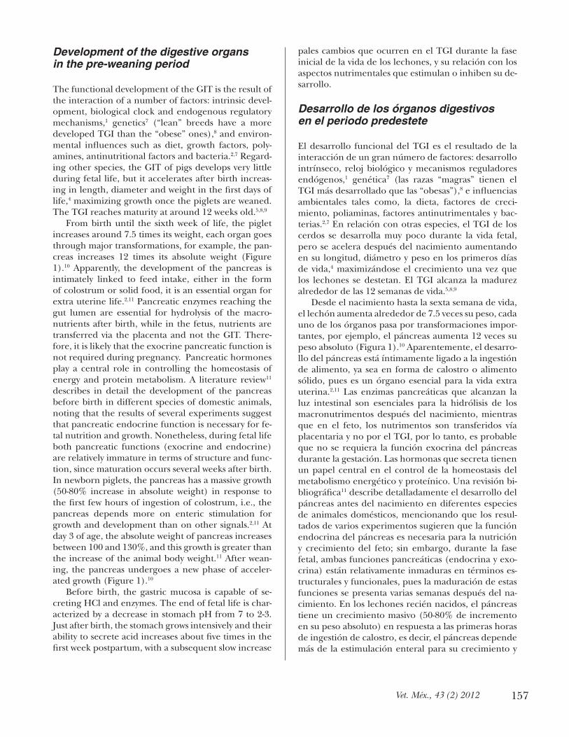
From birth until the sixth week of life, the piglet increases around 7.5 times its weight, each organ goes through major transformations, for example, the pan-creas increases 12 times its absolute weight (Figure 1).10 Apparently, the development of the pancreas is intimately linked to feed intake, either in the form of colostrum or solid food, it is an essential organ for extra uterine life.2,11 Pancreatic enzymes reaching the gut lumen are essential for hydrolysis of the macro-nutrients after birth, while in the fetus, nutrients are transferred via the placenta and not the GIT. There-fore, it is likely that the exocrine pancreatic function is not required during pregnancy. Pancreatic hormones play a central role in controlling the homeostasis of energy and protein metabolism. A literature review11 describes in detail the development of the pancreas before birth in different species of domestic animals, noting that the results of several experiments suggest that pancreatic endocrine function is necessary for fe-tal nutrition and growth. Nonetheless, during fetal life both pancreatic functions (exocrine and endocrine) are relatively immature in terms of structure and func-tion, since maturation occurs several weeks after birth. In newborn piglets, the pancreas has a massive growth (50-80% increase in absolute weight) in response to the first few hours of ingestion of colostrum, i.e., the pancreas depends more on enteric stimulation for growth and development than on other signals.2,11 At day 3 of age, the absolute weight of pancreas increases between 100 and 130%, and this growth is greater than the increase of the animal body weight.11 After wean-ing, the pancreas undergoes a new phase of acceler-ated growth (Figure 1).10
Before birth, the gastric mucosa is capable of se-creting HCl and enzymes. The end of fetal life is char-acterized by a decrease in stomach pH from 7 to 2-3. Just after birth, the stomach grows intensively and their ability to secrete acid increases about five times in the first week postpartum, with a subsequent slow increase
pales cambios que ocurren en el TGI durante la fase inicial de la vida de los lechones, y su relación con los aspectos nutrimentales que estimulan o inhiben su de-sarrollo.
Desarrollo de los órganos digestivos en el periodo predestete
El desarrollo funcional del TGI es el resultado de la interacción de un gran número de factores: desarrollo intrínseco, reloj biológico y mecanismos reguladores endógenos,1 genética7 (las razas “magras” tienen el TGI más desarrollado que las “obesas”),8 e influencias ambientales tales como, la dieta, factores de creci-miento, poliaminas, factores antinutrimentales y bac-terias.2,7 En relación con otras especies, el TGI de los cerdos se desarrolla muy poco durante la vida fetal, pero se acelera después del nacimiento aumentando en su longitud, diámetro y peso en los primeros días de vida,4 maximizándose el crecimiento una vez que los lechones se destetan. El TGI alcanza la madurez alrededor de las 12 semanas de vida.5,8,9
Desde el nacimiento hasta la sexta semana de vida, el lechón aumenta alrededor de 7.5 veces su peso, cada uno de los órganos pasa por transformaciones impor-tantes, por ejemplo, el páncreas aumenta 12 veces su peso absoluto (Figura 1).10 Aparentemente, el desarro-llo del páncreas está íntimamente ligado a la ingestión de alimento, ya sea en forma de calostro o alimento sólido, pues es un órgano esencial para la vida extra uterina.2,11 Las enzimas pancreáticas que alcanzan la luz intestinal son esenciales para la hidrólisis de los macronutrimentos después del nacimiento, mientras que en el feto, los nutrimentos son transferidos vía placentaria y no por el TGI, por lo tanto, es probable que no se requiera la función exocrina del páncreas durante la gestación. Las hormonas que secreta tienen un papel central en el control de la homeostasis del metabolismo energético y proteínico. Una revisión bi-bliográfica11 describe detalladamente el desarrollo del páncreas antes del nacimiento en diferentes especies de animales domésticos, mencionando que los resul-tados de varios experimentos sugieren que la función endocrina del páncreas es necesaria para la nutrición y crecimiento del feto; sin embargo, durante la fase fetal, ambas funciones pancreáticas (endocrina y exo-crina) están relativamente inmaduras en términos es-tructurales y funcionales, pues la maduración de estas funciones se presenta varias semanas después del na-cimiento. En los lechones recién nacidos, el páncreas tiene un crecimiento masivo (50-80% de incremento en su peso absoluto) en respuesta a las primeras horas de ingestión de calostro, es decir, el páncreas depende más de la estimulación enteral para su crecimiento y
158
desarrollo, que de otras señales.2,11 Al día 3 de vida, el peso absoluto del páncreas aumenta entre 100 y 130%, y este crecimiento es mayor que el aumento del peso vivo del animal.11 Después del destete, el páncreas pasa por una nueva fase de aceleración en su crecimiento (Figura 1).10
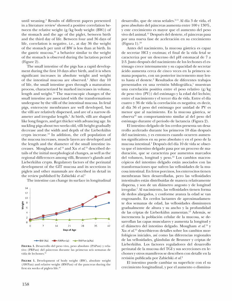
Antes del nacimiento, la mucosa gástrica es capaz de secretar HCl y enzimas; el final de la vida fetal se caracteriza por un descenso del pH estomacal de 7 a 2-3. Justo después del nacimiento de los lechones el es-tómago crece intensamente y su capacidad de secretar ácido aumenta cerca de cinco veces en la primera se-mana posparto, con un posterior incremento muy len-to hasta el destete.2 Resultados de diferentes trabajos presentados en una revisión bibliográfica,1 muestran una correlación positiva entre el peso relativo (g/kg de peso vivo (PV)) del estómago y la edad del lechón; entre el nacimiento y el tercer día de vida. Entre el día cuatro y 36 de vida la correlación es negativa; es decir, al día 36 el peso del estómago por unidad de PV es menor que al nacimiento. En la mucosa gástrica, se observa10 un comportamiento similar al del peso del estómago durante el periodo de lactancia (Figura 2).
El intestino delgado de los cerdos presenta un desa-rrollo acelerado durante los primeros 10 días después del nacimiento, y es entonces cuando ocurren aumen-tos significativos en su peso absoluto y en el peso de la mucosa intestinal.1 Después del día 10 de vida se obser-va que el intestino delgado pasa por un proceso de ma-duración, que se caracteriza por aumentos marcados del volumen, longitud y peso.12 Los cambios macros-cópicos del intestino delgado están asociados con las transformaciones que sufren las vellosidades de la mu-cosa intestinal. En fetos porcinos, los enterocitos tienen membranas bien desarrolladas, pero las vellosidades intestinales están distribuidas de manera relativamente dispersa, y son de un diámetro angosto y de longitud irregular.1 Al nacimiento, las vellosidades tienen forma de dedos alargados, y conforme avanza la edad se van engrosando. En cerdos lactantes de aproximadamen-te dos semanas de edad, las vellosidades disminuyen gradualmente de altura y su ancho y la profundidad de las criptas de Lieberkühn aumentan.13 Además, se incrementa la población celular de la mucosa, se de-sarrollan las capas musculares y aumenta la longitud y el diámetro del intestino delgado. Mougham et al.14 y Xu et al.15 describieron detalles sobre los cambios mor-fológicos iniciales, así como las diferencias regionales de las vellosidades, glándulas de Brunner y criptas de Lieberkühn. Los factores reguladores del desarrollo perinatal de la mucosa del TGI y sus secreciones en le-chones y otros mamíferos se describen con detalle en la revisión publicada por Zabielski et al.2
El intestino puede cambiar su superficie con el su crecimiento longitudinal, y por el aumento o disminu-
until weaning.2 Results of different papers presented in a literature review1 showed a positive correlation be-tween the relative weight (g/kg body weight (BW)) of the stomach and the age of the piglet, between birth and the third day of life. Between four and 36 days of life, correlation is negative, i.e., at day 36 the weight of the stomach per unit of BW is less than at birth. In the gastric mucosa,10 a behavior similar to the weight of the stomach is observed during the lactation period (Figure 2).
The small intestine of the pigs has a rapid develop-ment during the first 10 days after birth, and it is when significant increases in absolute weight and weight of the intestinal mucosa are observed.1 After day 10 of life, the small intestine goes through a maturation process, characterized by marked increases in volume, length and weight.12 The macroscopic changes of the small intestine are associated with the transformations undergone by the villi of the intestinal mucosa. In fetal pigs, enterocyte membranes are well developed, but the villi are relatively dispersed, and are of a narrow di-ameter and irregular length.1 At birth, villi are shaped like long fingers, and get thicker with advancing age. In suckling pigs about two weeks old, villi height gradually decrease and the width and depth of the Lieberkühn crypts increase.13 In addition, the cell population of the mucosa increases, muscle layers are developed and the length and the diameter of the small intestine in-creases. Mougham et al.14 and Xu et al.15 described de-tails of the initial morphological changes, as well as the regional differences among villi, Brunner's glands and Lieberkühn crypts. Regulatory factors of the perinatal development of the GIT mucosa and its secretions in piglets and other mammals are described in detail in the review published by Zabielski et al.2
The intestine can change its surface in longitudinal
Figura 1. Desarrollo del peso vivo, peso absoluto (PAPan) y rela-tivo (PRPan) del páncreas durante las primeras seis semanas de vida de lechones.10
Figure 1. Development of body weight (BW), absolute weight (AWPan) and relative weight (RWPan) of the pancreas during the first six weeks of piglets life.10
159Vet. Méx., 43 (2) 2012
ción de la altura de las vellosidades. El acortamiento y la fusión de vellosidades propiciarán en una pérdida de la superficie de digestión y absorción de los alimen-tos.16 Para que la digestión y la absorción de los nutri-mentos se lleven a cabo de una manera satisfactoria, es necesario que se mantenga la integridad de la mucosa intestinal, la cual depende del recambio y de la reno-vación de sus células. La función digestiva (secreción de carbohidrasas y peptidasas) de los enterocitos y sus microvellosidades comienza solamente cuando se completa la diferenciación estructural de la mucosa, lo que generalmente ocurre durante el periodo de mi-gración celular hacia el primer tercio de la vellosidad. La absorción de azúcares y aminoácidos empieza cuan-do el enterocito migra hacia la mitad de la vellosidad y continúa aumentando hasta que sale por descamación en la punta de ésta.16
Efecto del destete
El destete en condiciones naturales se lleva a cabo va-rias semanas después del nacimiento, y es un proceso durante el cual el sistema digestivo del lechón se adap-ta progresivamente a un menor consumo de leche y mayores cantidades de alimento sólido. Desde finales de la década de los setenta se practica el destete “tem-prano” con el objetivo de que las cerdas reinicien su ciclo reproductivo de manera prematura. Eso significó que se pasara de un destete gradual, realizado por la misma cerda entre las 15-22 semanas de edad de los lechones, a un destete brusco y ultra precoz alrededor de los 7 días de edad.17 En la actualidad, el destete se realiza en la mayor parte del mundo entre los 21 y 28 días de edad. Contrariamente a lo que ocurre en con-diciones naturales, en el destete temprano los factores psicológicos, sociales y nutrimentales inherentes a esta etapa, interfieren de manera importante en el desa-rrollo de los animales, particularmente del TGI.18 Las alteraciones generadas por el destete temprano no se presentan en ningun otra etapa del crecimiento del cerdo y, debido al cambio radical en la alimentación, los animales se someten a un severo estrés nutrimen-tal.19 La leche materna, líquida, altamente digestible y muy bien acoplada a las enzimas del tubo digestivo del lechón, se reemplaza por una dieta sólida, elaborada a base de cereales (fuente de almidón) y de proteínas de origen vegetal.19 Los lechones recién destetados po-seen un menor grado de maduración de la función digestiva, pues su TGI aún no produce todas las enzi-mas necesarias para la digestión de alimentos sólidos.20 Además, las secreciones digestivas no son suficientes,21 y el epitelio intestinal pasa por cambios morfológicos muy drásticos, por lo que la absorción de los nutrimen-tos se reduce. En consecuencia, se observa en el lechón
growth, and the increase or decrease in villi height. The shortening and fusion of villi foster a loss of diges-tion and absorption surface for food.16 For digestion and absorption of nutrients to take place in a satisfac-tory way, it is necessary to maintain the integrity of the intestinal mucosa, which depends on cells replace-ment and renewal. The digestive function (secretion of carbohydrases and peptidases) of enterocytes and microvilli begins only with full structural differentia-tion of the mucosa, which usually occurs during cell migration into the first third of the villus. The absorp-tion of sugars and amino acids begins when the entero-cyte migrates to half of the villi and continues increas-ing until it comes out scaling off the tip.16
Weaning effect
The natural weaning conditions take place several weeks after birth, and is a process during which the pig's digestive system is adapted progressively adapted to lower consumption of milk and greater amounts of solid feed. Since the late 70's, weaning is practiced "early" in order to restart prematurely the sows repro-ductive cycle. That meant a shift from a gradual wean-
Figura 2. Desarrollo del peso vivo, peso absoluto (PAMuc) y re-lativo (PRMuc) de la mucosa cardiaca del estómago durante las primeras seis semanas de vida de lechones (a)10 y (b).17
Figure 2. Development of body weight (BW), absolute weight (AW-Muc) and relative weight (RWMuc) of stomach mucosa during the first six weeks of piglets life (a)10 and (b).17

160
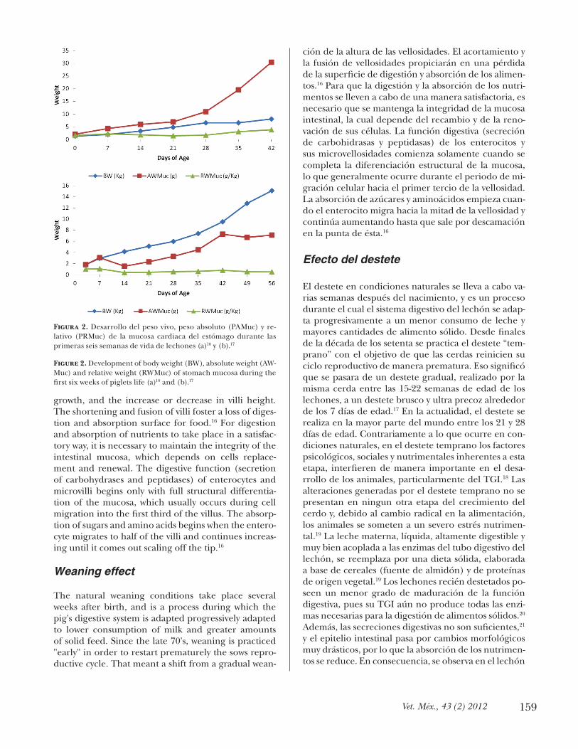
recién destetado una capacidad limitada de digestión y de absorción de los alimentos, sobre todo respecto a las proteínas vegetales,22 lo que repercute en el con-sumo de alimento.23-26 La primera semana posdestete suele ser en la que más consecuencias negativas tiene en el TGI. En los dos o tres primeros días posdestete se observa una disminución del peso de los órganos di-gestivos, aunque en algunos casos, el peso permanece constante en este periodo (Figuras 3 y 4).27,28
Efecto del consumo de alimento
La nutrición enteral representa una de las influencias más significativas en el desarrollo del TGI y del pán-creas, y depende de la naturaleza del alimento ingeri-do (calostro versus leche materna) y de la diversidad de los ingredientes que componen el alimento.2 El efecto del consumo de alimentos sólidos sobre el TGI es am-biguo, pues por un lado es benéfico y estimulador del desarrollo (mucosa gástrica, páncreas y flora microbia-na no patógena); por otro lado, es adverso debido a las alteraciones estructurales de la mucosa del intestino delgado y al desarrollo de una flora microbiana pató-gena. Así, dependiendo del órgano digestivo conside-rado, los efectos en el desarrollo son distintos.
Estómago. El crecimiento del estómago se asocia con el desarrollo de la mucosa gástrica, lo que provoca una mayor producción de HCl y de pepsina en res-puesta al estímulo físico provocado por el aumento de la masa alimenticia ingerida y al efecto trófico de la hormona gastrina.10 Durante las cuatro semanas pos-destete, se observó1 que el crecimiento del estómago fue positivamente alométrico, de tal manera que el peso estomacal aumentó de 4.9 a 6.3 g/kg en relación con el peso corporal. El peso absoluto de la mucosa gástrica se incrementa de manera relevante durante las dos primeras semanas posdestete (Figura 2);10,17 sin embargo, el desarrollo parece estabilizarse en las se-
ing, by the sow herself between 15-22 weeks of age to an ultra sharp and early weaning on about 7 day-old piglets.17 Currently, weaning takes place in most of the world between 21 and 28 days old. Contrary to what occurs in natural conditions, during early weaning the psychological, social and nutritional factors in this stage, significantly interfere in the development of an-imals, particularly of the GIT.18 Disturbances created by early weaning are not present in any other growth stage of the pig and, due to the radical change in diet, animals are exposed to severe nutritional stress.19 Milk, a highly digestible fluid, well coupled to the enzymes of the digestive tract of the pig, is replaced by a solid diet, made from grain (starch source) and vegetable proteins.19 The newly weaned piglets have a lower de-gree of maturation of digestive function, as its GIT does not yet produce all the enzymes necessary for digestion of solid feed.20 In addition, digestive secre-tions are not sufficient,21 and the intestinal epithelium passes through drastic morphological changes, so that the absorption of nutrients is reduced. Consequently, a limited capacity for food digestion and absorption is observed in the weaned piglet, especially in regard to plant protein,22 which affects feed intake.23-26 The first week after weaning is usually the most negative in consequences on the GIT. In the first two or three days after weaning there is a decrease of the weight
Figura 3. Peso absoluto de los órganos digestivos de lechones en diferentes días posdestete (a)27 y (b).28
Figure 3. Absolute weight of the digestive organs of piglets at dif-ferent days after weaning (a)27 and (b).28
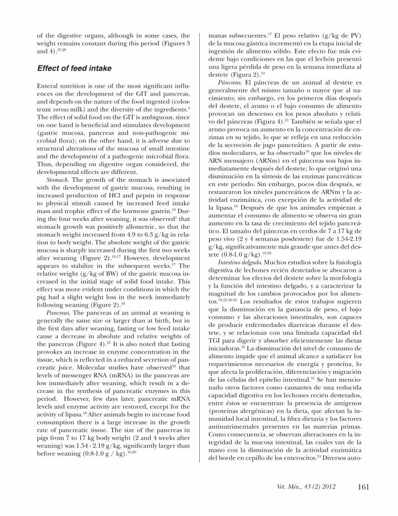
Figura 4. Peso relativo del páncreas de lechones en diferentes días posdestete.21
Figure 4. Relative weight of the pancreas of piglets at different days after weaning21
161Vet. Méx., 43 (2) 2012
of the digestive organs, although in some cases, the weight remains constant during this period (Figures 3 and 4).27,28
Effect of feed intake
Enteral nutrition is one of the most significant influ-ences on the development of the GIT and pancreas, and depends on the nature of the food ingested (colos-trum versus milk) and the diversity of the ingredients.2 The effect of solid food on the GIT is ambiguous, since on one hand is beneficial and stimulates development (gastric mucosa, pancreas and non-pathogenic mi-crobial flora); on the other hand, it is adverse due to structural alterations of the mucosa of small intestine and the development of a pathogenic microbial flora. Thus, depending on digestive organ considered, the developmental effects are different.
Stomach. The growth of the stomach is associated with the development of gastric mucosa, resulting in increased production of HCl and pepsin in response to physical stimuli caused by increased feed intake mass and trophic effect of the hormone gastrin.10 Dur-ing the four weeks after weaning, it was observed1 that stomach growth was positively allometric, so that the stomach weight increased from 4.9 to 6.3 g/kg in rela-tion to body weight. The absolute weight of the gastric mucosa is sharply increased during the first two weeks after weaning (Figure 2).10,17 However, development appears to stabilize in the subsequent weeks.17 The relative weight (g/kg of BW) of the gastric mucosa in-creased in the initial stage of solid food intake. This effect was more evident under conditions in which the pig had a slight weight loss in the week immediately following weaning (Figure 2).10
Pancreas. The pancreas of an animal at weaning is generally the same size or larger than at birth, but in the first days after weaning, fasting or low feed intake cause a decrease in absolute and relative weights of the pancreas (Figure 4).21 It is also noted that fasting provokes an increase in enzyme concentration in the tissue, which is reflected in a reduced secretion of pan-creatic juice. Molecular studies have observed18 that levels of messenger RNA (mRNA) in the pancreas are low immediately after weaning, which result in a de-crease in the synthesis of pancreatic enzymes in this period. However, few days later, pancreatic mRNA levels and enzyme activity are restored, except for the activity of lipasa.18 After animals begin to increase food consumption there is a large increase in the growth rate of pancreatic tissue. The size of the pancreas in pigs from 7 to 17 kg body weight (2 and 4 weeks after weaning) was 1.54 - 2.19 g/kg, significantly larger than before weaning (0.8-1.0 g / kg).10,29
manas subsecuentes.17 El peso relativo (g/kg de PV) de la mucosa gástrica incrementó en la etapa inicial de ingestión de alimento sólido. Este efecto fue más evi-dente bajo condiciones en las que el lechón presentó una ligera pérdida de peso en la semana inmediata al destete (Figura 2).10
Páncreas. El páncreas de un animal al destete es generalmente del mismo tamaño o mayor que al na-cimiento; sin embargo, en los primeros días después del destete, el ayuno o el bajo consumo de alimento provocan un descenso en los pesos absoluto y relati-vo del páncreas (Figura 4).21 También se señala que el ayuno provoca un aumento en la concentración de en-zimas en su tejido, lo que se refleja en una reducción de la secreción de jugo pancreático. A partir de estu-dios moleculares, se ha observado18 que los niveles de ARN mensajero (ARNm) en el páncreas son bajos in-mediatamente después del destete; lo que originó una disminución en la síntesis de las enzimas pancreáticas en este periodo. Sin embargo, pocos días después, se restauraron los niveles pancreáticos de ARNm y la ac-tividad enzimática, con excepción de la actividad de la lipasa.18 Después de que los animales empiezan a aumentar el consumo de alimento se observa un gran aumento en la tasa de crecimiento del tejido pancreá-tico. El tamaño del páncreas en cerdos de 7 a 17 kg de peso vivo (2 y 4 semanas posdestete) fue de 1.54-2.19 g/kg, significativamente más grande que antes del des-tete (0.8-1.0 g/kg).10,29
Intestino delgado. Muchos estudios sobre la fisiología digestiva de lechones recién destetados se abocaron a determinar los efectos del destete sobre la morfología y la función del intestino delgado, y a caracterizar la magnitud de los cambios provocados por los alimen-tos.19,23,30-32 Los resultados de estos trabajos sugieren que la disminución en la ganancia de peso, el bajo consumo y las alteraciones intestinales, son capaces de producir enfermedades diarreicas durante el des-tete, y se relacionan con una limitada capacidad del TGI para digerir y absorber eficientemente las dietas iniciadoras.32 La disminución del nivel de consumo de alimento impide que el animal alcance a satisfacer los requerimientos necesarios de energía y proteína, lo que afecta la proliferación, diferenciación y migración de las células del epitelio intestinal.31 Se han mencio-nado otros factores como causantes de una reducida capacidad digestiva en los lechones recién destetados, entre éstos se encuentran: la presencia de antígenos (proteínas alergénicas) en la dieta, que afectan la in-munidad local intestinal, la fibra dietaria y los factores antinutrimentales presentes en las materias primas. Como consecuencia, se observan alteraciones en la in-tegridad de la mucosa intestinal, las cuales van de la mano con la disminución de la actividad enzimática del borde en cepillo de los enterocitos.23 Diversos auto-

162
Small intestine. Many studies on the digestive physi-ology of weaned pigs were dedicated to determine the effects of weaning on the morphology and function of the small intestine, and to characterize the magnitude of the changes caused by food.19,23,30-32 The results of these studies suggest that the decrease in weight gain, low intake and intestinal disorders, are capable of pro-ducing diarrheic diseases at weaning, and are associat-ed with a limited GIT capacity to digest and efficiently absorb starter diets.32 The intake decrease prevents the animal to satisfy the requirements of energy and protein, which affects the proliferation, differentiation and migration of intestinal epithelial cells.31 Other fac-tors have been cited as causing a reduced digestive ca-pacity in newly weaned piglets, among them are: the presence of antigens (allergenic proteins) in the diet, which affect local intestinal immunity, dietary fiber and the antinutritional factors present in raw materials. As a result, there are alterations in the intestinal mucosal integrity, which go hand in hand with decreased en-zyme activity of the brush border of enterocytes.23 Sev-eral authors17,21,30 agree that the villi can reduce 75% of its height during the first 24 hours after weaning, due to the loss of mature enterocytes. This decrease is re-flected in a morphological change of the villi, long like a finger in the newborn and suckling pigs, to shorter and wider as a leaf or tongue, in the newly weaned.31 Simultaneously, the lactase activity decreases24,33 due to the presence of immature enterocytes in the short-ened villi, which can not express its maximum capac-ity of enzyme synthesis. The activity of peptidases in the membranes and cytosol of enterocytes, which are responsible for the final hydrolysis of peptides, de-creases from birth until the fifth week of age.1 It also may explain the reduced activity of intestinal enzymes by the erratic food consumption observed during the first days after weaning, resulting in a decrease of sub-strates in the intestinal lumen.21 For example, lactase activity decreases as a result of reduced intake of lac-tose.34 The structural and functional changes observed in the small intestine occur within the first 24 to 36 hours after weaning and result in a decrease of 20 to 30% of intestinal weight.18 Renewed growth of the villi is observed from day three after weaning,19,23,31,35 which has been linked to the presence of food in the diges-tive tract.
Influence of feed composition on the GIT
The intrinsic development and differentiation of new-born piglet GIT are deeply dependent on the interac-tion between dietary components and the microbial flora, food having a decisive effect. Colostrum and milk contain high concentrations of growth factors
res17,21,30 coinciden en que las vellosidades pueden dis-minuir 75% de su altura durante las primeras 24 horas posdestete, debido a la pérdida de enterocitos madu-ros. Esta disminución se manifiesta en un cambio mor-fológico de las vellosidades, de largas como un dedo en los recién nacidos y cerdos lactantes, a más cortas y anchas como una hoja o una lengua, en los recién destetados.31 Simultáneamente, la actividad de la lacta-sa disminuye,24,33 debido a la presencia de enterocitos inmaduros en las vellosidades acortadas, los cuales no pueden expresar su máxima capacidad de síntesis de enzimas. Las peptidasas presentes en las membranas y en el citosol de los enterocitos, y que se encargan de la hidrólisis final de los péptidos, disminuyen su actividad desde el nacimiento hasta la quinta semana de edad.1 También se puede explicar la reducción de la actividad de las enzimas intestinales, por el consu-mo de alimento errático que se observa durante los primeros días posdestete, lo que provoca una baja de sustratos en la luz intestinal.21 Por ejemplo, la actividad de la lactasa disminuye a consecuencia de la reducción en la ingestión de lactosa.34 Los cambios funcionales y estructurales que se observan en el intestino delgado suceden dentro de las primeras 24 a 36 horas posdes-tete y traen como consecuencia una disminución del 20 al 30% del peso intestinal.18 La recuperación del crecimiento de las vellosidades se observa desde el día tres posdestete,19,23,31,35 lo cual se ha relacionado con la presencia de alimento en el tubo digestivo.
Influencia de la composición del alimento sobre el TGI
El desarrollo y la diferenciación intrínseca del TGI del lechón recién nacido dependen profundamente de la interacción entre los componentes dietarios y la flora microbiana, teniendo el alimento un efecto determi-nante. El calostro y la leche contienen altas concen-traciones de factores de crecimiento que aceleran la proliferación y la maduración del intestino de los ani-males recién nacidos.26
Después del destete, la composición del alimento iniciador tiene un papel importante en los cambios morfofisiológicos del TGI. La manera de formular las dietas iniciadoras ha cambiado en las últimas décadas, pasando de dietas simples (dos ingredientes) a dietas más complejas, en las cuales el número de ingredien-tes es muy amplio.36 La dieta simple se formula estricta-mente con productos vegetales, por ejemplo, cereales y pasta de soya. Las dietas complejas están compues-tas por productos y subproductos lácteos, tales como: leche en polvo descremada, caseína y suero seco de leche; harinas de origen animal, al igual que una can-tidad mínima de cereales y pasta de soya, con la finali-

163Vet. Méx., 43 (2) 2012
that accelerate the proliferation and development of the intestine of newborn animals.26
After weaning, composition of starter feed has an important role in the GIT morphophysiological chang-es. The way of formulating starter diets has changed in recent decades, from simple diets (two ingredients) to more complex diets, in which the number of ingredi-ents is very wide.36 The simple diet is strictly formulated with plant products, such as cereals and soybean meal. Complex diets consist of dairy products and subprod-ucts such as skim milk powder, casein and dry whey, an-imal meals, along with a minimum amount of grain and soybean meals, in order to stimulate feed intake and maintain intestinal integrity.19 When making a change from a single to a complex diet, the piglet has already passed the stress of weaning and adjusted its consump-tion according to their physical ability, so that its diges-tive system is ready to receive higher amounts of starch and vegetable protein. Lactase, which is the main in-testinal enzyme for digestion of carbohydrates in the lactation period, gradually declines after weaning. The enzymes maltase and sucrase have a reversed behav-ior, as they are expressed and activated by consump-tion of solid food, containing starches and sucrose.1
Similarly, the intestinal mucosa appears to be quite sensitive to diet composition. A group of researchers37 compared two types of diets: a typical American, con-taining animal proteins and growth promoters (zinc oxide, copper sulfate and antibiotics) and the other a typical European diet, based on vegetable proteins without growth promoters. As a result it was found that consumption of the European diet caused a reduction of around 20% in the villi height and increased crypt depth, compared to piglets fed the North American diet, reducing the intestinal-absorption surface. This is probably caused by a reduction in the rate of mitosis or increased rate of apoptosis, or both processes.37 The development of the pancreas is directly related to the type of diet the animal eats, because if animals have access to rich protein food, fat or carbohydrates, this organ must perform its functions in accordance with such substrates.31 The main dietary components that affect the development of the GIT are the sources of lactose, protein and cereals.31
Dairy ingredients are a source of lactose and high quality protein and are key feeds during the first days after weaning, because weight gain is faster, since its presence in the starter diet will make the diet change less abrupt for piglets, due to the taste and nutritional value of these products. Besides, its GIT is adapted (enzyme secretions, motility and absorption) to this ingredients from birth.38 An important factor is the continuity of the presence of substrate in the gastro-intestinal tract after weaning, and this has an effect on the activity of intestinal lactase enzyme. The presence
dad de estimular el consumo de alimento y mantener la integridad intestinal.19 Al momento de realizar el cambio de una dieta compleja a una simple, el lechón ya ha superado el estrés del destete y ha ajustado su consumo de acuerdo con su capacidad física, de modo que su aparato digestivo se encuentra preparado para recibir mayores cantidades de almidón y de proteína de origen vegetal. La lactasa, que es la principal enzi-ma intestinal para la digestión de carbohidratos en el periodo de lactancia, declina paulatinamente después del destete; en el caso de las enzimas maltasa y sacarasa el comportamiento es a la inversa, pues se expresan y se activan con el consumo del alimento sólido, que contiene almidones y sacarosa.1 De igual forma, la mu-cosa intestinal parece ser bastante sensible a la com-posición de las dietas. Un grupo de investigadores37 comparó dos tipos de dietas: una típica estadunidense, que contenía proteínas de origen animal y promoto-res de crecimiento (óxido de zinc, sulfato de cobre y antibióticos); y la otra, una dieta típica europea, a base de proteínas vegetales, sin promotores de crecimien-to. Como resultado se encontró que el consumo de la dieta europea causó una reducción de alrededor de 20% en la altura de las vellosidades, y un aumento en la profundidad de la cripta, en comparación con los lechones alimentados con la dieta estadunidense, re-duciéndose la superficie de absorción intestinal. Esto probablemente se originó por una reducción en la tasa de mitosis o al aumento en la tasa de apoptosis, o por ambos procesos.37 El desarrollo del páncreas también está directamente relacionado con el tipo de dieta que consuma el animal, ya que si los animales tienen acceso a alimentos ricos en proteínas, grasas o carbohidratos, este órgano tendrá que desarrollar sus funciones de acuerdo con dichos sustratos.31 Los prin-cipales componentes de la dieta que afectan el desa-rrollo del TGI son las fuentes de lactosa, los alimentos proteínicos y los cereales.31
Los ingredientes lácteos son fuente de lactosa y de proteína de alta calidad y son alimentos clave durante los primeros días posdestete, pues la recuperación de peso es más rápida, ya que su presencia en la dieta ini-ciadora hará que el cambio de alimentación sea menos brusco para el lechón, por el sabor y el valor nutritivo de estos productos, además de que su TGI está adap-tado (secreciones enzimáticas, motilidad y absorción) a sus componentes desde el nacimiento.38 Un factor importante es la continuidad de la presencia de sus-trato en el tracto gastrointestinal después del destete, y ésta tiene un efecto sobre la actividad de la enzima lactasa intestinal. La presencia de lactosa en la dieta, independientemente de la fuente (lactosa cristalina o suero de leche), incrementó la actividad de la lacta-sa (68%) y el peso del intestino delgado durante los primeros 14 días posdestete.34 Otra ventaja es que los
164
of lactose in the diet, regardless of the source (crystal-line lactose or whey), increased lactase activity (68%) and weight of the small intestine during the first 14 days after weaning.34 Another advantage is that dairy products have growth factors that stimulate the GIT development in weaned animals.26
Protein sources. The quantity, source and digestibility of the dietary protein determine the availability of ami-no acids and peptides in the lumen of small intestine, which may influence recovery of intestinal morphol-ogy after weaning.39 Results of different works suggest that glutamine is an amino acid that serves as very im-portant energy substrate for enterocytes.31 Piglets fed diets with added exogenous glutamine showed higher villi and deeper crypts in the first week after weaning than pigs that did not receive glutamine.40
The quality of the protein source included in the diet for piglets has an important role in GIT develop-ment. That quality largely depends on the origin of protein sources (animal or vegetable). Animal protein sources are used frequently in starter diets for its high protein content, high digestibility and biological val-ue,41 and by the absence of antinutritional factors, but are expensive. Until the appearance of bovine spongi-form encephalitis, the most traditionally used ingredi-ents in the diets of weaned piglets were blood and meat meals and blood plasma (bovine), which substantially improved consumption, growth and conversion rate, as well as the immune status of the animal. Currently, these ingredients are banned in several countries, and dairy, pig plasma and fishmeal are used in larger scale, whose biggest problem is quality variability.26
Vegetable protein sources, mainly soybean meal, are often economic and constitute a higher protein intake during the growing and finishing phases of the pig. The excellent amino acid profile of soybean meal is well known, but there is variation in the nutritional value of different varieties,42 due to factors such as level of crude protein and the amount and type of antinu-tritional factors. The presence of certain components that often cause digestive problems in piglets is rele-vant in soybeans, especially when it is not processed correctly,43 so its use is limited in the starter phase. Among the the antinutritional components present in soybean, inhibiting factors (chymotrypsin and tryp-sin), antivitamins and lectins are included.44 Some pro-tein fractions (β-conglycinin and glycinin) can damage the intestinal villi, due to a transient hypersensitivity process that occurs in the intestinal mucosa and, con-sequently, decrease protein digestion.45, 46 Lectins are glycoproteins resistant to proteolytic digestive enzymes and by binding to carbohydrate molecules of the mem-branes of epithelial cells of the intestinal mucosa can damage the intestinal villi; toxicity depends on the magnitude of this link. Thus, antinutritional factors
subproductos lácteos poseen factores de crecimiento, que estimulan el desarrollo del TGI en los animales recién destetados.26
Fuentes de proteína. La cantidad, la fuente y la diges-tibilidad de la proteína dietaria determinan la dispo-nibilidad de aminoácidos y péptidos en el lumen del intestino delgado, lo que puede influir en la recupe-ración de la morfología intestinal después del deste-te.39 Resultados de diferentes trabajos sugieren que la glutamina es un aminoácido que sirve como sustrato energético muy importante para los enterocitos.31 Le-chones alimentados con dieta adicionadas con glu-tamina exógena presentaron vellosidades más altas y criptas más profundas en la primera semana después del destete, que cerdos que no recibieron glutamina.40
La calidad de la fuente de proteína incluida en la dieta para lechones tiene un papel importante en el de-sarrollo del TGI. Esa calidad depende en gran medida del origen de las fuentes proteínicas (animal o vegetal). Las fuentes proteínicas de origen animal se emplean con gran frecuencia en las dietas iniciadoras por su alto contenido de proteína, valor biológico y alta digestibi-lidad,41 así como por la ausencia de factores antinutri-mentales; sin embargo, tiene un costo elevado. Hasta la aparición de la encefalitis espongiforme bovina, los ingredientes animales más utilizados tradicionalmente en las dietas de lechones destetados, se encontraban las harinas de sangre y de carne y el plasma sanguíneo (bovino), que mejoraban sustancialmente el consumo, el crecimiento y el índice de conversión, así como el estado inmunitario del animal. En la actualidad, estos ingredientes están prohibidos en varios países, y se uti-lizan en mayor escala los productos lácteos, el plasma porcino y la harina de pescado, cuyo mayor problema radica en la variabilidad de su calidad.26
Las fuentes proteínicas de origen vegetal, princi-palmente la pasta de soya, suelen resultar económicas y constituyen un mayor aporte de proteína durante las fases de crecimiento y finalización del cerdo. El exce-lente perfil de aminoácidos de la pasta de soya es muy reconocido, pero existe una variación en el valor nutri-mental de sus distintas variedades,42 debido a factores como el nivel de proteína cruda y la cantidad y tipo de factores antinutrimentales. En la soya es relevante la presencia de ciertos componentes que suelen provo-car problemas digestivos en los lechones, especialmen-te cuando no se procesa correctamente,43 por lo que su uso es limitado en la fase inicial de crianza. Entre los componentes antinutrimentales presentes en la pasta de soya se incluyen los factores inhibidores de pro-teasas (quimotripsina y tripsina), antivitaminas y lec-tinas.44 Algunas fracciones proteínicas (β-conglicinina y glicinina) pueden dañar las vellosidades intestinales, debido a un proceso de hipersensibilidad transitoria que ocurre en la mucosa intestinal y, consecuente-
165Vet. Méx., 43 (2) 2012
can cause growth inhibition, decreased feed efficiency, pancreatic hypertrophy, hypoglycemia and liver dam-age in ruminant animals, depending on the species, age, size, sex, health status and feeding plane.43 The mucosal lesions caused by soybean meal may vary de-pending on the level of inclusion in the diet,47 or the variety used. To increase the nutritional value, soybean varieties have been selected with a higher concentra-tion of crude protein or a lesser amount of trypsin in-hibitors and lectin.43 Fortunately, many antinutritional factors are heat labile and are destroyed during appli-cation of heat treatments. Oligosaccharides (stachyose and raffinose) are other undesirable components in soybean, which are not removed in processing.48 These represent 4 to 6% of dry matter and can cause flatu-lence, diarrhea and stress in non-ruminants, due to their inability to digest these substances because they do not synthesize the enzyme α-1, 6-galactosidase, which is responsible for their digestion. In this case oligosaccharides are left free as a substrate for bacte-rial fermentation processes in the posterior intestinal tract, which contains anaerobic bacteria, and that can produce hydrogen, carbon dioxide and small amounts of methane.46
Currently, there are varieties of soybeans contain-ing 70 to 90% less oligosaccharides than conventional soybean meal. By its use, a greater digestibility of most amino acids of the soybean meal is observed with lower level of oligosaccharides in relation to the usual, and an energy digestibility comparable to conventional soybean meal;50 so probable, soybean meal low in oli-gosaccharides has a less negative impact on the intes-tinal integrity.
The feed industry has developed other soy prod-ucts of high biological value, such as concentrates and isolated soy protein. These feeds reduce the transient hypersensitivity reaction that occurs when using soy-bean meal, which is manifested in less damage of in-testinal villi, higher digestibility of nutrients and better weight gain. However, these highly digestible feeds do not stimulate the growth of intestinal villi.51
Other sources of vegetable protein are also evalu-ated as potential alternatives to soybean meal. The inclusion of 10% of natural canola or pelleted canola in diets consumed for 14 days after weaning did not affect intestinal villus height.52 Comparing diets con-taining soybean meal or sesame paste, no differences were seen in the morphology of intestinal villi or crypts or after a period of 25-day consumption.53 In another study,54 pigs fed for 22 days after weaning with diets based on soybean or sesame showed no differences in the relative weight of digestive organs (Table 1).
Cereals. Cereals are the ingredients present in great-er proportion in starter diets (50 to 70%), but the consequences of its use on intestinal integrity55 are not
mente, disminuyen la digestión de proteínas.45,46 Las lectinas son glicoproteínas resistentes a las enzimas digestivas proteolíticas y al ligarse a las moléculas de hidratos de carbono de las membranas de las células epiteliales de la mucosa intestinal pueden dañar las vellosidades intestinales; la toxicidad que causa de-pende de la magnitud de este enlace.44 Así, los facto-res antinutrimentales pueden causar la inhibición del crecimiento, disminución de la eficiencia alimentaria, hipertrofia pancreática, hipoglicemia y daño hepático en animales no ruminantes, en función de la especie, edad, tamaño, sexo, estado de salud y plano de alimen-tación.43 Las lesiones de la mucosa causadas por la pas-ta de soya pueden variar según el nivel de inclusión en las dietas,47 o la variedad utilizada. Para aumentar el va-lor nutrimental de la soya se han seleccionado varieda-des con una mayor concentración de proteína cruda o con una menor cantidad de inhibidores de tripsina y lectinas.43 Felizmente, muchos de los factores antinu-trimentales son termolábiles y se destruyen durante la aplicación de tratamientos térmicos. Otros componen-tes no deseables en la soya, que no son eliminados con el procesamiento,48 son los oligosacáridos (estaquiosa y rafinosa); éstos representan de 4 a 6% de su mate-ria seca y pueden causar flatulencia, diarrea y estrés a los animales no rumiantes, debido a su incapacidad para digerir estas sustancias, ya que no sintetizan la enzima α-1,6 galactosidasa, que es la responsable de su digestión, dejándolas como sustrato para los pro-cesos fermentativos bacterianos en el tracto intestinal posterior, que alberga las bacterias anaerobias, y que pueden producir hidrógeno, dióxido de carbono y pe-queñas cantidades de metano.49
Actualmente, existen variedades de soya que con-tienen de 70 a 90% menos oligosacáridos que la pasta de soya convencional. Por su uso se observa una ma-yor digestibilidad de la mayoría de los aminoácidos de la pasta de soya con menor nivel de oligosacáridos en relación con la convencional, y una digestibilidad de energía comparable a la pasta de soya convencional,50 por lo que probablemente la pasta de soya baja en oli-gosacáridos, tenga un menor impacto negativo en la integridad intestinal.
La industria de alimentos ha desarrollado otros subproductos de la soya con alto valor biológico, como es el caso de los concentrados y aislados de proteína de soya, éstos disminuyen la reacción de hipersensibi-lidad pasajera que se presenta al utilizar pasta de soya, lo que se manifiesta en un menor daño de las vellosida-des intestinales, mayor digestibilidad de los nutrimen-tos y mejores ganancias de peso. Sin embargo, estos alimentos altamente digestibles no son estimuladores del crecimiento de las vellosidades intestinales.51
Otras fuentes de proteína vegetal también son eva-luadas como posibles alternativas a la pasta de soya.

166
well determined. According to some authors,56 tannins and non-starch polysaccharides (fiber) in cereals55-58 could adversely affect the structure of the small intes-tinal mucosa.57 However, other authors35 found no sig-nificant differences in absolute or relative weight (g/kg body weight) of the small intestine or in villi height and depth of the intestinal crypts from animals fed, in the first 14 days after weaning, with diets containing corn or sorghum (low fiber) or barley (rich in fiber). The level of tannins in sorghum included in the diets did not affect the morphology of the intestinal villi and the crypts (Table 1).35-59
Other ingredients of the diet. In order to reduce the impact of weaning on pig growth and health, some in-gredients are added (functional foods) to mitigate or prevent gastrointestinal problems, impeding the pro-liferation of pathogenic bacteria, improving digestive function and replacing antibiotics. Functional foods are compounds that either provide or not nutrients, have positive effects on one or more functions of the organism and foster animal welfare.60 These include: antibiotics, probiotics, prebiotics, synbiotics, plant ex-tracts, organic acids, antioxidants, non-digestible oli-gosaccharides (FOS and MOS), structural lipids, etc.. There is relatively little information on the effect of adding these ingredients to starter diets on the devel-opment of the GIT and the improvement observed in the intestinal health of piglets. It appears to be relat-ed to changes in the microbiota and its fermentation products. Some authors agree that the use of probiot-ics or antimicrobial additives does not affect the intes-tinal structure. The addition of yeast alone or with add-ed citric acid,61 or potato protein (Solanum tuberosum L. cv. Golden Valley) and certain plant extracts62,63 in diets for piglets, did not cause a positive effect on the height of the intestinal villi. However, the use of pre-biotics, such as quito-oligosaccharides added to after-weaning diets during 21 days, increased the villi height in jejunum and ileum.64
La inclusión de 10% de canola natural o canola pele-tizada en dietas que se consumieron durante 14 días después del destete no afectó la altura de las vellosi-dades intestinales.52 Comparándose dietas formuladas con pasta de soya o pasta de ajonjolí no se observaron diferencias en la morfología de las vellosidades o de las criptas intestinales después de un periodo de 25 días de consumo.53 En otro trabajo,54 lechones alimentados durante 22 días después del destete con dietas a base de pasta de soya o de ajonjolí no presentaron diferen-cias en el peso relativo de los órganos digestivos (Cua-dro 1).
Cereales. Los cereales son los ingredientes presentes en mayor proporción en las dietas de iniciación (entre 50 y 70%), pero no están bien determinadas las con-secuencias de su uso sobre la integridad intestinal.55
Según algunos autores,56 los taninos y los polisacáridos no amiláceos (fibra) de los cereales55-58 podrían afectar negativamente la estructura de la mucosa del intestino delgado.57 Sin embargo, otros autores35 no observaron diferencias significativas en el peso absoluto o relati-vo (g/kg de peso vivo) del intestino delgado o en la altura de las vellosidades y en la profundidad de las criptas intestinales entre animales que consumieron, en los primeros 14 días posdestete, dietas con maíz o sorgo (pobres en fibra) o con cebada (rica en fibra). El nivel de taninos de los sorgos incluidos en las dietas tampoco afectó la morfología de las vellosidades ni de las criptas intestinales (Cuadro 1).35-59
Otros ingredientes de la dieta. Para reducir el impac-to del destete sobre el crecimiento y en la salud del lechón se adicionan a las dietas algunos ingredientes (alimentos funcionales) para atenuar o evitar proble-mas gastrointestinales, previniendo la proliferación de bacterias patógenas, mejorando la función digestiva y sustituyendo a los antibióticos. Los alimentos funcio-nales son compuestos que, ya sea que aporten, o no, nutrimentos, tienen efectos positivos en una o varias funciones del organismo y propician bienestar en los
Cuadro 1
Peso relativo de algunos órganos digestivos de lechones recién destetados
Relative weight of some digestive organs of weanling piglets
Weight (g/kg BW)Protein source of the diets* Cereal of the diets**
Soybean meal Sesame meal Sorghum LT Sorghum HT
Pancreas 2 2.1 1.4 1.4
Stomach 9.2 9.2 7 6
Liver 26 25 24 24
Small intestine 42 47 32 33
Large intestine 11 13 – –
BW = body weight; LT = low in tannins; HT = high in tannins.*From pigs slaughtered at 21 days after weaning.52
**From pigs slaughtered at 16 days after weaning.59
167Vet. Méx., 43 (2) 2012
Organic acids are compounds used as additives in the diet to prevent diarrhea, by controlling the prolif-eration of undesirable bacteria in the GIT.65 Some data suggest that supplementation of milk formula with sodium butyrate may contribute to the further devel-opment of the intestinal mucosa of neonatal piglets.66 The protective action of organic acids can be of great value when changing from milk to the starter diet is abrupt. The use of butyrate in starter diets increased the thickness of the gastric mucosa, and the number of gastric parietal cells per gland and neuroendocrine cells, which is probably related to its action on mucosal maturation and cellular differentiation.67
Influence of microbial fermentation on the GIT
At the time of birth the gastrointestinal tract is ster-ile, because in non-pathological conditions, piglets are free of microorganisms inside the uterus of the sow. A few hours after birth, bacterial colonies from the sow can be found (mainly from feces and birth canal), or from the motherhood environment, so that, at 12 hours of age, 108-109 bacteria/g of feces are detected in the faeces of piglets. These bacteria seek the most appropriate niche, where they compete and interact with each other, eventually forming a relatively stable and complex population representing the saprophytic intestinal flora. After the initial microbial colonization in newborn animals, the number of GI bacterial popu-lations remain fairly stable during the period of lac-tation, but qualitative changes can occur.68 However, solid food consumption causes greater qualitative and quantitative alterations in the flora, increasing strict anaerobic bacteria such as Bacteroides and decreasing facultative microorganisms at the same time. There-fore, the weaning process may leave young animals vulnerable to the presence of potentially pathogenic microorganisms.69 Undigested nutrients that are not absorbed and present in the intestinal lumen serve as substrate for enteropathogenic bacteria, promoting their proliferation70 and leading to acute diarrhea.
Although enzymatic digestion and the absorption process are very effective in the small intestine, there is a constant contribution of nutrients to the large intes-tine from undigested dietary components, endogenous enzymes and desquamated intestinal epithelium cells. Microbial enzymes facilitate the use of the undigested food such as resistant starch and non-starch polysaccha-rides.71 Colonic bacteria metabolize available carbohy-drates generating energy for growth and maintenance (including motility, enzyme synthesis, maintenance of ionic and osmotic gradient and active transport).69
Fermentation occurs more intensely in the large intestine In non-ruminant animals, mainly by the in-
animales.60 Entre ellos destacan: los antibióticos, pro-bióticos, prebióticos, simbióticos, extractos de plantas, ácidos orgánicos, antioxidantes, oligosacáridos no di-gestibles (FOS y MOS), lípidos estructurales, etc. Exis-te relativamente poca información sobre el efecto de la adición de estos alimentos a las dietas iniciadoras so-bre el desarrollo del TGI y la mejoría observada en la salud intestinal de los lechones parece estar relaciona-da con la modificación de la microbiota y los produc-tos de la fermentación que realizan. Algunos autores coinciden en que la utilización de aditivos probióticos o antimicrobianos no afecta la estructura intestinal. La inclusión de levaduras solas o adicionadas con ácido cítrico,61 o proteína de papa (Solanum tuberosum L. cv. Golden valley) y ciertos extractos de plantas,62-63 en die-tas para lechones, no provocó un efecto positivo en la altura de las vellosidades intestinales. Sin embargo, el uso de prebióticos, como los quito-oligosacáridos, adicionados a dietas posdestete durante 21 días, incre-mentó la altura de las vellosidades de yeyuno e íleon.64
Los ácidos orgánicos son compuestos que se utili-zan como aditivos en las dietas para prevenir diarreas, al controlar la proliferación de bacterias indeseables en el TGI.65 Algunos datos sugieren que la comple-mentación de una fórmula láctea con butirato de sodio puede contribuir a un mayor desarrollo de la mucosa intestinal en lechones neonatales.66 La acción protectora de los ácidos orgánicos puede ser de gran valor cuando el cambio de leche a la dieta iniciadora es abrupto. El uso de butirato en dietas de iniciación incrementó el espesor de la mucosa gástrica, así como el número de células parietales por glándula gástrica y de células neuroendocrinas, lo que probablemente esté relacionado con su acción en la maduración de las mucosas y diferenciación celular.67
Influencia de la fermentación microbiana en el TGI
El tubo digestivo en el momento del nacimiento es estéril, pues en condiciones no patológicas, los lecho-nes se encuentran exentos de microorganismos en el útero de la cerda. Pasadas algunas horas después del nacimiento, ya se encuentran colonias de bacterias procedentes de la propia cerda (fundamentalmen-te a partir de las heces y del canal del parto), o bien del ambiente de la maternidad, de tal manera que, a las 12 horas de vida, ya se detecta en las heces de los lechones una cifra de 108-109 bacterias/g de heces. Estas bacterias buscan el nicho más adecuado, donde compiten e interactúan entre sí, constituyendo final-mente una población relativamente estable y compleja que representa a la flora intestinal saprófita. Después de la colonización microbiana inicial en los animales

168
creased transit time in this part of the GIT. While the time it takes to move bowel contents along the small intestine is only two to four hours in humans, the in-testinal contents in the large intestine takes between twenty and eighty hours, this allows development and activity of the microflora.69,71 The quantity and compo-sition of substances that reach the large intestine can be easily modified depending on the diet, being most important the fraction of carbohydrates resistant to di-gestion in the small intestine. It is likely that carbohy-drates could stimulate the growth of certain microor-ganisms, which produce volatile fatty acids (VFA) and using ammonia (NH3) as their nitrogen source. Some specific compounds can have more precise effects on specific bacterial species; for example, it is known that substances such as mannose72 and galactose73, block the adhesion of pathogenic strains of E. coli.
Anaerobic fermentation of carbohydrates has been summarized by the following equation:74
57.5 C6H12O6 + 45 H2O → 6 Acetate + 20 propionate + 15 n-butyrate + 140 H2 + 95 CO2 + 288 ATP
The colon absorbs quickly the VFA created, con-serving energy and reducing the osmotic pressure. The VFA, especially the butyric acid, are an important energy source for colonocytes. All VFA are absorbed, but only acetic acid appears in considerable quanti-ties in the systemic circulation via the liver, leading to the muscles where it can be metabolized. The butyric acid is metabolized by colonocytes. Thus, microbial fermentation in the GIT affects the intestinal health of the animal host,75 for the VFA formed during the fermentation of carbohydrates are important for the maintenance of morphological and functional integ-rity of the epithelium of the colon.
As sources of carbohydrates (starches and other fer-mentable carbohydrates) are reduced due to fermen-tation in the large intestine, nitrogen (N) in the cecum decreases and becomes a more proteolytic fermenta-tion.76 Therefore, some of the intestinal VFA may de-rive from polypeptides, which, apparently, are the main source of most of the branched VFA (isobutyric, vale-ric and isovaleric), which are formed from metabolism of branched amino acids, such as valine, leucine and isoleucine.77 A proteolytic fermentation can also lead to the formation of potentially toxic metabolites such as NH3, amines, volatile phenols and indoles,77-79 which are found in small amounts in a healthy colon,80 but in high quantities in pathological processes, which gener-ates diarrhea.75 In fact, the deamination of amino acids of both the endogenous as the dietary protein, is the main source of NH3 in the colon.81 The NH3 gener-ated in the colon passes through the intestinal wall, gaining access to other body tissues. NH3 can affect the development of the intestinal mucosa82 and it has been
recién nacidos, el número de las poblaciones bacte-rianas del TGI permanecen bastante estables durante el periodo de lactancia, pero pueden ocurrir cambios cualitativos.68 Sin embargo, el consumo de alimento sólido causa una mayor alteración tanto cualitativa como cuantitativa en la flora, aumentando las bacte-rias anaeróbicas estrictas, como los Bacteroides y dis-minuyendo concomitantemente los microorganismos facultativos. Por lo tanto, el proceso de destete puede dejar a los animales jóvenes vulnerables a la presencia de microorganismos potencialmente patógenos.69 Los nutrimentos no digeridos y no absorbidos, presentes en la luz intestinal, sirven de sustrato para las bacterias enteropatógenas, propiciando su proliferación70 y con-duciendo al animal a cuadros diarreicos.
A pesar de que la digestión enzimática y el proceso de absorción son muy efectivos en el intestino delga-do, existe un constante aporte de nutrimentos al intes-tino grueso proveniente de componentes dietarios no digeridos, enzimas endógenas y células del epitelio in-testinal descamadas. Las enzimas microbianas facilitan la utilización de los alimentos no digeridos como el almidón resistente y los polisacáridos no almiláceos.71 Las bacterias del colon metabolizan los carbohidratos disponibles generadores de energía para su crecimien-to y mantenimiento (incluyendo motilidad, síntesis de enzimas, mantenimiento del gradiente iónico y osmó-tico y transporte activo).69
En los animales no rumiantes la fermentación ocurre de forma más intensa en el intestino grueso, principalmente por el mayor tiempo de tránsito en este tramo del TGI. Mientras que el tiempo que tar-da en pasar el contenido intestinal a lo largo del in-testino delgado es solamente de dos a cuatro horas en el ser humano, en el intestino grueso el conteni-do intestinal tarda entre veinte y ochenta horas; esto quiere decir que hay mucho tiempo para el desarro-llo y la actividad de la microflora.69,71 La cantidad y la composición de las sustancias que llegan al intestino grueso pueden modificarse fácilmente en función de la dieta, siendo la fracción más importante la de los carbohidratos resistentes a la digestión en el intestino delgado. Es probable que los carbohidratos puedan estimular el crecimiento de ciertos microorganismos, los cuales producen ácidos grasos volátiles (AGV) y uti-lizan amoniaco (NH3) como su fuente de nitrógeno. Algunos compuestos específicos pueden tener efectos más precisos en especies bacterianas particulares; por ejemplo, se sabe que substancias tales como la mano-sa72 y la galactosa,73 bloquean la adherencia de cepas patógenas de E. coli.
La fermentación anaeróbica de los carbohidratos ha sido resumida mediante la siguiente ecuación ge-neral:74
169Vet. Méx., 43 (2) 2012
57.5 C6H12O6 + 45 H2O → 65 acetato + 20 propionato + 15 n-butirato + 140 H2 + 95 CO2 + 288 ATP
El colon absorbe rápidamente los AGV formados, conservando la energía y reduciendo la presión osmó-tica. Los AGV, especialmente el ácido butírico, son un combustible importante para los colonocitos. Todos los AGV son absorbidos, pero solamente el ácido acé-tico aparece en la circulación sistémica, vía hepática, en cantidades considerables, llegando a los músculos en donde puede ser metabolizado; el ácido butírico se metaboliza en los colonocitos. Así, la fermentación mi-crobiana en el TGI repercute en la salud intestinal del animal hospedero,75 pues los AGV formados durante la fermentación de los carbohidratos son importantes para el mantenimiento de la integridad morfológica y funcional del epitelio del colon.
Como las fuentes de carbohidratos (almidones y otros carbohidratos fermentables) se van reduciendo debido a la fermentación en el intestino grueso, el ni-trógeno (N) en el ciego disminuye y la fermentación se vuelve más proteolítica.76 Por lo tanto, algunos de los AGV intestinales pueden provenir de polipéptidos, los cuales, aparentemente, son la principal fuente de la mayoría de los AGV ramificados (isobutírico, valé-rico e isovalérico), los cuales se forman a partir del metabolismo de los aminoácidos ramificados, como valina, leucina e isoleucina.77 Una fermentación pro-teolítica también puede conducir a la formación de metabolitos potencialmente tóxicos, como NH3, ami-nas, fenoles volátiles e indoles,77-79 los cuales se encuen-tran en pequeñas cantidades en un colon saludable,80 pero en elevadas cantidades en procesos patológicos, lo que genera los cuadros diarreicos.75 De hecho, la desaminación de los aminoácidos tanto de la proteína endógena como de la dietaria, es la principal fuente de NH3 en el colon.81 El NH3 generado en el colon pasa a través de la pared intestinal, ganando acceso a otros tejidos del cuerpo. El NH3 puede afectar el de-sarrollo de la mucosa intestinal82 y se ha determina-do que está negativamente correlacionado con la al-tura de las vellosidades intestinales. El NH3 absorbido debe ser excretado en forma de urea, con un costo de energía de 7% del total del gasto energético, tanto en animales no rumiantes como en rumiantes.83 Los niveles subagudos de NH3 pueden influir en el meta-bolismo del huésped y resultar en una disminución en el rendimiento del animal.76 Las aminas generadas por los microbios pueden alterar el nivel de ácido y bicarbonato agregado por parte de la mucosa intesti-nal y la velocidad de tránsito a través del intestino. Sin embargo, la presencia de carbohidratos fermentables estimula el crecimiento microbiano. Ello conlleva a un aumento en la demanda de aminoácidos por parte de los microorganismos,84 como está indicado a través
determined that it is negatively correlated with villi height. Once absorbed, NH3 must be excreted as urea, with an energy cost of 7% of total energy expenditure, in both ruminants and non-ruminants.83 Sub-acute lev-els of NH3 can affect the metabolism of the host and result in decreased animal performance.76 The amines generated by microbes may alter the level of acid and bicarbonate added by the intestinal mucosa and the speed of transit through the gut. However, the pres-ence of fermentable carbohydrates stimulates micro-bial growth. This leads to an increased demand of amino acids by microorganisms,84 as indicated by the reduction of protein catabolism in the intestine and, consequently, will have lower concentrations of NH3 and urinary nitrogen excretion.85 Therefore, the pro-duction of potentially harmful compounds such as ska-tole derived from tryptophan catabolism, would be less likely with the presence of carbohydrate sources. Simi-larly, there is evidence that N excretion in the form of urea through the urine can be displaced by the excre-tion of N as bacterial protein in the feces by means of the inclusion of fibrous compounds in the diet.86
Conclusions
The ontogeny of the gastrointestinal tract of piglets depends on several physiological, environmental and nutritional factors. The different phases of the diges-tive system appear to be adapted to the animal's needs for nourishment and protection from pathogens that constantly threaten their survival, because of their im-mature digestive and immune functions. Intrauterine nutrition is the responsibility of the mother, which re-quires no further development of digestive structures. After birth, the digestive system goes through a period of intense development, but the digestive organs are morphophysiologically adapted to dairy (colostrum and milk), allowing high performance of animals dur-ing lactation. Weaning is a period which abruptly stops the harmony achieved in lactation and the digestive system suffers a delay in their development during the first week after weaning, resulting in a transient period of undernourishment in the piglet. After a further pe-riod of adaptation to the new feed and new environ-ment, it recovers the pace of development of all living systems, especially the digestive tract. In this process, the amount of feed provided to weaned pigs and raw materials that make up their feed, play an important role in the maturation of the gastrointestinal tract.
Referencias
1. CRANWELL PD. Development of the neonatal gut and enzyme systems. In: VARLEY MA, editor. Wallingford UK: CAB International, 1995: 99-154.
170
de la reducción del catabolismo de las proteínas en el intestino grueso y, consecuentemente, habrá menores concentraciones de NH3 y excreción de nitrógeno uri-nario.85 Por lo tanto, la producción de compuestos po-tencialmente perjudiciales, como el escatol derivado del catabolismo del triptófano a, sería menos probable con la presencia de fuentes de carbohidratos. De ma-nera similar, existe evidencia de que la excreción de N en forma de urea a través de la orina puede ser despla-zada por la excreción de N como proteína bacteriana en las heces por medio de la inclusión de compuestos fibrosos en la dieta.86
Conclusiones
La ontogénesis del tracto gastrointestinal de los lecho-nes depende de diferentes factores fisiológicos, ambien-tales y nutrimentales. Las diferentes fases del aparato digestivo parecen estar adaptadas a las necesidades del animal para nutrirse y protegerse de los patógenos que amenazan constantemente su supervivencia, debido a la inmadurez de sus funciones digestivas e inmunológicas. La nutrición intrauterina está bajo la responsabilidad de la madre, lo que no requiere de un mayor desarrollo de las estructuras digestivas. Después del nacimiento, el sistema digestivo pasa por un periodo de intenso desa-rrollo, pero los órganos digestivos están morfofisiológi-camente adaptados a la alimentación láctea (calostro y leche), permitiendo un alto desempeño de los animales durante la fase de lactación. El destete es un periodo durante el cual se interrumpe bruscamente la armonía alcanzada en la lactancia y, el aparato digestivo, sufre un revés en su desarrollo durante la primera semana posdestete, ocasionando un periodo de subnutrición transitoria en el lechón. Tras un nuevo periodo de adap-tación a la nueva alimentación y al nuevo ambiente, éste recupera el ritmo de desarrollo de todos sus sistemas vitales, principalmente del sistema digestivo. En este proceso, la cantidad de alimento que se proporciona al cerdo recién destetado y las materias primas que com-ponen su alimento, desempeñan un papel importante en la maduración del tracto gastrointestinal.
2. ZABIELSKI R, LE HUËRON-LURON I, GUILLOTEAU P. Development of gastrointestinal and pancreatic functions in mammalians (mainly bovine and porcine species): influence of age and ingested food. Reprod Nutr Dev 1999; 39:5-26.
3. DIRKZWAGER A, VELDMAN B, BIKKER P. A nutri-tional approach for the prevention of Post Weaning Syndrome in piglets. Anim Res 2005; 54:231-236.
4. LE DIVIDICH J, SÈVE B. Effects of underfeeding during the weaning period on growth, metabolism, and hormonal adjustments in the piglet. Domest Anim Endocrinol 2000; 19:63-74.
5. QUINIOU N, NOBLET J. Prediction of tissular body composition from protein and lipid deposition in growing pigs. J Anim Sci 1995; 73: 1567-1575.
6. NYACHOTI CM, DE LANGE CFM, SCHULZE H. Estimating endogenous amino acid flows at the terminal ileum and true ileal amino acid digestibilities in feedstuffs for growing pigs using the homoarginine method. J Anim Sci 1997; 75: 3206-3213.
7. DROZDOWSKI LA, CLANDININ T, THOMSON ABR. Ontogeny, growth and development of the small intestine: Understanding pediatric gastroenterology. World J Gastroenterol 2010; 16: 787-799.
8. KOONG LJ, NIENABER JA, MERSMANN J. Effects of plane of nutrition on organ size and fasting heat production in genetically obese and lean pigs. J Nutr 1983; 113: 1626-1631.
9. BIKKER P, VERSTEGEN MWA, KEMPS B, BOSH MW. Performance and body composition of finishing gilts (45 to 85 kilograms) as affected by energy intake and nutrition in earlier life: I. Growth of the body and components. J Anim Sci 1996; 74: 806-816.
10. LINDEMAN MD, CORNELIUS SG, EL KANDELGY SM, MOSER RL. Effect of Age, Weaning and diet on digestive enzyme levels in the piglet. J Anim Sci 1986; 62:1298-1307.
11. SANGILD PT. Biology of the pancreas before birth. In: PIERZYNOWSKI SG AND ZABIELSKI, editors. Biology of the pancreas in growing animals. Amsterdan, The Netherlands. Elsevier Science BV, 1999:1-13.
12. SEVE B. Elévage et sevrage des porcelets. In: PÉREZ JM, MORNET P, RÉRAT A, editors. Le porc et son élevage, bases scientifiques et techniques. France: Editorial Maloine, 1986: 403.
13. CERA KR, MAHAN DC, CROSS RF, REINHART GA, WHITMOYER RE. Effect of age, weaning and postweaning diet on small intestinal growth and jejunal morphology in young swine. J Anim Sci 1988; 66:574-584.
14. MOUGHAM PJ, BIRTLES MJ, CRANWELL PD, SMITH WC, PEDRAZA M. The piglet as a model for studing aspects of digestion and absorption in milk-fed human infants. World Rev Nutr Diet 1992; 67:40-113.
15. XU RJ, TUNGTHANATHANICH P, BIRTLES MJ, REYNOLDS GW, SIMPSON HV. Growth and morphological changes in the small and large intestine in piglets during the first three days after birth. J Dev Physiol 1992; 18:161-172.
16. VAN DIJK AJ, MARGRY CF, VAN DER LEE AG, HEMKE G, BEYNEN AC. Growth performance and Health status in weanling piglets fed spray-dried proccine plasma under typical Northen european conditions. J Anim Physiol Anim Nutr 2002; 86:17-25.
17. JENSEN MS, JENSEN SK, JAKOBSEN K. Development of digestive enzymes in pigs with emphasis on lypolitic activity in the stomach and pancreas. J Anim Sci 1997 75:437-445.
18. LALLÈS JP, BOUNDRY G, FAVIER C, LE FLOC’H N, LURON I, MONTAGNE L et al. Gut function and dysfunction in young pigs: physiology. Anim Res 2004; 53:301-316.

171Vet. Méx., 43 (2) 2012
19. PLUSKE JR, WILLIAMS IH, AHERNE FX. Nutrition of the neonatal pig. In: VARLEY MA, editor. The neonatal pig: development and survival. Wallingford, UK: CAB International, 1995:187-235.
20. FOWLER WR. The nutrition of weaned pigs. Pigs News Info 1980, 1:11.
21. MAKKINK CA, BERNTSEN PJM, OP DEN KAMP BML, KEMP B, VERSTEGEN WA. Gastric protein breakdown and pancreatic enzyme activities in response to two different dietary protein sources in newly weaned pigs. J Anim Sci 1994; 72:2843-2850.
22. LALLÈS JP, TOULLEC R. Digestion des proteins vègètales et hypersensibilitè digestive chez le veau prèrumiant. INRA. Prod Anim 1996; 9:255-264.
23. HAMPSON D. Alterations in the pig small intestinal structure at weaning. Res Vet Sci 1986; 40:32-40.
24. MILLER BG, JAMES PS, SMITH MW, BOURNE FJ. Effect of weaning on the capacity of pig intestinal villi to digest and absorb nutrients. J Agric Sci Camb 1986; 107:579-589.
25. KELLY D, SMYTH JA, MCCRACKEN KJ. Digestive development of the early-weaned pig. 2. Effect of level of food intake on digestive enzyme activity during the immediate post-weaning period. Br J Nutr 1991; 65:181-188.
26. KELLY D, KING TP. Digestive physiology and development pigs. In: VARLEY MA, WISEMAN J, editors. The weaner pig, nutrition and Management, 2001: 179-206.
27. AGUILERA BA, SOUZA RTC, MARISCAL LG, JUÁREZ GY. Desarrollo de los órganos digestivos hasta la cuarta semana posdestete en lechones alimentados con subproductos lácteos. Memorias del XI Congreso de la AMENA y I del CLANA; 2003 agosto 18-23; Cancún, Quintana Roo, México. Querétaro, Querétaro: Cuarón JA, Cortez M, Fernández D., Labandero E, Pérez VG, Quintana M, editores, 2003:335-336.
28. ESCOBAR GK, REIS DE SOUZA TC, GUERRERO MJC, MARISCAL LG, AGUILERA BA, BERNAL MGS. Morfología del intestino delgado de lechones destetados alimentados con diferentes fuentes de proteína. Memorias del III Congreso CLANA; 2008 Noviembre 18-21; Cancún, Quintana Roo, México. CELIS A, GARCÍA M, editores, 2008:513-516.
29. OWSLEY WF, ORR DE, TRIBBLE LF. Effects of age and diet on the development of the pancreas and synthesis and secretion of pancreatic secretions in the young pig. J Anim Sci 1986; 63: 497-504.
30. EFIRD RC, ARMSTRONG WD, HERMAN DL. The development of digestive capacity in the young pigs: effect of weaning regimen and dietary treatment. J Anim Sci 1982; 55: 1371-1379.
31. PLUSKE JR, HAMPSON DJ, WILLIAM IH. Factors influencing the structure and function of the small intestine in the weaning pig: a review. Livest Prod Sci 1997; 51: 215-236.
32. XIANHONG G, DEFA L, RUIPING S. Effect of weaning on small intestinal structure and function in the piglet. Arch Anim Nutr 2002; 56:275-286.
33. HAMPSON DJ, KIDDER DE. Influence of creep feeding and weaning on brush border enzyme activities in the piglet small intestine. Res Vet Sci 1986; 40:24-31.
34. REIS DE SOUZA TC, MARISCAL LG, AGUILERA BA. Empleo de dos fuentes de lactosa en la dieta de lechones y sus efectos en el aparato digestivo. Tec Pec Méx 2002; 40:299-308.
35. REIS DE SOUZA TC, GUERRERO MJC, AGUILERA BA, MARISCAL LG. Efecto de diferentes cereales sobre la morfología intestinal de lechones recién destetados. Tec Pec Méx 2005; 43:309:321.
36. MEDEL P, LATORRE MA, MATEOS GG. Nutrición y alimentación de lechones destetados precozmente. En: REBOLLAR PG, DE BLAS C, MATEOS GG, editores. XV Curso de especialización avances en nutrición y alimentación animal. Madrid, España: Fundación Española para el Desarrollo de la Nutrición Animal, 1999:145-196
37. PIVA A, GRILLI E, FABBRI L, PIZZAMIGLIO V, GATTA PP, GALVANO F et al. Intestinal metabolism of weaned piglets fed a typical United States or European diet with or without supplementation of tributyrin and lactitol. J Anim Sci 2008; 86:2952-2961.
38. ZIJLSTRA RT, WHANG KY, EASTER RA, ODLE J. Effect of feeding a milk replacer to early-weaned pigs on growth, body composition, and small intestinal morphology, compared with suckled littermates. J Anim Sci 1996; 74:2948-2959.
39. VENTE-SPREEUWENBERG MAM, VERDONK JMAJ, KOENINKX JFJG, BEYNEN AC, VERSTEGEN MWA. Dietary protein hydrolysates vs. intact proteins do not enhance mucosal integrity and growth performance in weaned piglets. Liv Prod Sci 2004; 85:151-164.
40. WU G, MEIER SA, KNABE DA. Dietary glutamine supplementation prevents jejunal atrophy in weaned pigs. J Nutr 1996; 126:2578-2584.
41. HANSEN JA, NELSSEN JL, GOODBAND RD, WEEDEN TL. Evaluation of animal protein supplements in diets of early-weaned pigs. J Anim Sci 1993; 71:1853-1862.
42. SMIRICKY MR, GRIESHOP CM, ALBIN DM, WUBBEN JE, GABERT VM, FAHEY GC Jr. The influence of soy oligosaccharides on apparent and true ileal amino acid digestibilities and fecal consistency in growing pigs. J Anim Sci 2002; 80:2433-2441.
43. PALACIOS MF, EASTER RA, SOLTWEDEL KT, PARSONS CM, DOUGLAS MW, HYMOWITZ T et al. Effect of soybean variety and processing on growth performance of young chicks and pigs. J Anim Sci 2004; 82:1108-1114.
44. LIENER IE. Factors affecting the nutritional quality of soya products. J Am Oil Chem Soc 1981; 58:406-415.
45. LI DF, NELSSEN JL, REDDY PG, BLECHA F, HANCOCK JD, ALLEE GL et al. Transient hypersen-sitivity to soybean meal in the early-weaned pig. J Anim Sci 1990; 68:1790-1799.
46. FRIESEN KG, NELSSEN JL, GOODBAND RD, BEHNKE KC, KATS LJ. The effect of pre and postweaning exposure to soybean meal on growth performance and on the immune response in the early-weaned pig. J Anim Sci 1993; 71:2089-2098.
172
47. DUNSFORD BR, KNABE DA, HAENSLY WE. Effect of dietary soybean meal on the microscopic anatomy of the small intestine in the early weaned pigs. J Anim Sci 1989; 67:1855-1863.
48. LESKE KL, JEVNE CJ, COON CN. Effect of oligosaccharide additions on nitrogen-corrected true metabolizable energy of soy protein concentrate. Poult Sci 1993; 72:664-668.
49. CUMMINGS JH, ENGLYST HN. Gastrointestinal effects of food carbohydrate. Am J Clin Nutr 1995; 61(suppl):938S-945S.
50. BAKER KM, STEIN HH. Amino acid digestibility and concentration of digestible and metabolizable energy in varieties of soybeans and fed to growing pigs soybean meal produced from conventional, high-protein, or low-oligosaccharide. J Anim Sci 2009; 87:2282-2290.
51. REIS DE SOUZA TC, AGUILERA MAB, AGUILERA AB, MARISCAL GL, CARRILLO MJG. Digestive tract morphology of piglets fed diets with isolated or concentrate soy protein. Arch Latinoam Prod Anim 2007; 15:134-140.
52. REIS STC, GUERRERO CMJ, MARISCAL LG, AGUILERA BA. Integridad intestinal en lechones alimentados con dietas complementadas con pasta de canola. XII Congreso Nacional AMENA; 2005 octubre 25-28; Puerto Vallarta, Jalisco, México. Querétaro, Querétaro: AMENA, 2005:1-2.
53. ESCOBAR GK, REIS DE SOUZA TC, GUERRERO MJC, MARISCAL LG, AGUILERA BA, BERNAL MGS. Morfología del intestino delgado de lechones destetados alimentados con diferentes fuentes de proteína. Memorias del III Congreso CLANA; 2008 Noviembre 18-21; Cancún, Quintana Roo, México. Celis A, García M, editores, 2008: 2008:513-516.
54. REIS TC, AGUILERA BA, MARISCAL LG, BALDERRAMA PVN, BERNAL SMG, ESCOBAR GK et al. Efecto de las pastas de ajonjolí y soya de sobre el tracto digestivo de lechones destetados. XLVI Reuniones Nacionales de Investigación e Innovación Agroalimentaria y Forestal en México; 2010 Noviembre 22-27; Campeche, Campeche, México. Guadalajara, Jalisco, México: Editores Prometeo SA de CV, 2010:249.
55. RIOPÉREZ J, SÁNCHEZ CP, CASTAÑO M. Estudio histopatológico del íleon de lechones precozmente destetados dependiente del cereal utilizado en su alimentación. Arch Zootec 1991; 40:261-271.
56. SALGADO P, FREIRE LPB, MOURATO M, CABRAL F, TOULLEC R, LALLÈS JP. Comparative effects of different legume protein sources in weaning piglets: Nutrient digestibility, intestinal morphology and digestive enzymes. Liv Prod Sci 2002; 74:191-202.
57. BEDFORD MR, CAMPBELL LD. Enzymes application for monogastric feeds a review. Can J Anim Sci 1992; 72:449-466.
58. COWAN WD, KORSBAK A, HASTRUP T, RASMUSSEN PB. Influence of added microbial enzymes on energy and protein availability of selected feed ingredients. Anim Feed Sci Technol 1996; 60:311-319.
59. GÓMEZ-SOTO JG, REIS DE SOUZA TC, MARISCAL-LANDÍN G, AGUILERA-BARREYRO A,
MONTEMAYOR HM. Efecto del nivel de taninos del sorgo sobre la morfofisiología digestiva en lechones destetados. XLVI Reuniones Nacionales de Investigación e Innovación Agroalimentaria y Forestal en México; 2010 Noviembre 22-27; Campeche, Campeche, México. Guadalajara, Jalisco, México: Editores Prometeo SA de CV, 2010:247.
60. FIGUEROA VJL, CHI MEE, CERVANTES RM, DOMINGUEZ VIA. Functional foods for weanling pigs. Vet Méx 2006; 37:117-136.
61. WHITE LA, NEWMAN MC, CROMWELL GL, LINDEMANN MD. Brewers dried yeast as a source of mannan oligosaccharides for weanling pigs. J Anim Sci 2002; 80:2619-2628.
62. JIN Z, YANG YX, CHOI JY, SHINDE PL, YOON SY, HAHNB TW et al. Effects of potato (Solanum tuberosum L. cv. Golden valley) protein having antimicrobial activity on the growth performance, and intestinal microflora and morphology in weanling pigs. Anim Feed Sci Tech 2008; 140:139-154.
63. MANZANILLA EG, PÉREZ JF, MARTÍN M, BLANDÓN JC, BAUCELLS F, KAMEL C et al. Dietary protein modifies effect of plant extracts in the intestinal ecosystem of the pig at weaning. J Anim Sci 2009; 87:2029-2037.
64. LIU P, PIAO XS, KIM SW, WANG L, SHEN YB, LEE HS et al. Effects of chito-oligosaccharide supplementation on the growth performance, nutrient digestibility, intestinal morphology, and fecal shedding of Escherichia coli and Lactobacillus in weaning pigs. J Anim Sci 2008; 86: 2609-2618.
65. MAVROMICHALIS I, PATON F. Nuevos ingredientes en la alimentación de cerdos. XX Curso de Especialización FEDNA: Avances en Nutrición y Alimentación Animal. En: REBOLLAR PG, DE BLAS C, MATEOS GG, editores. Fira de Barcelona, España: Fundación Española para el Desarrollo de la Nutrición Animal, 2004:125-147.
66. KOTUNIA A, WOLIÑSKI J, LAUBITZ D, JURKOWSKA M, ROMÉ V, GUILLOTEAU P et al. Effect of sodium butyrate on the small intestine development in neonatal piglets feed by artificial sow. J Physiol Pharm 2004; 55:59-68.
67. MAZZONI M, LE GALL M, DE FILIPPI S, MINIERI L, TREVISI P, WOLINSKI J et al. Supplemental Sodium Butyrate Stimulates Different Gastric Cells in Weaned Pigs. J Nutr 2008; 138: 1426-1431.
68. SNEL J, HARMSEN HJM, VAN DER WIELEN PWJJ, WILLIAMS BA. 2. Dietary strategies to influence the gastro-intestinal microflora of young animals, and its potential to improve intestinal health. In: BLOK MC, VAHL H A, DE LANGE L, VAN DE BRAAK AE, HEMKE G, HESSING M, editors. Nutrition and health of the gastrointestinal tract. Wageningen, The Netherlands: Wageningen Academic publishers, 2006:37-60.
69. WILLIAMS BA, VERSTEGEN MWA, TAMMINGA S. Fermentation in the large intestine of single-stomached animals and its relationship to animal health. Nutr Res Rev 2001; 14:207–227.
173Vet. Méx., 43 (2) 2012
70. MCCRACKEN KJ, KELLY D. Development of digestive function and nutrition/disease interactions in the weaned pig. In: FARRELL DJ, editor. Recent advances in animal nutrition in Australia. Armidale: Department of Biochemistry, Microbiology and Nutrition, University of New England, 1993:182-192.
71. BUGAUT M. Occurrence, absorption and metabolism of short chain fatty acids in the digestive tract of mammals. Comp Biochem Physiol 1987; 86B, 439-472.
72. OFEK I, MIRELMAN D, SHARON N, Adherence of Escherichia coli to human mucosal cells mediated by mannose receptors. Nature 1977; 265:623-625.
73. MATHEW AG, SUTTON AL, SCHEIDT AB, PATTERSON JA, KELLY DT, MEYERHOLZ KA. Effect of galactan on selected microbial populations and pH and volatile fatty acids in the ileum of the weanling pig, J Anim Sci 1993; 71:1503-1509.
74. EWING WN, COLE DJA. The Living Gut: An Introduction to Micro-organisms in Nutrition. Dunga-nnon, Ireland: Context: 1994:220.
75. REIS DE SOUZA TC, MARISCAL LANDÍN G, ESCOBAR GARCÍA K. Algunos factores fisiológicos y nutricionales que afectan la incidencia de diarreas posdestete en lechones. Vet Méx 2010; 41:275-288.
76. PIVA A, PANCIROLI A, MEOLA E, FORMIGONI A. Lactitol enhances short-chain fatty acid and gas production by swine microflora to a greater extent when fermenting low rather than high fibre diets. J Nutr 1995; 126: 280-289.
77. MACFARLANE GT, GIBSON GR, BEATTY E, CUMMINGS JH. Estimation of short-chain fatty acid production from protein by human intestinal bacteria based on branched-chain fatty acid measurements, FEMS Microbiol Ecol 1992; 101:81-88.
78. YOKOYAMA MT, TABORI C, MILLER ER, HOGBERG MG. The effects of antibiotic in the weanling pig diet on growth and the excretion of volatile phenolic and aromatic bacterial metabolites. Am J of Cl Nutr 1982; 35:1417-1424.
79. RUSSELL JB, SNIFFEN CJ, VAN SOEST PJ. Effect of carbohydrate limitation on degradation and utilization of casein by mixed rumen bacteria. J Dairy Sci 1983; 66:763-775.
80. RASMUSSEN HS, HOLTUG K, MORTENSEN PB. Degradation of amino to short-chain fatty acids in humans. Scand J Gastroenterol 1988; 23:178-182.
81. WRONG OM, VINCE AJ. Urea and ammonia metabolism in the human large intestine. Proc Nutr Soc 1984; 43:77-86.
82. VISEK WJ. Ammonia: its effects on biological systems, metabolic hormones, and reproduction. J Dairy Sci 1984; 67:481-498.
83. EISEMANN JH, NIENABER JA. Tissue and whole body oxygen uptake in fed and fasted steers. Br J Nutr 1990; 64:399-411.
84. HAWE SM, WALKER N, MOSS BW. The effects of dietary fibre, lactose and antibiotic on the levels of skatole and indole in faeces and subcutaneous fat in growing pigs. Anim Prod 1991; 54:413-419.
85. MISIR S, SAUER WC. Effect of starch infusion at the terminal ileum on N balance and apparent digestibilities of nitrogen and amino acids in pigs fed meat-and-bone and soybean meal diets. J Anim Sci 1982; 55:599-607.
86. KIRCHGESSNER M, KREUZER M, MACHMÜLLER A, ROTH-MAIER DA. Evidence for a high efficiency of bacterial protein synthesis in the digestive tract of adult sows fed supplements of fibrous feedstuffs. Anim Feed Sci Tech 1993; 46:293-306.