SEMINARIO 5 Y 6 - MAQUETAS 1 UNIDAD (1).pptx
32
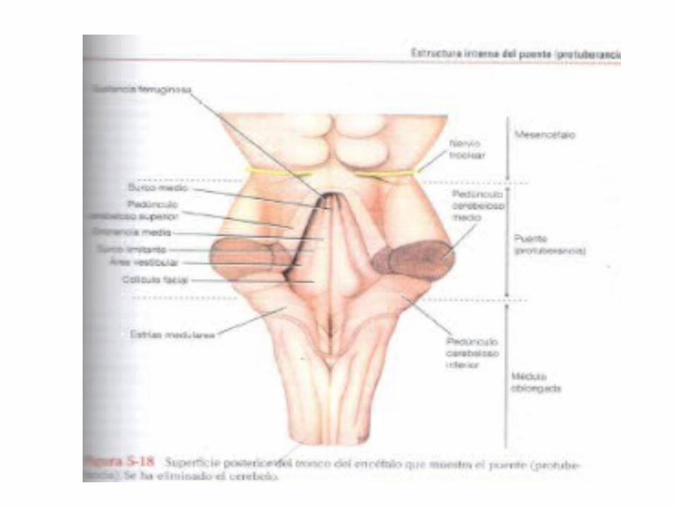
PROTUBERANCIA VIAS ASCENDENTES Y DESCENDENTES DEL TRONCO CEREBRAL MESENCEFALO DR ROBINSON LEON
Transcript of SEMINARIO 5 Y 6 - MAQUETAS 1 UNIDAD (1).pptx

PROTUBERANCIAVIAS ASCENDENTES Y DESCENDENTES DEL
TRONCO CEREBRALMESENCEFALO
DR ROBINSON LEON
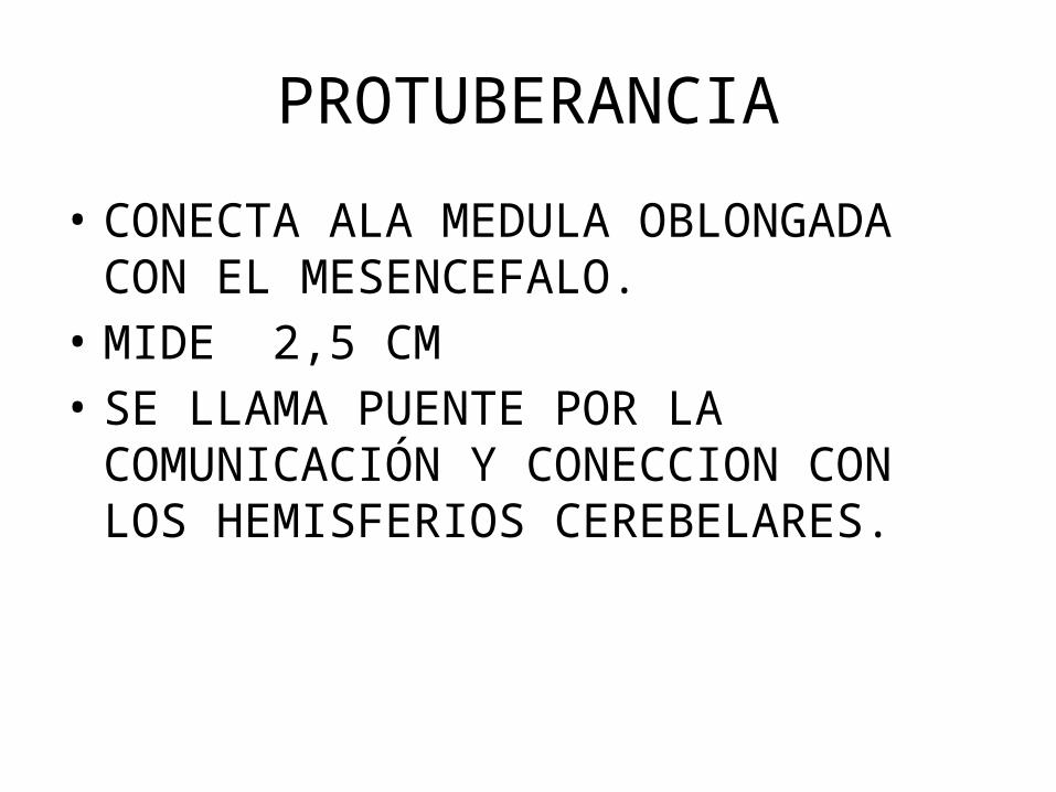
PROTUBERANCIA
• CONECTA ALA MEDULA OBLONGADA CON EL MESENCEFALO.
• MIDE 2,5 CM • SE LLAMA PUENTE POR LA COMUNICACIÓN Y
CONECCION CON LOS HEMISFERIOS CEREBELARES.
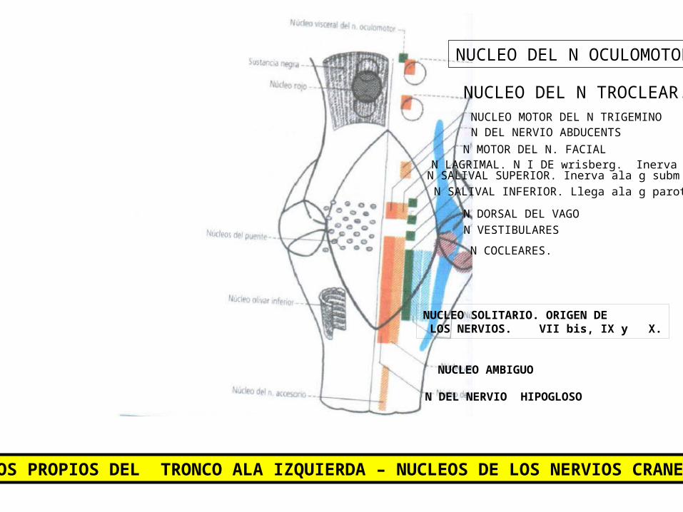
NUCLEOS PROPIOS DEL TRONCO ALA IZQUIERDA – NUCLEOS DE LOS NERVIOS CRANEALES
NUCLEO DEL N OCULOMOTOR
NUCLEO DEL N TROCLEAR.NUCLEO MOTOR DEL N TRIGEMINON DEL NERVIO ABDUCENTS
N MOTOR DEL N. FACIALN LAGRIMAL. N I DE wrisberg. Inerva ala gl.
N SALIVAL SUPERIOR. Inerva ala g subm y subl.N SALIVAL INFERIOR. Llega ala g parotida.
N DORSAL DEL VAGON VESTIBULARES
N COCLEARES.
NUCLEO SOLITARIO. ORIGEN DE LOS NERVIOS. VII bis, IX y X.
NUCLEO AMBIGUO
N DEL NERVIO HIPOGLOSO
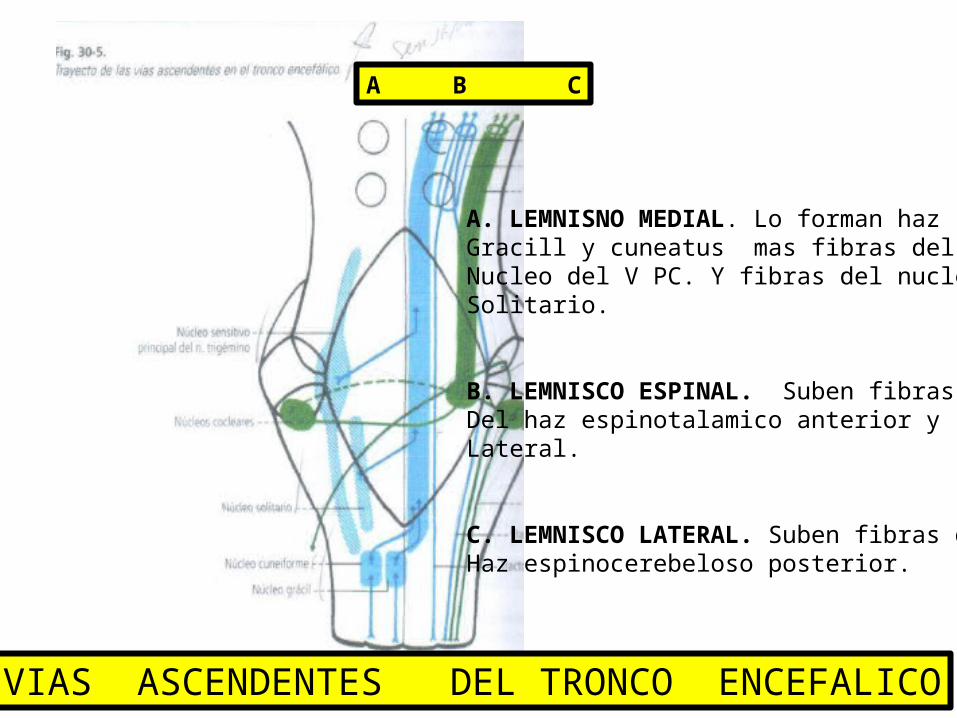
A. LEMNISNO MEDIAL. Lo forman hazGracill y cuneatus mas fibras delNucleo del V PC. Y fibras del nucleoSolitario.
B. LEMNISCO ESPINAL. Suben fibrasDel haz espinotalamico anterior yLateral.
C. LEMNISCO LATERAL. Suben fibras del Haz espinocerebeloso posterior.
A B C
VIAS ASCENDENTES DEL TRONCO ENCEFALICO

VIAS DESCENDENTES DEL TRONCO ENCEFALICOTRACTO CORTICONUCLEAR
NUCLEO ROJO
TRACTO CORTICOESPINAL ANTERIOR.
TRACTO OLIVOESPINAL
NUCLEOS VESTIBULARES
TRACTO VESTIBULOESPINAL
TRACTO CORTICOESPINAL
Division de la protuberancia
• Dos partes. Una ANTERIOR BASAL Y UNA POSTERIOR CAUDAL O TEGMENTO.
• SEPARADAS POR EL CUERPO TRAPEZOIDAL.
• Porción caudal pasa por el coliculo facial• Porción craneal pasan los nucleos trigeminos.
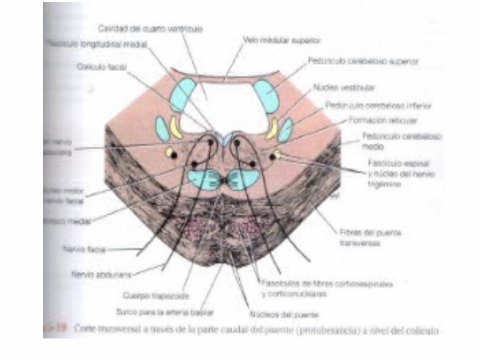
Corte a nivel de la porción caudal.
• El lemnisco medial. Rota al pasar desde la M.OBLONGADA AL PUENTE.
• EL COLICULO FACIAL. Se forma cuando la fibras del nervio FACIAL giran alrededor del NERVIO abducens.
• El núcleo del facial es posterior ala porción lateral del lemnisco medial.

• El fasciculo logitudinal medial. ESTA SITUADO debajo del suelo del IV ventriculo auno y otro lado dela linea media.
• El fasciculo longiutudinal une los nucleos vestibular y coclear con los nucleos controladores de los mus controladores.( III, IV Y VI)
• N VESTIBULAR MEDIAL. Situado lateral al nervio abducents.
• El cuerpo trapezoide. N cocleares y n del cuerpo trapezoidal.
Corte a nivel de la porción caudal.
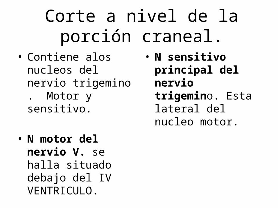
Corte a nivel de la porción craneal.
• Contiene alos nucleos del nervio trigemino . Motor y sensitivo.
• N motor del nervio V. se halla situado debajo del IV VENTRICULO.
• N sensitivo principal del nervio trigemino. Esta lateral del nucleo motor.
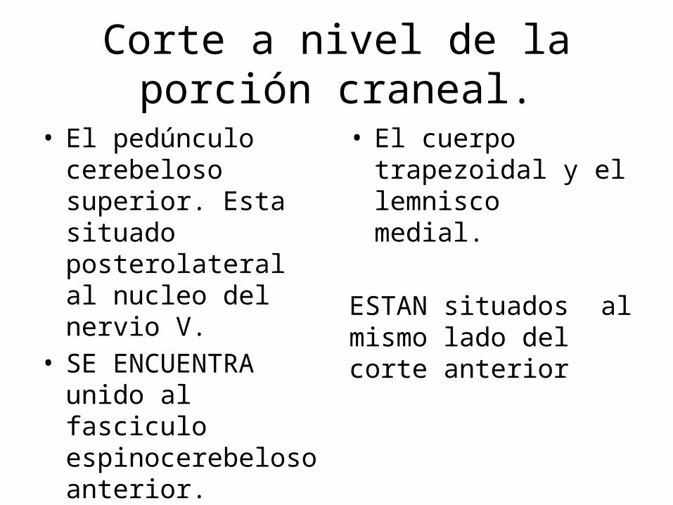
• El pedúnculo cerebeloso superior. Esta situado posterolateral al nucleo del nervio V.
• SE ENCUENTRA unido al fasciculo espinocerebeloso anterior.
• El cuerpo trapezoidal y el lemnisco medial.
ESTAN situados al mismo lado del corte anterior
Corte a nivel de la porción craneal.
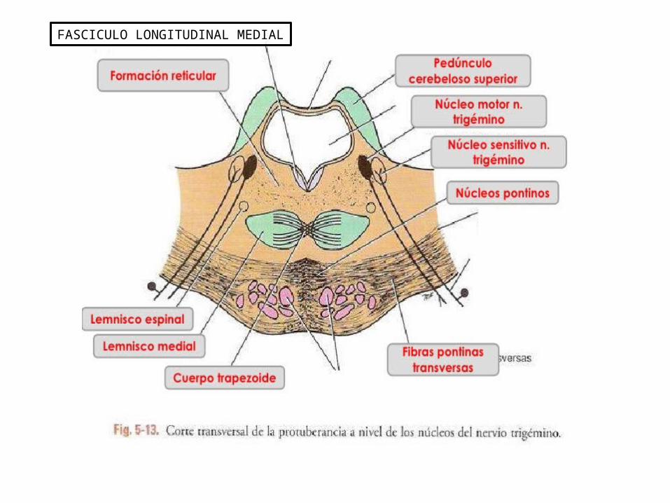
FASCICULO LONGITUDINAL MEDIAL

MESENCEFALO – MACROSCOPIASISTEMATIZACION
DR ROBINSON LEON
MESENCEFALO
• Comprende dos mitades laterales conocidas como pedunculos cerebrales.
• La parte posterior se le conoce como techo o tegmento.
• La parte anterior el pie peduncular.• Esta conectando El III – IV ventriculo atraves
del acueducto de silvio.
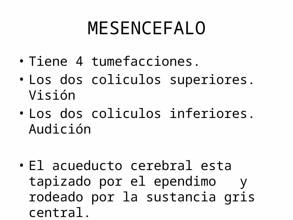
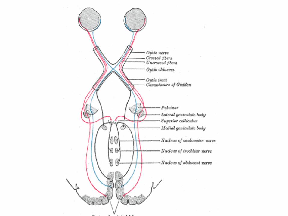
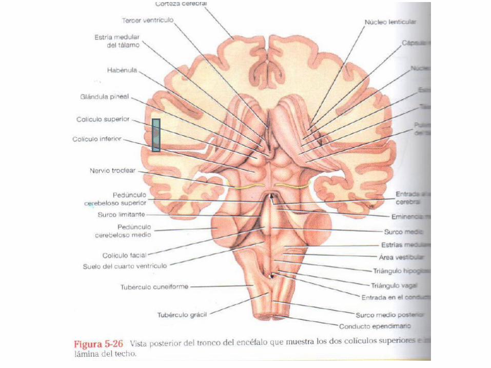
• Tiene 4 tumefacciones.• Los dos coliculos superiores. Visión• Los dos coliculos inferiores. Audición
• El acueducto cerebral esta tapizado por el ependimo y rodeado por la sustancia gris central.
MESENCEFALO
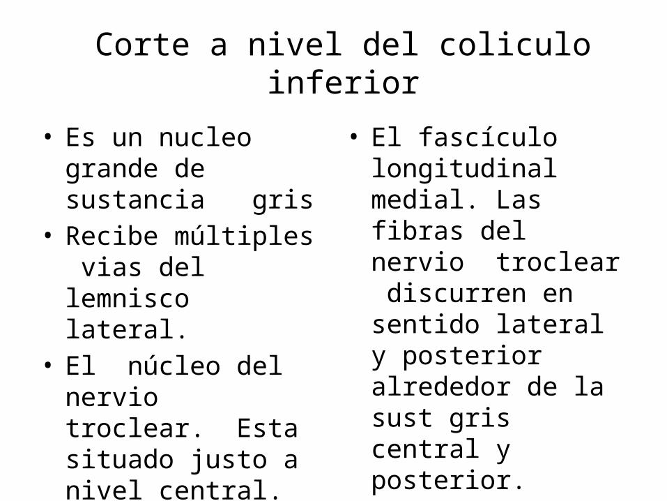
Corte a nivel del coliculo inferior
• Es un nucleo grande de sustancia gris
• Recibe múltiples vias del lemnisco lateral.
• El núcleo del nervio troclear. Esta situado justo a nivel central.
• El fascículo longitudinal medial. Las fibras del nervio troclear discurren en sentido lateral y posterior alrededor de la sust gris central y posterior.

Corte a nivel del coliculo inferior
• Forma parte de los reflejos visuales.• Nucleo gris situado debajo dela elevacion
superficial.• Recibe fibras aferentes del nervio optico ,
corteza visual y fasciculo espinotectal.• Las fibras eferentes forman los fasciculos
tecto espinal y tectomedular.
Corte a nivel del coliculo superior
• La via aferente para el reflejo luminico termnina en el nucleo pretectal.
• Luego la informacion pasa la nucleo accesorio de edinger westphal.
• Luego las fibras pasan al NERVIO OCULOMOTOR esta situado en la sust gris cerca al plano medio.
Corte a nivel del coliculo superior

LESION DEL BULBO RAQUIDEO
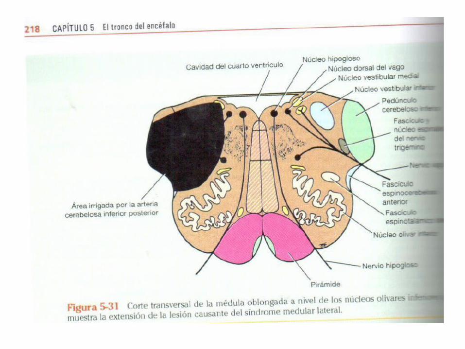
SD MEDULAR LATERAL DE WELLENBERG
• La parte lateral del bulbo es irrigada por la arteria cerebelosa inferior posterior, rama de la arteria vertebral.
• La trombosis de estas arterias produce en el paciente.
• CLINICA. DISFAGIA- DISARTRIA ( INERV POR n ambiguo), analgesia y termoalgesia. Por alteracion del nucleo y fasciculo espinal del V.
• VERTIGO, NAUSEAS , VOMITOS, NISTAGMUS. nucleos vestibulares.
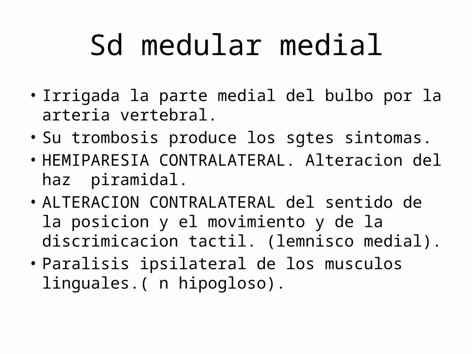
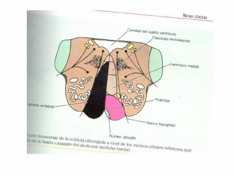
Sd medular medial
• Irrigada la parte medial del bulbo por la arteria vertebral.
• Su trombosis produce los sgtes sintomas.• HEMIPARESIA CONTRALATERAL. Alteracion del haz
piramidal.• ALTERACION CONTRALATERAL del sentido de la
posicion y el movimiento y de la discrimicacion tactil. (lemnisco medial).
• Paralisis ipsilateral de los musculos linguales.( n hipogloso).
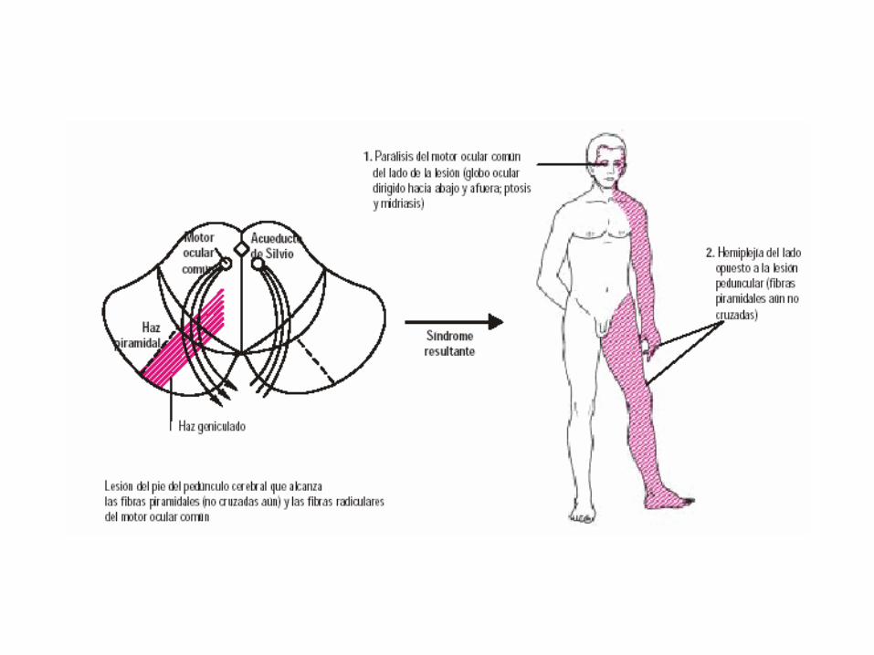
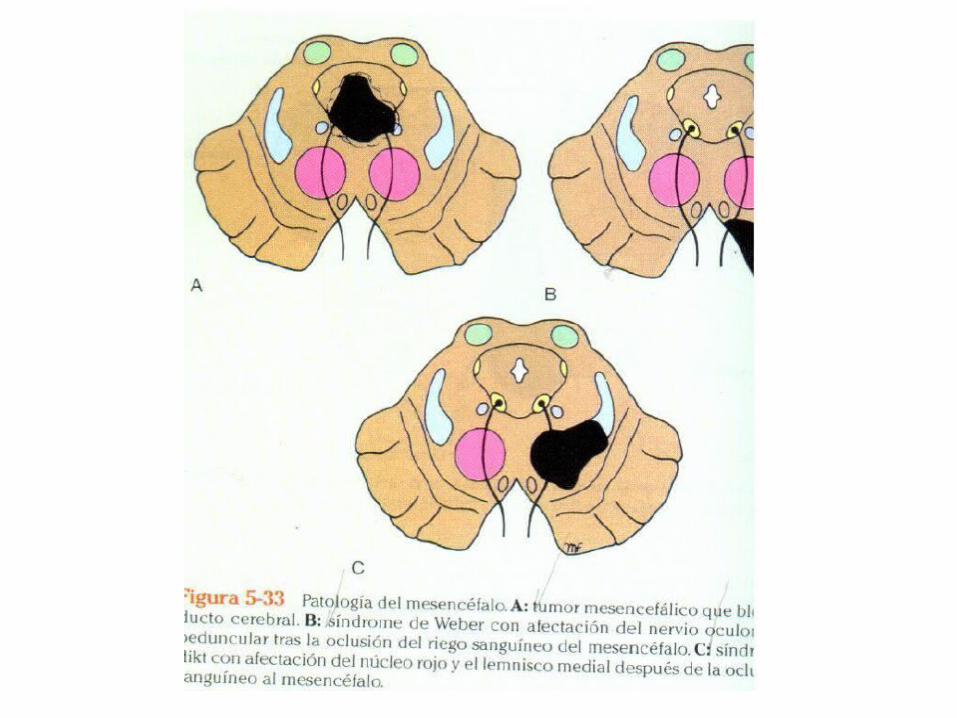
Patología del mesencefalo
• Sindrome de weber.
• Oclusion dela rama de la arteria cerebral posterior. Esta irriga al mesencefalo prod necrosis del tejido mesencefalico.
• Afecta al nervio oculomotor, y al pie peduncular.
Sd de weber
• Paciente presenta oftalmoplejia ipsilateral. Y paralisis inferior de los musculos de la cara . Lengua, brazo y pierna.
• Ojo se desvia hacia afuera por paralisis del musculo recto interno,
• Hay ptosis del parpado superior , pupila dilatada sin rta ala luz ni acomodacion.