UNIVERSIDAD COMPLUTENSE DE MADRID · A Coia, pequeña líder natural y alma de todos los grupos,...
271
UNIVERSIDAD COMPLUTENSE DE MADRID FACULTAD DE VETERINARIA DEPARTAMENTO DE FISIOLOGÍA ANIMAL TESIS DOCTORAL Effect of Origin and Culture Conditions on the Heterogeneity of Pluripotent Cell Populations Efecto del origen y las condiciones de cultivo en la heterogeneidad de poblaciones celulares pluripotentes MEMORIA PARA OPTAR AL GRADO DE DOCTORA PRESENTADA POR Priscila Ramos Ibeas Directores Alfonso Gutiérrez Adán Miguel Ángel Ramírez de Paz Madrid, 2014 ©Priscila Ramos Ibeas, 2014
Transcript of UNIVERSIDAD COMPLUTENSE DE MADRID · A Coia, pequeña líder natural y alma de todos los grupos,...
UNIVERSIDAD COMPLUTENSE DE MADRID FACULTAD DE VETERINARIA
DEPARTAMENTO DE FISIOLOGÍA ANIMAL
TESIS DOCTORAL
Effect of Origin and Culture Conditions on the Heterogeneity of Pluripotent Cell Populations
Efecto del origen y las condiciones de cultivo en la heterogeneidad de poblaciones celulares
pluripotentes
MEMORIA PARA OPTAR AL GRADO DE DOCTORA
PRESENTADA POR
Priscila Ramos Ibeas
Directores
Alfonso Gutiérrez Adán Miguel Ángel Ramírez de Paz
Madrid, 2014 ©Priscila Ramos Ibeas, 2014

UNIVERSIDAD COMPLUTENSE DE MADRID
FACULTAD DE VETERINARIA
Departamento de Fisiología Animal
EFFECT OF ORIGIN AND CULTURE CONDITIONS ON
THE HETEROGENEITY OF PLURIPOTENT CELL
POPULATIONS
EFECTO DEL ORIGEN Y LAS CONDICIONES DE CULTIVO
EN LA HETEROGENEIDAD DE POBLACIONES
CELULARES PLURIPOTENTES
MEMORIA PARA OPTAR AL GRADO DE DOCTOR
PRESENTADA POR
Priscila Ramos Ibeas
Madrid, 2014
DIRECTORES:
Alfonso Gutiérrez Adán
Miguel Ángel Ramírez de Paz


Los doctores Alfonso Gutiérrez Adán y Miguel Ángel Ramírez de Paz, Investigadores
Titulares del Departamento de Reproducción Animal del Instituto Nacional de
Investigación y Tecnología Agraria y Alimentaria (INIA) hacen constar:
Que la memoria de Tesis Doctoral presentada por la Licenciada en Veterinaria Priscila
Ramos Ibeas, con el título: “Effect of origin and culture conditions on the heterogeneity
of pluripotent cell populations / Efecto del origen y las condiciones de cultivo en la
heterogeneidad de poblaciones celulares pluripotentes”, ha sido realizada bajo nuestra
dirección y que tras su revisión consideramos que tiene la debida calidad para su
presentación y defensa.
Madrid, 25 de Abril 2014
Fdo.: D. Alfonso Gutiérrez Adán Fdo: D. Miguel Ángel Ramírez de Paz


EUROPEAN DOCTORATE MENTION
This thesis has been proposed for the European doctorate mention by virtue of
the following European research stay and thesis reports:
Research stay:
• The University of Nottingham (United Kingdom). Dr. Ramiro Alberio laboratory,
Animal Science Department, Sutton Bonington Campus; 3 months in 2013.
Thesis reports:
• Dr. András Dinnyés. Veterinary Science University, Budapest, Hungary
• Pat Lonergan. School of Agriculture & Food Science. University College Dublin,
Ireland
• Dr. María Arias Álvarez, Facultad de Veterinaria. Universidad Complutense de
Madrid, Spain


This thesis has been supported by the projects:
AGL2009-11358 (Ministry of Science and Innovation, Spain),
AGL2012-39652-C02-01 (Ministry of Economy and Competitiveness, Spain).
Priscila Ramos Ibeas was supported by
a Postgraduate Scolarship
“Formación de Personal Investigador (FPI)”
from Spanish Ministry of Science and Innovation.


Agradecimientos
Desde que me decidí a empezar con la aventura de la tesis, siempre me he
imaginado con ilusión este momento: escribir el capítulo de agradecimientos. Y es
que somos quienes somos gracias a la gente que nos rodea, nos aprecia y nos
apoya; y yo no habría llegado a este punto sin la ayuda de un montón de gente.
Espero no olvidarme de nadie.
A mi familia:
En primer lugar, mil gracias a mis padres. Creo que no se puede tener unos padres
mejores que los que yo tengo. Gracias por educarme, comprenderme y apoyarme
en todas mis decisiones, aunque no siempre haya sido fácil, y por ayudarme
siempre en todo lo que necesito. Sé que lo habitual es que sean los padres los que
se enorgullezcan de sus hijos, pero vosotros hacéis que me sienta muy orgullosa;
hasta mis amigos me dicen: “Pris, cómo molan tus padres”. Sois los mejores.
Gracias a mi hermano Óscar, por ser mi compañero de juegos y aventuras cuando
éramos pequeños. Siempre me reiré recordando cómo hacíamos el tonto en casa
por las noches sacando de quicio a mamá. Me hace feliz saber que siempre estás
ahí aunque nos veamos de ciento en viento.
A mis abuelos; los que ya no están y los que espero que sigan por muchos años,
por cuidarme y consentirme siempre que papá y mamá no estaban, sobre todo a
mi yaya Concha, que ha sido como una segunda madre. Gracias también a mis tíos
y primos, en especial a mi tía Conchi. A mis vecinos, por ser tan cercanos durante
toda mi vida en Burgos.
Gracias a los animales que han pasado por mis manos, desde los cangrejos-
mascota salvados de la cazuela, grillos y pajaritos huérfanos y rescatados de
accidentes, hasta los que quiero y he querido con toda el alma: Piti, Joey, y
especialmente Dana. Porque todos ellos despertaron mi vocación y por ello
estudié veterinaria.
A los responsables de esta tesis:
A Alfonso, por ser la persona más eficaz que he conocido. Tu ritmo de trabajo me
ha motivado a dar lo mejor de mí y a seguir adelante. Me siento muy afortunada
por haber podido pasar estos años bajo tu sabia dirección. Gracias por tu apoyo y
dedicación, y por animarme siempre a trabajar duro.
A Miguel, por el entusiasmo que le pones a cada experimento, por enseñármelo
todo sobre el mundo celular, por ser tan cercano, y por estar siempre dispuesto a
ayudar. Gracias también por tu apoyo y dedicación durante estos años.
To the researchers that accepted me during my research stays: Bhanu Telugu
(University of Maryland) and Ramiro Alberio (The University of Nottingham), for
giving me the opportunity of working and learning in their laboratories. To all the
people that I met there and helped me to feel more like at home, especially to
Haixin and to Choulia.
A todo el personal del laboratorio, porque da gusto trabajar en un ambiente así:
A Pablo, porque para mí la estancia en Maryland marcó un antes y un después.
Gracias por hacerme creer en mí, por enseñarme tantísimas cosas en el
laboratorio (y en los campos americanos) y por cuidarme como un hermano
mayor. Por estar siempre ahí con una solución para todo y por tu ayuda en este
tramo final de la tesis.
A Eva, gran compañera de sufrimientos celulares y consejera; no sé si alguna vez
te lo habré dicho pero siempre he valorado mucho tu punto de vista. Porque
siempre se puede confiar en ti para todo, muchas gracias por tu apoyo.
A Sandra, la alegría del laboratorio, porque todas las conversaciones contigo
acaban siendo entretenidas. A Raúl, porque siempre transmites calma y
serenidad, gracias por los consejos de motorista. A Toñi, por enseñarme todo del
animalario y por ser siempre tan alegre, y al resto de gente del animalario. A
Miriam. A Alberto, por los desayunos, los eventos deportivos y los festivos; se te
echa de menos. A los de bovino: Richard, Meriem y Dimitrios.
To the nice Belgian people that came to Madrid to make PCRs and to enjoy the
Spanish lifestyle, and helped me to practice my English: Jessie and Veerle.
No es que me haya olvidado del resto, sólo os he cambiado de apartado.
A mis amigos:
A Angy, una de las personas más importantes en mi vida y con la que he
compartido tantas cosas. Gracias por llegar al laboratorio e intentar convertirme
en una princesita, por estar ahí siempre dispuesta a escuchar y a repartir abrazos,
por alimentarme tan bien y tan rico, por las clases de baile y por tus modelos para
la tesis. Te quiero mucho, aunque hasta hace poco me dijeras que ya no :P.
A Coia, pequeña líder natural y alma de todos los grupos, que sepas que esto no
es lo mismo desde que te fuiste. Espero que tu aventura te traiga pronto de vuelta
a Madrid, y esta vez para quedarte.
A Fati, por ser tan especial, tan bonita y tan manchega. Por aparecer en mi vida
hace relativamente poco y haberte convertido en una imprescindible. A ti también
te quiero mucho!
A Ricardo, porque siempre estás dispuesto a apuntarte a un bombardeo, por tus
comentarios inoportunos que dejan silencios incómodos de los que nos podemos
reír después, y porque sé que siempre puedo contar contigo.
A Vero, por ser tan dulce y a la vez sacar el genio cuando hace falta, y por
escucharme siempre en los ratos de análisis y tertulia en el laboratorio. A Beto,
por los partidos del Barça y los viajes.
A todos los “Pacerditos” y agregados: a Mariana y a los productos Procter, mucha
suerte por Ginebra! A Amaia, por haber sido tan buena sustituta de Ricardo. A
Jeorgea, porque ya se queda pasadas las 12 de la noche. A Eka, por la tortilla de
patata y los viajes a Bilbao. A Yosu, por ser tan buena persona y transmitir tanta
alegría. A Celia, por no perderse nunca ningún evento de cañas.
Al equipo Esencia Norte, por los ratos de fútbol y sobre todo por tantos buenos
momentos de fuera de los campos.
Al Colegio Mayor San Juan Evangelista, el “Johnny”, y a todo lo que allí viví
durante toda la carrera. Gracias por darme esa oportunidad de conocer a tanta
gente tan diferente, y de hacer tantos y tan buenos amigos con los que pasé una
de las mejores épocas de mi vida. A Patri, Ro, Leti, Judith, Ali, Vir, MariÁngeles,

Charlie, Adolf… A Ali y a Vir, que os merecéis que os nombre otra vez porque a
pesar de que pase el tiempo y de que nos veamos poco, siento que puedo seguir
contando con vosotras igual que antes. A Luisa, porque cuando empecé con esta
tesis te imaginaba apareciendo en este capítulo, y me alegra que al final sea así.
A Iván, por ser un amigo de los buenos, de los que perdura; por seguir
manteniendo el contacto aunque esporádicamente, y porque buena parte de mis
méritos académicos durante la carrera y los de más de media promoción se deben
a tus apuntes.
A las musiqueras burgalesas: Patri, Rachel, Viole, Lore y Bea. Por aquellos años de
Conservatorio que tanto disfruté, y porque aunque andemos cada una en una
punta del planeta sigamos teniendo al menos una comida navideña en el Morito
cada año.
A Cris:
Mi pequeñi y mi todo, por aguantar mis rollos de científicos y la distancia que a
veces nos ha tocado, por apoyarme y escucharme siempre, por cuidarme y
quererme tanto, por llenarme de alegría cada vez que llegas a casa, por disfrutar
de las pequeñas cosas, por acompañarme en los viajes y sobretodo en el día a día,
porque todo es más fácil a tu lado. Te quiero “mutíísimo”.

A mis padres.
Por sentar las bases de lo que soy hoy
y por ser un apoyo constante.


A Cris.
Lo que nos depare el mañana nos hará más fuertes.


Index / Índice
I
Index / Índice

Index / Índice
II

Index / Índice
III
Abbreviations / Abreviaturas ............................................................................................ VII
Abstract ................................................................................................................................ XI
Resumen ........................................................................................................................... XVII
Introduction .......................................................................................................................... 1
Discovering pluripotency, a brief historical perspective ................................................... 3
1. From the discovery of pluripotent cells to pluripotency capture in vitro. ................ 3
2. Different theories for the origin of embryonic stem cells ......................................... 6
Pluripotency tests and hallmarks ...................................................................................... 8
Influence of cellular or embryonic source on epigenetics and pluripotency .................... 9
1. Epigenetic modifications ........................................................................................... 9
1.1. Genomic imprinting .......................................................................................... 10
1.2. X chromosome inactivation (XCI) ..................................................................... 12
2. Influence of embryonic source on ESCs epigenetics and pluripotency ................... 12
3. Influence of the cellular source on iPSCs epigenetics and pluripotency ................. 14
Influence of culture conditions on pluripotency ............................................................. 15
1. Influence of culture conditions on pluripotency capture in vitro ............................ 15
2. Influence of culture conditions on epigenetics and pluripotency maintenance ..... 17
Pluripotency in adult tissues ........................................................................................... 18
Pluripotency and germline common markers ................................................................. 20
1. Germline specification and development ............................................................... 20
2. Expression of germline markers in pluripotent cells ............................................... 21
Trophectoderm specification and pluripotency .............................................................. 25
1. Specie-specific differences in trophectoderm specification ................................... 25
2. Bovine trophoblast cell lines derivation .................................................................. 26
Introducción ........................................................................................................................ 29
El descubrimiento de la pluripotencia ............................................................................. 31
1. Del descubrimiento de las células pluripotentes a la captura de la pluripotencia in vitro .............................................................................................................................. 31
2. Diferentes teorías sobre el origen de las ESCs ........................................................ 34
Señales y pruebas de pluripotencia ................................................................................. 36

Index / Índice
IV
Influencia de la fuente embrionaria o celular en la epigenética y la pluripotencia de las
ESCs.................................................................................................................................. 38
1. Modificaciones epigenéticas ................................................................................... 38
1.1. La impronta genómica ...................................................................................... 38
1.2. La inactivación del cromosoma X ..................................................................... 40
2. Influencia de las características del embrión en la epigenética y la pluripotencia de las ESCs ........................................................................................................................ 40
3. Influencia de las características de la célula de origen en la epigenética y la pluripotencia de las iPSCs ............................................................................................ 43
Influencia de las condiciones de cultivo en la epigenética y en la pluripotencia ............ 44
1. Influencia de las condiciones de cultivo en la captura de la pluripotencia in vitro . 44
2. Influencia de las condiciones de cultivo en la estabilidad epigenética y el mantenimiento de la pluripotencia ............................................................................. 46
Pluripotencia en tejidos adultos ...................................................................................... 47
Marcadores comunes de pluripotencia y de linaje germinal .......................................... 49
1. Especificación y desarrollo del linaje germinal ........................................................ 49
2. Marcadores de linaje germinal expresados en células pluripotentes ..................... 50
Especificación del trofoectodermo ................................................................................. 54
1. Diferencias interespecíficas ..................................................................................... 54
2. Aislamiento de líneas celulares de trofoblasto bovino ........................................... 55
Bibliography/Bibliografía ................................................................................................... 57
Objectives ............................................................................................................................ 71
Objetivos ............................................................................................................................. 75
Chapter I / Capítulo I ........................................................................................................... 79
Intracytoplasmic sperm injection using DNA-fragmented sperm in mice negatively affects
embryo-derived ES cells, reduces the fertility of male offspring and induces heritable
changes in epialleles
Chapter II / Capítulo II ...................................................................................................... 105
Germ Cell culture conditions facilitate the reprogramming to produce mouse ESCs
Chapter III / Capítulo III .................................................................................................... 131
Dazl-GFP mice model generated by a two-step ESC-based strategy to identify pluripotent
and germ cells

Index / Índice
V
Chapter IV / Capítulo IV .................................................................................................... 157
An efficient system to establish biopsy-derived trophoblastic cell lines from bovine
embryos
General discussion ............................................................................................................ 187
Discusión general .............................................................................................................. 197
Bibliography / Bibliografía ................................................................................................ 209
Conclusions ....................................................................................................................... 217
Conclusiones ..................................................................................................................... 221
Curriculum vitae ................................................................................................................ 225

VI

Abbreviations / Abreviaturas
VII
Abbreviations / Abreviaturas

Abbreviations / Abreviaturas
VIII
Abbreviations / Abreviaturas
IX
2i Two kinase inhibitors
ALH Amplitude of lateral head displacement
ART Assisted reproductive technologies
ASC Adult stem cell
BBT Bovine biopsied trophoblast
BEF Bovine embryonic fibroblast
BES Base excision repair
bFGF Basic fibroblast growth factor
BM Bone marrow
BMP Bone morphogenetic protein
BNC Binucleate cell
BOEC Bovine oviductal epithelial cell
COC Cumulus oocyte complex
Ct Cycle threshold
DFS DNA fragmented sperm
DMEM Dulbecco´s modified Eagle medium
DMR Differentially methylated region
DNA Deoxyribonucleic acid
Dnmt DNA methyltransferase
Dpc Days post coitum
EB Embryoid body
ECC Embryonal carcinoma cell
eCG Equine chorionic gonadotropin
EDTA Ethylenediaminetetraacetic acid
EF Embryonic fibroblast
EGC Embryonic germ cell
EGF Epidermal growth factor
EGFP Enhanced green fluorescent protein
EpiSC Epiblast stem cell
ESC Embryonic stem cell
ExE Extraembryonic ectoderm
FCS Foetal calf serum
FGF4 Fibroblast Growth Factor 4
GDNF Glial cell-derived neurotrophic factor
GSC Germline stem cell
GSK3 Glycogen synthase kinase 3
hAFSC Human amniotic fluid stem cell
hCG Human chorionic gonadotropin
HSC Hematopoietic stem cell
IAP Intracisternal-A particle
ICM Inner cell mass
Abbreviations / Abreviaturas
X
ICSI Intracytoplasmic sperm injection
iPSC Induced pluripotent stem cell
ISAS Integrated semen analysis system
IVC In vitro cultured
IVF In vitro fertilized
KSR Knockout serum replacement
LIF Leukemia inhibitory factor
LTR Long terminal repeat
MAPK/MEK Mitogen-activated protein kinase
MEF Mouse embryonic fibroblast
MEG Maternally expressed gene
mGSC Multipotent germline stem cell
MNC Mononucleate cell
MSC Mesenchymal stem cell
NES Nucleotide excision repair
PBS Phosphate-buffered saline
PGC Primordial germ cell
PI Propidium iodide
PMSG Pregnant mare serum gonadotropin
RNA Ribonucleic acid
RT-PCR Reverse transcription-polymerase chain reaction
SCF Stem cell factor
SCNT Somatic cell nuclear transfer
SNP Single nucleotide polymorphism
SOF Synthetic oviduct fluid
SPV Smoothed path velocity
SSC Spermatogonial stem cell
STR Straightness ratio of VSL/VAP
TE Trophectoderm
TET Ten-eleven translocation
TSC Trophoblast stem cell
TUNEL Terminal deoxynucleotidyl transferase dUTP nick end labeling
TV Track velocity
Vs. Versus
VSELs Very small embryonic stem cells
XCI X chromosome inactivation

Abstract
XI
Abstract

Abstract
XII
Abstract
XIII
Pluripotent cells have fascinated the society since they were first discovered, and much
research has been performed on them as they constitute a powerful tool for regenerative
medicine or genetic manipulation. However, basic research is necessary to determine the
optimal conditions for their identification, isolation and in vitro culture. Thus, in this
thesis, several crucial aspects for cell lines derivation, such as the effect of embryonic
source or culture conditions, or the origin of pluripotent cells, have been analyzed.
Embryonic stem cells (ESCs) have been widely used for research, and ESCs isolation
techniques and culture systems have evolved in the last years improving derivation
efficiency. Nevertheless, it remains controversial whether embryonic characteristics have
an influence over the process. Some studies have described that modifications or
alterations present in the original embryonic cells can be transmitted to their
corresponding ESCs lines. However, in other circumstances, embryonic characteristics are
not reflected in ESCs lines. In chapter I, the potential to derive ESCs lines from bad quality
embryos was analyzed, and it was investigated whether these ESCs lines reflect some of
the characteristics previously observed in such embryos.
Assisted reproductive technologies (ARTs) have been widely performed in humans and
animals. However, embryo manipulation and in vitro culture is associated with
perturbations of the embryonic ultrastructure and genetic and epigenetic alterations that
may result in long-term effects, causing syndromes and diseases during adulthood.
Especially worrying is the case of intracytoplasmic sperm injection (ICSI), as it bypasses
the natural selection of spermatozoa during fertilization, allowing sperm with fragmented
or damaged DNA to fertilize an oocyte. In this perspective, ICSI using DNA-fragmented
sperm (DFS-ICSI) in mice was used as a model to generate bad quality embryos for ESCs
lines derivation in chapter I of this thesis. DFS-ICSI embryos showed a reduced potential
to generate ESCs lines compared to in vivo-produced embryos. Furthermore, during early
passages, these DFS-ICSI-ESCs differed from in vivo-ESCs in the expression of certain
genes related to pluripotency and epigenetic repression, DNA damage and repair, and de
novo DNA methyltransferases and histone deacetylases. Gene deregulation in ESCs could
reflect the alterations previously described in DFS-ICSI-generated embryos and adult
animals However, at late passages, DFS-ICSI and in vivo-ESCs adopted similar expression
profiles. Consequently, ESCs retain some memory of the embryos from which they were
derived, although continuous passaging directs them to adopt similar profiles.
Furthermore, in chapter I it was observed that DFS-ICSI reduces sperm production and
fertility in the male progeny, and affects the postnatal expression of a defined
epigenetically sensitive allele, and that this modification may be inherited across
generations.
Abstract
XIV
Culture conditions constitute another main factor affecting cell lines derivation and
features. In chapter II, the effect of culture conditions was analyzed during ESCs lines
derivation. First, the effect of Leukemia inhibitory factor (LIF) supplementation during
preimplantational embryo culture up to the blastocyst stage was analyzed. LIF, which is
an essential factor for ESCs derivation and maintenance, could favor the transition from
embryonic cells to ESCs. Although the embryos supplemented with LIF showed a lower
total cell number, the ratio of inner cell mass (ICM)/total cells in blastocysts was
significantly higher. Furthermore, these embryos proved to be more suitable for ESCs
isolation, as their ESCs derivation efficiencies were higher. Additionally, blastocysts were
cultured over a feeder layer of mouse embryonic fibroblasts (EF) in medium
supplemented with: fetal calf serum (FCS) and LIF (ES medium); FCS, LIF and germline
stem cells (GSCs)-related growth factors (GS medium); or FCS, LIF and MEK and GSK3β
inhibitors (2i medium). We could observe an improvement in ESCs derivation efficiency
when GS medium was used compared to ES medium. ESCs lines derived after LIF
supplementation up to blastocyst stage and posterior culture in GS medium (LIF+GS)
fulfilled pluripotency criteria and presented a higher expression of the imprinted gene
Meg3, whose expression levels have been associated with chimeric animals formation
ability in induced pluripotent stem cells (iPSCs). Meg3 overexpression correlated to higher
ESCs derivation efficiency and better chimeric animals formation ability. Thus, a possible
synergy between LIF supplementation during embryo culture and the posterior addition
of growth factors present in GS medium was observed that favours ESCs derivation.
Therefore, ESCs derivation efficiency is highly dependent on culture medium. In the same
way, transcriptome is defined by culture conditions, as gene expression in ESCs lines
varied according to the different culture media employed for their derivation (ES, GS and
2i). Indeed, it has been reported that culture conditions are the major aspect determining
gene expression, over embryonic origin and derivation procedure.
Historically, murine ESCs were equated to ICM cells because they were first produced
from 3.5 dpc mouse blastocysts. However, several evidences based on the similarities
existing among ESCs and embryonic germ cells (EGCs), the expression of key pluripotency
genes in primordial germ cells (PGCs), or the fact that germ-cell markers are expressed in
pluripotent cells indicate a different origin for ESCs. One of the most interesting theories
states that a particular subpopulation of epiblast cells predisposed to differentiate
towards the germ cell lineage is selected during the derivation process, giving rise to
ESCs.
In chapter II, blastocysts were cultured in a germ cell-specific (GS) medium to favor ESCs
lines derivation through a possible germ cell-like intermediate state. ESCs derivation
efficiency was significantly higher in GS medium than in regular ES medium. The
expression of germ cell specific genes was detected in all conditions, although there was

Abstract
XV
no evidence for a shift towards germ cell specification induced by GS medium, as we
found germ cell-specific genes expression in all culture conditions (GS, ES and 2i). The
appearance of this common germ cell-like intermediate state has been reported in other
articles that used standard FCS culture conditions for ESCs derivation, but it seems to be
facultative for the stabilization of pluripotency in vitro, since culture in 2i conditions
without FCS or EFs enables the effective direct recruitment of ESCs skipping this step.
Consequently, it could be possible that the intermediate germ cell-like state is induced by
FCS or by other factors secreted by murine EFs.
Multipotent stem cells could also present this germ cell-like state, as it has been
demonstrated that RNA processing pattern in certain stem cells is similar to the testicle,
and the expression of germline-specific genes has been detected in adult tissues
containing multipotent cells populations. In chapter III, we studied the expression of the
germline-specific gene Dazl throughout development by using a Dazl-eGFP-transgenic
mouse. Preimplantational embryos, foetal, neonatal and adult tissues were analyzed for
Dazl-driven-eGFP expression that could indicate the presence of pluripotent cells. During
preimplantational embryo development, Dazl-eGFP was detected from zygote to
blastocysts. Although Dazl-eGFP expression was localized mainly in the gonads during
fetal development and in adulthood, it was also detected in other tissues as intestine and
bone marrow. Interestingly, different multipotent cells populations reside in these
tissues, such as intestinal stem cells and bone marrow mesenchymal stem cells.
Supporting our results, other studies have reported the expression of germline-specific
genes in mouse and human bone marrow. Thus, multipotent cells could share a common
germ cell-like origin with other in vitro cultured pluripotent populations, and Dazl-eGFP
transgene could be used to explore the presence of multipotent cells in different tissues.
In chapter IV, a system to derive bovine biopsy-derived trophoblastic cell lines was
established. Two critical aspects for bovine trophoblastic cell lines establishment are
embryo or biopsy adhesion velocity to the culture plate and a suitable culture medium.
To date, most of the bovine trophoblastic cell lines have been derived by co-culture with
mouse embryonic fibroblasts. We have developed a microdrop culture system over a
gelatinized surface to enhance fast adhesion, and we have analyzed culture media
conditioned by different cell lines as an alternative to co-culture, avoiding the risk of
contamination with other cell types. Conditioned media from mouse embryonic
fibroblasts (Cm), bovine embryonic fibroblasts (Cb) and bovine oviductal cells (Co) were
assayed. Except for Cb, conditioned media improved derivation efficiency, being Cm the
most efficient medium for trophoblastic cell lines derivation. High variability in gene
expression patterns was observed in trophoblastic cell lines derived in the same
conditions. These different gene expression patterns should be due to the embryonic
source, confirming that trophoblastic cell lines derivation, like ESCs derivation, is affected

Abstract
XVI
by embryonic characteristics. In the same way, transcriptome seems to be affected by
long term culture as gene expression patterns varied along time in culture, indicating that
trophoblastic cell lines are dynamic populations. Trophoblastic cell lines mimicked in vivo
trophectoderm behaviour and showed characteristics previously described by other
authors as mononucleate and binucleate cells presence and trophoblastic-specific genes
expression. Furthermore, cell lines were able to proliferate for more than two years, and
pluripotency-related genes expression was detected, revealing certain self-renewal
capacity and the presence of a population of multipotent cells.

Resumen
XVII
Resumen

Resumen
XVIII
Resumen
XIX
Desde su descubrimiento, las células pluripotentes han fascinado a la sociedad y han sido
ampliamente utilizadas en investigación debido a que constituyen una poderosa
herramienta para la medicina regenerativa y para la manipulación genética. Existen
todavía muchas lagunas sobre las condiciones óptimas para su identificación, aislamiento
y cultivo in vitro. Por ello, en esta tesis se han analizado varios aspectos determinantes
para la obtención de líneas celulares, como el efecto de la fuente embrionaria, las
condiciones de cultivo o el origen de las células pluripotentes.
Las células troncales embrionarias (“embryonic stem cells”, ESCs) son uno de los tipos de
células pluripotentes más empleado, y las técnicas para su aislamiento y cultivo han
evolucionado en los últimos años para mejorar su eficiencia de obtención, pero aún se
desconoce si las características embrionarias influyen en su aislamiento. Estudios previos
han descrito que las modificaciones o alteraciones presentes en las células embrionarias
originales pueden ser transmitidas, en algunos casos, a sus correspondientes líneas de
ESCs; sin embargo, en otras circunstancias no se ven reflejadas en las ESCs. En el capítulo I
de esta tesis se ha analizado el potencial de embriones de “mala calidad” para dar lugar a
líneas de ESCs, y si estas líneas de ESCs reflejan algunas de las características previamente
observadas en dichos embriones.
Las técnicas de reproducción asistida (“assisted reproductive technologies”, ARTs) han
sido extensamente utilizadas en humanos y en animales. Sin embargo, la manipulación y
el cultivo in vitro de los embriones se ha asociado con la aparición de alteraciones
genéticas, epigenéticas y en la ultraestructura de los embriones que pueden dar lugar a
síndromes y enfermedades durante la edad adulta. El caso de la técnica de inyección
intracitoplasmática de espermatozoides (“intracytoplasmic sperm injection”, ICSI) es en
algunos casos especialmente preocupante, ya que franquea las barreras de selección
natural del esperma, permitiendo que espermatozoides con ADN dañado o fragmentado
fertilicen el ovocito. Por lo tanto, la técnica de ICSI utilizando espermatozoides con ADN
fragmentado (“DNA-fragmented sperm”, DFS-ICSI) en el ratón se ha utilizado como un
modelo para generar embriones de “mala calidad” para la obtención de líneas de ESCs en
el capítulo I de esta tesis. Los embriones generados por DFS-ICSI mostraron un menor
potencial para generar líneas de ESCs que los embriones producidos in vivo. Además, en
pases tempranos, estas líneas obtenidas mediante DFS-ICSI mostraron diferencias en la
expresión de ciertos genes relacionados con la pluripotencia y la represión epigenética, el
daño y la reparación del ADN, ADN metil-transferasas de novo y deacetilasas de histonas,
en comparación con las ESCs procedentes de embriones producidos in vivo. Sin embargo,
en pases tardíos las ESCs in vivo y las procedentes de DFS-ICSI adoptaron perfiles de
expresión génica similares. Consecuentemente, las ESCs retienen cierta memoria de los
embriones de los que proceden, aunque el cultivo a largo plazo hace que adopten perfiles
similares.
Resumen
XX
Además, en el capítulo I se observaron otras alteraciones en los animales obtenidos
mediante DFS-ICSI, como una reducción en la producción de esperma y en la fertilidad de
los machos de la descendencia, y ciertas alteraciones en la expresión postnatal de un
alelo sensible definido epigenéticamente, siendo esta modificación heredada
transgeneracionalmente.
Las condiciones de cultivo son otro factor crítico que afecta a la obtención de líneas
celulares y a sus posteriores características. En el capítulo II se ha analizado el efecto de
las condiciones de cultivo durante la obtención de líneas de ESCs. En primer lugar se
analizó el efecto de suplementar el medio con el factor inhibidor de leucemia (“Leukemia
inhibitory factor”, LIF) durante el cultivo del embrión preimplantacional hasta el estadio
de blastocisto. LIF, que es esencial para las ESCs de ratón, podría favorecer la transición
de las células embrionarias a ESCs. Aunque los embriones suplementados con LIF
mostraron un menor número total de células, la proporción entre el número de células de
la masa celular interna (ICM) y las células totales del blastocisto fue significativamente
mayor. Además, estos embriones fueron más aptos para el aislamiento de ESCs, ya que
mostraron una mayor eficiencia de obtención. Posteriormente, los blastocistos fueron
cultivados sobre una monocapa de fibroblastos embrionarios (“embryonic fibroblasts”,
EF) murinos en medio suplementado con: suero fetal bovino (“fetal calf serum”, FCS) y LIF
(medio ES); FCS, LIF y factores de crecimiento utilizados para el cultivo de células
troncales de linaje germinal (“germline stem cells”, GSCs) (medio GS); o FCS, LIF e
inhibidores de MEK y GSK3β (medio 2i). Pudimos observar un incremento en la eficiencia
de obtención de ESCs en el medio GS en comparación con el medio ES. Las líneas
obtenidas tras la adición de LIF durante el cultivo hasta blastocisto y el posterior empleo
de medio GS (LIF+GS) presentaron un buen patrón de pluripotencia y una mayor
expresión del gen de imprinting Meg3, cuyos niveles de expresión se han asociado con la
capacidad de formación de animales quiméricos de las células de pluripotencia inducida
(“induced pluripotent stem cells”, iPSCs). Dicha sobre-expresión de Meg3 correlacionó
con una mayor eficiencia de obtención de ESCs y con una mayor capacidad de formación
de ratones quiméricos. Se observó por lo tanto una posible sinergia entre la
suplementación con LIF durante el cultivo embrionario in vitro y la adición posterior de
otros factores de crecimiento presentes en el medio GS que favorece la creación de
nuevas líneas de ESCs. Por ello, la eficiencia de obtención de ESCs depende en gran
medida del medio de cultivo. Del mismo modo, el transcriptoma está determinado por las
condiciones de cultivo, ya que la expresión génica de las líneas de ESCs varió de acuerdo a
los diferentes medios de cultivo empleados para su obtención (ES, GS y 2i). De hecho, se
ha descrito que las condiciones de cultivo son el factor más condicionante para la
expresión génica de las células pluripotentes, por encima del origen embrionario o el
procedimiento de obtención.

Resumen
XXI
Históricamente las ESCs se han considerado equivalentes a las células de la ICM porque se
obtuvieron por vez primera a partir de blastocistos murinos de día 3,5. Sin embargo,
varias evidencias como la similitud existente entre las ESCs y las células germinales
embrionarias (“embryonic germ cells”, EGCs), la expresión de genes de pluripotencia en
células primordiales germinales (“primordial germ cells”, PGCs), o el hecho de que en las
células pluripotentes se expresen marcadores de células germinales, indican que las ESCs
podrían tener un origen diferente. Una de las teorías más interesantes acerca de ello
indica que una subpoblación de células del epiblasto en concreto, predispuesta a
diferenciarse hacia el linaje germinal, sería seleccionada durante el proceso para dar lugar
a las ESCs.
En el capítulo II se cultivaron blastocistos en un medio específico de células germinales
(medio GS) para favorecer la obtención de líneas de ESCs a través de este estado
intermedio predispuesto a diferenciarse hacia el linaje germinal, y la eficiencia de
obtención fue significativamente mayor en medio GS que en el medio ES tradicional. Sin
embargo, no hubo resultados que indicaran una diferenciación más pronunciada hacia el
linaje germinal en estas células inducida por el medio GS en concreto, ya que pudimos
encontrar expresión de genes específicos del linaje germinal en todas las condiciones de
cultivo (ES, GS y 2i), indicando la aparición de este estado similar a una célula germinal
durante el proceso de obtención de ESCs en todas las condiciones analizadas. La aparición
de este estado se ha descrito en otros artículos en los que se utilizaron condiciones de
cultivo estándares con FCS para la obtención de ESCs; aunque parece no ser indispensable
para la estabilización de la pluripotencia in vitro, ya que el cultivo en condiciones 2i (con
los inhibidores de MEK y GSK3β) sin el uso de FCS o EFs, permite el reclutamiento directo
y efectivo de ESCs saltándose este estado. Consecuentemente, podría ser posible que el
estado intermediario similar a una célula germinal esté inducido por el FCS o por otros
factores secretados por los EFs.
Por otra parte, las células multipotentes podrían mostrar también este estado similar a
una célula germinal, ya que se ha demostrado que el patrón de procesamiento de RNA de
ciertas células multipotentes es similar al del testículo; además, se ha encontrado
expresión de genes específicos del linaje germinal en algunos tejidos adultos que
contienen poblaciones de células multipotentes. En el capítulo III hemos estudiado el gen
específico del linaje germinal Dazl mediante un ratón transgénico Dazl-eGFP, analizando
el desarrollo embrionario preimplantacional y los tejidos fetales, neonatales y adultos en
busca de expresión de eGFP inducida por Dazl que pudiera indicar la presencia de células
pluripotentes. Durante el desarrollo embrionario preimplantacional, Dazl-eGFP fue
detectado desde el estadio de cigoto hasta el de blastocisto. Aunque la expresión de Dazl-
eGFP se localizó principalmente en las gónadas durante el desarrollo fetal y en la edad
adulta, también se detectó en otros tejidos como el intestino y la médula ósea.
Resumen
XXII
Curiosamente, en estos tejidos existen diferentes poblaciones de células multipotentes
como las células troncales intestinales y las células troncales mesenquimales de la médula
ósea. Otros estudios han descrito la expresión de genes específicos del linaje germinal en
la médula ósea en humanos y en el ratón. Por ello, las células multipotentes podrían
compartir un origen común similar a una célula germinal con otras poblaciones de células
pluripotentes cultivadas in vitro, y el transgen Dazl-eGFP podría ser usado para explorar la
presencia de células multipotentes en diferentes tejidos.
En el capítulo IV de esta tesis se ha establecido un sistema para obtener líneas celulares
trofoblásticas a partir de biopsias embrionarias bovinas. Dos aspectos críticos para el
establecimiento de líneas celulares trofoblásticas bovinas son la velocidad de adhesión
del embrión o de la biopsia a la placa de cultivo, y el empleo de un medio de cultivo
adecuado. Hasta la fecha, la mayoría de las líneas de células trofoblásticas bovinas han
sido obtenidas mediante co-cultivo sobre una monocapa de fibroblastos embrionarios
murinos. Nosotros hemos desarrollado un sistema de cultivo en microgota sobre una
superficie gelatinizada para facilitar una rápida adhesión, y hemos analizado medios
condicionados por diferentes líneas celulares como alternativa al cocultivo, evitando así el
riesgo de contaminación con otros tipos celulares. Se ha analizado la eficiencia de
obtención de líneas celulares en medios de cultivo condicionados por fibroblastos
embrionarios murinos (Cm), fibroblastos embrionarios bovinos (Cb) y células oviductales
ovinas (Co). A excepción del medio Cb, los medios condicionados mejoraron la eficiencia
de obtención, siendo Cm el medio más eficiente para el aislamiento de líneas de células
trofoblásticas. Se observó una alta variabilidad en los patrones de expresión génica entre
las líneas de células trofoblásticas obtenidas en las mismas condiciones. Dichas
diferencias en la expresión génica podrían estar debidos a la fuente embrionaria,
confirmando que la obtención de líneas de células trofoblásticas, al igual que la obtención
de ESCs, se encuentra afectada por las características embrionarias. Del mismo modo, el
cultivo a largo plazo parece afectar al transcriptoma de estas líneas celulares, ya que los
patrones de expresión génica variaron a lo largo de diferentes pases, lo que también
indica que las líneas de células trofoblásticas son poblaciones dinámicas. Las líneas de
células trofoblásticas generadas mostraron un comportamiento similar al del
trofectodermo in vivo y características similares a las descritas previamente por otros
autores como la presencia de células mononucleadas y binucleadas, y la expresión de
genes específicos del trofoblasto. Además, las líneas celulares fueron capaces de
proliferar durante más de dos años y se detectó expresión de genes relacionados con la
pluripotencia, revelando cierta capacidad de auto-renovación o la presencia de una
población de células multipotentes.

Introduction
1
Introduction

Introduction
2
Introduction
3
Discovering pluripotency, a brief historical
perspective
1. From the discovery of pluripotent cells to pluripotency capture in vitro. Pluripotency and stem cells have fascinated both biologists and clinicians for over a
century. The term stem cell was first employed in the scientific literature as early as 1868
by the eminent german biologist Ernst Haeckel, who used the term ‘‘Stammzelle’’ to
describe the ancestor unicellular organism from which he presumed all multicellular
organisms evolved (Haeckel 1868). Later, he proposed that the fertilized egg should also
be called stem cell (Haeckel 1877). Thus, according to Haeckel, the term stem cell was
used in two senses: as the unicellular ancestor of all multicellular organisms and as the
fertilized egg that gives rise to all cells of the organism (Ramalho-Santos and Willenbring
2007).
In 1892, another german scientist, Theodor Boveri, took Haeckel’s definition of stem cell
as the fertilized egg one step further: he proposed the term stem cell for the earliest
germline originated in animal embryos, which would presumably carry the germ-plasm
and would differentiate later into germ cells (Boveri 1892). Therefore, in these early
studies, the term stem cell referred to what we today call germline, or primordial germ
cells.
Four years later, the term was popularized in the English language by Edmund B. Wilson,
an American scientist who reviewed Boveri’s studies in his famous book The Cell in
Development and Inheritance (Wilson 1896). This book was inspirational to embryologists
and geneticists of the time, and Wilson was generally credited as having coined the term
stem cell.
Around the same time, research on the development and regeneration of the
hematopoietic system was going on, and a group of scientists believed that a cell existed
that represented the common origin of the various cell types of the blood. Some of them
began to use the term stem cell to refer this common precursor (Pappenheim 1896,
Ramalho-Santos and Willenbring 2007).
Thus, the first interpretations of stem cells in the late 19th century concerned
fundamental questions in embryology: the continuity of the germline and the origin of
the blood system.
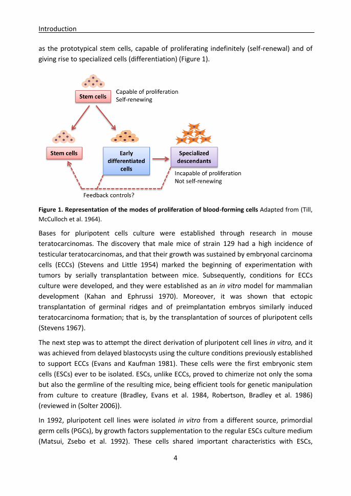
The existence of hematopoietic stem cells was demonstrated afterwards (Till and Mc
1961, Becker, Mc et al. 1963, Till, McCulloch et al. 1964) and these cells were established
Introduction
4
as the prototypical stem cells, capable of proliferating indefinitely (self-renewal) and of
giving rise to specialized cells (differentiation) (Figure 1).
Figure 1. Representation of the modes of proliferation of blood-forming cells Adapted from (Till,
McCulloch et al. 1964).
Bases for pluripotent cells culture were established through research in mouse
teratocarcinomas. The discovery that male mice of strain 129 had a high incidence of
testicular teratocarcinomas, and that their growth was sustained by embryonal carcinoma
cells (ECCs) (Stevens and Little 1954) marked the beginning of experimentation with
tumors by serially transplantation between mice. Subsequently, conditions for ECCs
culture were developed, and they were established as an in vitro model for mammalian
development (Kahan and Ephrussi 1970). Moreover, it was shown that ectopic
transplantation of germinal ridges and of preimplantation embryos similarly induced
teratocarcinoma formation; that is, by the transplantation of sources of pluripotent cells
(Stevens 1967).
The next step was to attempt the direct derivation of pluripotent cell lines in vitro, and it
was achieved from delayed blastocysts using the culture conditions previously established
to support ECCs (Evans and Kaufman 1981). These cells were the first embryonic stem
cells (ESCs) ever to be isolated. ESCs, unlike ECCs, proved to chimerize not only the soma
but also the germline of the resulting mice, being efficient tools for genetic manipulation
from culture to creature (Bradley, Evans et al. 1984, Robertson, Bradley et al. 1986)
(reviewed in (Solter 2006)).
In 1992, pluripotent cell lines were isolated in vitro from a different source, primordial
germ cells (PGCs), by growth factors supplementation to the regular ESCs culture medium
(Matsui, Zsebo et al. 1992). These cells shared important characteristics with ESCs,
Introduction
5
including morphology, pluripotency and germline transmission in chimera formation.
They were called embryonic germ cells (EGCs), to distinguish them from ESCs derived
from blastocysts.
Since murine ESCs were isolated for the first time, it took seventeen years until the
isolation of human ESCs was announced (Thomson, Itskovitz-Eldor et al. 1998). This was
probably due to the developmental differences existing between these species. However,
pluripotency of these cells was lower than their murine counterparts, as they were not
competent to contribute to blastocyst chimeras under standard culture conditions (it has
been recently discovered that culture medium supplementation with certain factors
increases human ESCs pluripotency, making them equivalent to murine ESCs and able to
contribute to chimeras (Gafni, Weinberger et al. 2013)). Moreover, human ESCs formed
flat colonies, resembling cell lines derived from mouse late epiblasts, termed epiblast
stem cells (EpiSCs) (Tesar, Chenoweth et al. 2007), in contrast to dome-shaped colonies
formed by mouse ESCs. Therefore, two states of pluripotency were proposed: naïve
pluripotency, comprising rodent ESCs depending on LIF/Stat3 signaling, and primed
pluripotency, comprising primate ESCs and rodent EpiSCs depending on Fgf/ERK signaling
(Nichols and Smith 2009).
Although human ESCs derivation was recognized as a great medical advance, several
ethical concerns raised about the employment of human embryos for research. These
concerns were overcame by the discovery of the reprogramming factors (Oct4, Sox2, Klf4
and c-Myc) by Yamanaka et al., which allowed the reprogramming of somatic cells into
pluripotent cells, generating mouse induced pluripotent stem cells (iPSCs) (Takahashi and
Yamanaka 2006). Subsequently, human iPSCs were obtained omitting c-Myc and using
LIN28 as the fourth factor (Takahashi, Tanabe et al. 2007).
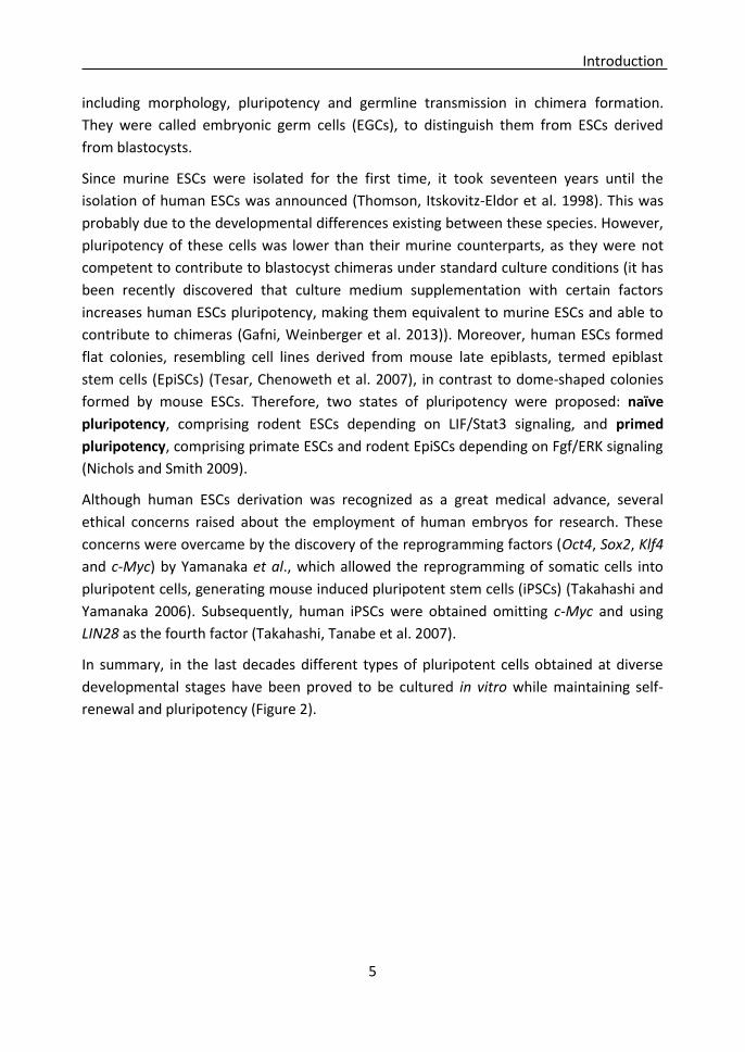
In summary, in the last decades different types of pluripotent cells obtained at diverse
developmental stages have been proved to be cultured in vitro while maintaining self-
renewal and pluripotency (Figure 2).

Introduction
6
Figure 2. Pluripotent cell types. During preimplantation development, ESCs can be derived from
early epiblasts from delayed blastocysts. Later, EpiSCs can be derived from late epiblasts from
implanted embryos. Afterwards, EGCs can be isolated from PGCs in foetus, and during adulthood
iPSCs can be obtained through somatic cell reprogramming and ECCs can be isolated from
teratocarcinomas.
2. Different theories for the origin of embryonic stem cells During development, a totipotent zygote gives rise to billions, even trillions, of
differentiated cells in a coordinated manner to form an organism (Niwa and Fujimori
2010).
In vertebrates, the evolution of the embryo gives rise to both extraembryonic lineages
and proembryonary cells. Proembryonary cells are pluripotent cells that maintain an
undifferentiated state and show a high mitotic rate (Johnson 2001), and these properties
lead directly to their ability to derive ESCs. According to the taxonomy and to the
developmental stage, proembryonary cells can have different names. They are called
inner cell mass in mice and later epiblast (Rossant 2001). In birds they are known as germ,
then blastoderm and later epiblast (Eyal-Giladi and Kochav 1976), and their equivalent in
marsupials is pluriblast and later epiblast (Johnson and Selwood 1996).
Morphology and distribution of ICM and TE depend on taxonomy. In marsupials both ICM
and TE are superficial, establishing an unilaminar blastocyst. After hypoblast
differentiation the blastocyst becomes bilaminar, being the epiblast and the TE
superficial. In birds, the X stage of Hamburger and Hamilton is composed of two
differentiated regions: the area pellucida that will form the embryo and a surrounding
dark area (area opaca) that will form the extraembryonic yolk sac (Eyal-Giladi and Kochav
1976). The formation of the hypoblast starts in the X stage as well, and in the XI stage, the
Introduction
7
area pellucida is composed of both the epiblast and the hypoblast. Thus, the X stage is
used in birds for ESCs isolation and for gene targeting to produce chimeric animals. In
zebrafish, medakafish and goldfish, after the tenth division (mid-blast), the zygotic
genome is activated giving rise to the first three cell lineages. Two of them are
extraembryonic lineages: the yolk sincitial layer, and the surrounding external layer, and
the third layer is the pluriblast (inner cell layer) that will form the embryo (Fan, Crodian et
al. 2004). In these three fish species, the mid-blast stage has been used for ESCs isolation,
and the germinal ridge in zebrafish as well.
In mice, at 3.5 dpc (days post coitum), the blastocyst is composed of two lineages, the
ICM and the trophectoderm (TE). The ICM gives rise to the primitive ectoderm or epiblast
and to the primitive endoderm or hypoblast, while the TE contributes just to the placental
tissues (Rossant 2001). After the differentiation of the hypoblast, the residual ICM turns
into the early postimplantation epiblast, and these pluripotent cells quickly differentiate
into the primary germ layers during the gastrulation. Historically, ESCs have been equated
to ICM cells because they were first produced from 3.5 dpc mouse blastocysts. However,
growing evidence indicates a different origin for mouse ESCs. Some years ago, a theory
arose that a particular subpopulation of epiblast cells, selected during the derivation
process, gives rise to ESCs, and an attractive candidate for this subpopulation were
epiblast cells predisposed to differentiate towards the germ cell lineage (Zwaka and
Thomson 2005). Supporting this thought, on one hand primordial germ cells (PGCs) can
be induced to generate pluripotent cell lines (EGCs), which are indistinguishable from
ESCs (Matsui, Zsebo et al. 1992, Resnick, Bixler et al. 1992), and on the other hand, among
all lineages that develop from the epiblast, only germ cells recover the expression of
pluripotency-related genes during their specification, such as Oct4, Nanog and Sox2
(Durcova-Hills and Surani 2008, Chu, Surani et al. 2011). Furthermore, germ-cell
specification factor Blimp1 and other germ-cell markers, such as Dppa3 (Stella) and
Prdm14, were shown to be activated in blastocysts explants cultured in regular ESCs
conditions (medium supplemented with fetal calf serum and LIF) during ESC derivation
(Chu, Surani et al. 2011), suggesting that cells committed to become ESCs transiently
activate a transcriptional program specific for PGCs (Hochedlinger 2011). In fact, when
cells upregulating Blimp1 were sorted from ICM outgrowths and transplanted into 8.5 dpc
germ cell-deficient embryos, they migrated to the genital ridges and upregulated the
germline maturation marker Mvh. Moreover, these sorted cells gave rise to ESCs lines
nine times more efficiently than bulk ICM cells did. Nevertheless, a germ-cell biased
reprogramming does not seem to be strictly necessary for ESCs derivation, as ESCs lines
could be derived from blastocysts deficient for Blimp1 (Chu, Surani et al. 2011).
Furthermore, ESCs derivation in 2i culture system (further explained afterwards in the
introduction) does not result in Blimp1 upregulation; thus, it does not involve a transitory

Introduction
8
germ cell program, but directly captures epiblast cells self-renewal potential
(Hochedlinger 2011).
Pluripotency tests and hallmarks
Pluripotent cells are defined by two characteristics: the capacity to divide indefinitely
while maintaining the undifferentiated state or self-renewal, and the ability to
differentiate towards any of the three germ layers (endoderm, mesoderm and ectoderm).
Several in vitro and in vivo techniques are regularly used to validate pluripotency.
In vitro, naïve pluripotent cells grow as round dome-shaped colonies, while flat colonies
are characteristic of primed pluripotent cells or of differentiation (Nichols, Silva et al.
2009, Nichols and Smith 2009).
One of the most reliable protocols used to detect pluripotency is alkaline phosphatase
staining, as undifferentiated pluripotent cells show elevated levels of this enzime.
Furthermore, a panel of biochemical and molecular markers has been identified that are
specific to pluripotent cells and fundamental for maintaining the undifferentiated state.
In mice, the main pluripotent markers are Oct4, Nanog, Sox2 and Fut4 (Ssea1) (Marti,
Mulero et al. 2013).
Furthermore, in vivo and in vitro differentiation tests can be performed. When allowed to
differentiate in vitro through embryoid bodies (EBs), pluripotent cells form round
compact cellular aggregates that grow in suspension and generate the three primitive
embryonic layers (ectoderm, mesoderm and endoderm). In the same way, when injected
into ectopic sites in host animals, pluripotent cells produce teratomas, which contain
multiple types of differentiated tissue, representative of the three primitive embryonic
layers in vivo. It is remarkable that not only fully pluripotent cells differentiate into the
three embryonic layers, as human ESCs and murine EpiSCs generate teratomas and EBs as
well (Garcia-Lavandeira et al. 2012).
The golden pluripotency hallmark lies in the generation of germline-competent chimaeras
by combining host embryos with pluripotent cells. Chimaeras can be generated by the
tetraploid complementation assay or by pluripotent cells microinjection. Tetraploid
embryos are produced by fusing the two cells of an embryo at the two-cell stage by an
electrical current. These embryos can develop normally to the blastocyst stage and
tetraploid cells can form the extra-embryonic tissue; however, a proper fetus rarely
develops. In the tetraploid complementation assay, a tetraploid embryo, either at the
morula or blastocyst stage, is combined with diploid pluripotent cells, and the embryo
then develops normally, being the fetus exclusively derived from the pluripotent cells,

Introduction
9
while the extra-embryonic tissues are established by the tetraploid cells. The ability of
contributing to the generation of chimaeras can be also tested after pluripotent cells
microinjection into a host embryo in morulla or blastocyst stage (Ramirez, Fernandez-
Gonzalez et al. 2009) or after pluripotent cells aggregation with an 8-cell embryo
(Ramirez, Pericuesta et al. 2007). Only truly pluripotent cells are able to integrate into the
embryo and to contribute to the formation of all organs of the animal and, more
important, to the germline in order to be transmitted to following generations, and this
property is characteristic of naïve pluripotent cells. On the contrary, primed pluripotent
cells as mouse EpiSCs and ESCs in species as pig, bovine and human, are unable to
contribute to chimeric animals, demonstrating that they are not fully pluripotent (Brevini,
Antonini et al. 2008, Alvarez, Garcia-Lavandeira et al. 2012). However, some authors have
described the generation of chimeric animals from naïve pluripotent cells in pig (Fujishiro,
Nakano et al. 2013), human (Gafni, Weinberger et al. 2013) and sheep (Sartori,
DiDomenico et al. 2012).
Influence of cellular or embryonic source on
epigenetics and pluripotency
1. Epigenetic modifications
Epigenetic modifications comprise all changes in the chromatin that modify gene
expression without altering the nucleotide sequence. The main epigenetic modifications
are DNA methylation, which involves the addition of a methyl group to the 5 position of
the cytosine, and different histone modifications.
DNA methylation at a gene promoter reveals a repressive chromatin environment that
typically does not allow gene expression, while DNA demethylation allows it. This event
mostly occurs in CpG dinucleotides, and is catalyzed by de novo DNA methyltransferases
Dnmt3a and Dnmt3b, and maintained by Dnmt1 (Bird 2002). Some important
mechanisms regulated by DNA methylation are genomic imprinting and X chromosome
inactivation.
Introduction
10
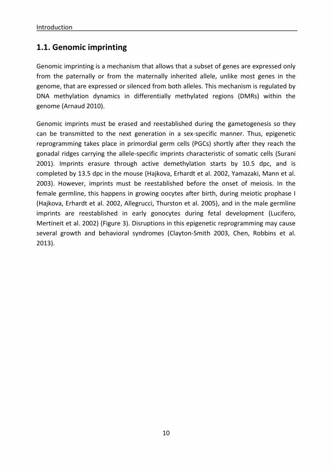
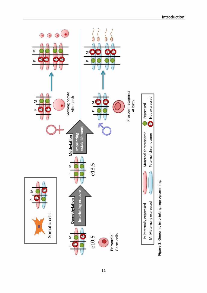
1.1. Genomic imprinting
Genomic imprinting is a mechanism that allows that a subset of genes are expressed only
from the paternally or from the maternally inherited allele, unlike most genes in the
genome, that are expressed or silenced from both alleles. This mechanism is regulated by
DNA methylation dynamics in differentially methylated regions (DMRs) within the
genome (Arnaud 2010).
Genomic imprints must be erased and reestablished during the gametogenesis so they
can be transmitted to the next generation in a sex-specific manner. Thus, epigenetic
reprogramming takes place in primordial germ cells (PGCs) shortly after they reach the
gonadal ridges carrying the allele-specific imprints characteristic of somatic cells (Surani
2001). Imprints erasure through active demethylation starts by 10.5 dpc, and is
completed by 13.5 dpc in the mouse (Hajkova, Erhardt et al. 2002, Yamazaki, Mann et al.
2003). However, imprints must be reestablished before the onset of meiosis. In the
female germline, this happens in growing oocytes after birth, during meiotic prophase I
(Hajkova, Erhardt et al. 2002, Allegrucci, Thurston et al. 2005), and in the male germline
imprints are reestablished in early gonocytes during fetal development (Lucifero,
Mertineit et al. 2002) (Figure 3). Disruptions in this epigenetic reprogramming may cause
several growth and behavioral syndromes (Clayton-Smith 2003, Chen, Robbins et al.
2013).
Introduction
11
Figu
re 3
. Ge
no
mic
imp
rin
tin
g re
pro
gram
min
g

Introduction
12
1.2. X chromosome inactivation (XCI) X-chromosome inactivation (XCI) is a complex epigenetic mechanism that is required to
ensure that most X-linked genes are expressed equally in both sexes (Lyon 1961). The
process compensates the double dosage of X-linked genes by the transcriptional silencing
of one of the X chromosomes in females. This silencing occurs randomly in the maternal
or paternal inherited X chromosome early in embryogenesis, while the extra-embryonic
tissues inactivate only the paternal X chromosome in mice (Bermejo-Alvarez, Ramos-Ibeas
et al. 2012).
XCI is initiated by the expression of the RNA Xist (X-inactive specific transcript), which
recruits several proteins that coat the X chromosome by diverse epigenetic marks. The
most characteristic is the trimethylation of histone H3 at lysine 27 (H3K27me3), a
reppresive mark that impedes transcription (Bermejo-Alvarez, Ramos-Ibeas et al. 2012).
In the early epiblast of the blastocyst, both X chromosomes are active in females. Thus, it
would be expected that ESCs derived from these epiblasts show an absence of XCI.
However, human ESCs are highly heterogeneous in their XCI status, and presence of both
X chromosomes in an active state has been proposed as a hallmark of ground-state
pluripotency and a quality marker for female ESCs. In the same way, XCI reversal in
female somatic cell reprogramming is a key event to achieve naïve pluripotency
(Bermejo-Alvarez, Ramos-Ibeas et al. 2012).
2. Influence of embryonic source on ESCs epigenetics and pluripotency ESCs lines can be derived from embryos that may have different characteristics, and in
some cases these characteristics, or even alterations, can be transmitted to their
corresponding ESCs lines.
Disease-carrying human ESCs have been obtained from affected embryos following
preimplantation genetic diagnosis. In this way, hESC lines were derived carrying
mutations for myotonic dystrophy type 1, cystic fibrosis, Huntington´s disease (Mateizel,
De Temmerman et al. 2006), adrenoleukodystrophy, Duchenne and Becker muscular
dystrophy and thalassaemia (Verlinsky, Strelchenko et al. 2005). These cell lines provide a
powerful in vitro tool for modelling disease progression, identifying molecular
mechanisms that may prevent this progression, and investigating drugs in vitro toxicity
and efficacy (Stephenson, Mason et al. 2009). In the same way, human iPSC lines have
been obtained from patients with a variety of neurological diseases and used to produce
a number of neuronal subtypes (Juopperi, Song et al. 2011).

Introduction
13
However, in other circumstances, embryonic characteristics are not reflected by their
derived ESCs lines. Before iPSCs were first derived, somatic cell nuclear transfer (SCNT)
allowed for the derivation of ESCs lines from somatic cells of diseased individuals that
could be differentiated into a host of cell types for cell replacement therapy. In the
beginning, it was rejected as a valid technology in humans because of the severely
abnormal phenotypes observed in tissues of cloned animals (Brambrink, Hochedlinger et
al. 2006). However, it was later demonstrated that these abnormal phenotypes were not
carried by SCNT-derived ESCs lines, as SCNT- and fertilization-derived ESCs lines were
functionally and transcriptionally indistinguishable. In contrast to SCNT-derived animals,
the process of SCNT-ESCs lines derivation could select for those cells that have erased the
“epigenetic memory” of the donor nucleus (Brambrink, Hochedlinger et al. 2006, Ding,
Guo et al. 2009).
ESCs lines derivation involves the manipulation and in vitro culture of blastocysts. These
embryos are epigenetically dynamic and very sensitive to environmental variations, so
epigenetic alterations can be induced by their manipulation and culture in non-
physiological conditions, which can persist during adulthood and cause disorders such as
obesity, infertility and behavior and growth alterations (Ramirez, Pericuesta et al. 2006,
Fernandez-Gonzalez, Moreira et al. 2008, Calle, Miranda et al. 2012).
It has been demonstrated that in vitro embryo culture can produce imprinting alterations,
resulting in biallelic expression of some genes as H19, Igf2 and Igf2r; and these alterations
are transmitted to the ESC lines. In some cases, they can be corrected by continuous
culture (Ramirez, Pericuesta et al. 2007, Horii, Yanagisawa et al. 2010); however, some
studies indicate that they persist after differentiation producing aberrant genetic
expression patterns (Feinberg, Ohlsson et al. 2006). Most of the imprinted genes regulate
growth and cell proliferation, so their epigenetic deregulation could have oncogenic
effects (Morison, Ramsay et al. 2005). Actually, chimeric mice generated from ESCs with
global loss of imprinting develop multiple tumors (Holm, Jackson-Grusby et al. 2005).
Especially worrying is the case of human ESCs, because all cell lines are derived from
embryos donated by patients undergoing assisted reproductive techniques (ARTs), and
are believed to be unsuitable for use or cryopreservation (Stephenson, Mason et al.
2009). In the last years, it has been reported a significantly increased risk of birth defects
in infants conceived by ARTs, although possibly this increased risk may be due to the
underlying infertility of the couples pursuing ARTs, and not to ARTs themselves (Wen,
Jiang et al. 2012, Wen, Jiang et al. 2012, Hansen, Kurinczuk et al. 2013, Vermeiden and
Bernardus 2013). Abnormalities found in ART-produced embryos could be maintained in
ESCs lines derived from them, as it was demonstrated by Horii et al., who derived ESCs
lines from and in vitro fertilized (IVF)-embryos, and although derivation efficiency was not
significantly different to in vivo-produced embryos, they observed abnormal genomic
Introduction
14
imprinting and expression patterns of methylation-related genes in IVF-derived ESCs lines
at early passages. In contrast, there was no significant difference at later passages (Horii,
Yanagisawa et al. 2010).
Intracytoplasmic sperm injection (ICSI) is currently the most commonly used method to
overcome male infertility; however, some studies have suggested that ICSI bypasses
natural selection barriers, allowing sperm with damaged DNA to fertilize an oocyte
(Schultz and Williams 2002, Hourcade, Perez-Crespo et al. 2010). Furthermore, 40% of the
infertile men following ICSI treatment have high proportions of DNA strand breaks or
other types of DNA damage in their sperm (Lopes, Jurisicova et al. 1998, Sergerie, Laforest
et al. 2005, Zini, Meriano et al. 2005). In mice, it has been reported that embryos
produced by ICSI using DNA-fragmented sperm are genetically and epigenetically altered.
However, some of them implant and develop into animals that show aberrant growth,
premature ageing, abnormal behavior, and mesenchymal tumors (Fernandez-Gonzalez,
Moreira et al. 2008). Consequently, ICSI using DNA fragmented sperm could be used as a
model to analyze if ESCs lines derived from these embryos display the same alterations
observed in the animals, and if pluripotency is affected. In this way, the generation of
animals for the study of ARTs secondary effects could be avoided.
3. Influence of the cellular source on iPSCs epigenetics and pluripotency After the discovery of induced pluripotency, some reports suggested that the
reprogrammed iPSCs retained some epigenetic memory of the cell type of origin, as they
maintained the expression of some transcripts characteristisc from the original cell, and
this was associated with variability in their differentiation capacity (Kim, Doi et al. 2010,
Polo, Liu et al. 2010). Kim et al. compared SCNT-derived ESCs, in vivo fertilized embryo-
derived ESCs and low passage iPSCs derived from fibroblasts or blood. They observed an
intriguing difference in differentiation propensities; while both SCNT- and in vivo embryo-
derived ESCs could differentiate readily down every lineage, iPSCs preferentially
differentiated towards a specific lineage linked to their cell of origin; the blood lineage
and the osteogenic pathway. DNA methylation analysis in DMRs showed that whereas
SCNT- and in vivo embryo-derived ESCs were very similar, iPSC lines were dissimilar to
each other and differed to ESCs (Kim, Doi et al. 2010).
Stadtfeld et al. realized that some iPSCs lines that lacked the full development potential
of ESCs also showed silencing of some imprinted genes. They showed that a few
transcripts encoded within the imprinted Dlk1-Dio3 cluster were aberrantly silenced in
iPSCs clones that contributed poorly to chimaeras and failed to support the development
Introduction
15
of entirely iPSC-derived animals by tetraploid complementation assay. However, this
locus could be reactivated just by treatment with a histone deacetylase inhibitor, rescuing
the ability to support full-term development of all-iPSC mice (Stadtfeld, Apostolou et al.
2010).
Polo et al. showed that the differences among iPSCs derived from different somatic cell
types became more obvious when epigenetic analysis was extended to histone
modifications. Excitingly, this study demonstrates that continuous passaging of iPSCs
leads to the erasure of the differences observed, as early passage (passage 4) iPSCs were
different transcriptionally, epigenetically and on their differentiation potential; but by
passage 16, these differences were abrogated. However, the observed silencing of the
imprinted Dlk1-Dio3 cluster was not modified by passaging of cells, suggesting that not all
epigenetic modifications are reset (Polo, Liu et al. 2010).
Influence of culture conditions on pluripotency
1. Influence of culture conditions on pluripotency capture in vitro Despite the fact that culture conditions shown in literature are not always properly
detailed, and that ESCs-specific markers are limited and have not been described for most
species, it is remarkable that there are more similitudes than differences in culture
conditions used for ESCs derivation in different vertebrates.
Different cell types have been used as feeder layers for ESCs culture in diverse species,
including homologous and heterologous embryonic fibroblasts (EFs). Homologous EFs
have been successfully employed for ESCs derivation in mink, marsupial, human and
porcine. In species as sheep, cow and chicken, the use of homologous EFs has failed to
support ESCs (Familari and Selwood 2006). It is possible that EFs in these species are not
able to produce the essential factors for self-renewal, or perhaps the gestational stage in
which these feeder layers were obtained was not equivalent to the functional stage in
which murine EFs are derived (mid-gestation). Some of the heterologous feeder layers
employed for ESCs derivation are Buffalo rat liver cells in mice (Smith and Hooper 1987);
bovine umbilical cord cells in equine (Saito, Ugai et al. 2002); human fetal lung fibroblasts
in cow (Gjorret and Maddox-Hyttel 2005); and rainbow trout spleen fibroblasts in
zebrafish (Fan, Crodian et al. 2004). However, murine EFs or STO (a transformed murine
fibroblasts cell line) allow for the derivation of ESCs in most mammals and are nowadays
used.

Introduction
16
Mouse ESCs grow as round compact colonies of small cells, which depend on the
LIF/STAT3 pathway. Traditionally, culture conditions consisted on a “feeder” cell layer of
mitotically inactivated mouse EFs, and medium supplemented with foetal calf serum
(FCS) and leukemia inhibitory factor (LIF) (Evans and Kaufman 1981). LIF is a cytokine
produced by the endometrium, which allows blastocyst implantation (Pera and Tam
2010). In ESCs, LIF binds to Gp130 receptor and maintains self-renewal and pluripotency
by phosphorylating STAT3 (Williams, Hilton et al. 1988, Niwa, Burdon et al. 1998). The
effect of LIF on the in vitro development of embryos has been widely studied but results
are often contradictory. Some studies have demonstrated that LIF has the capacity to
enhance blastocyst formation and to decrease embryo fragmentation in mouse (Tsai,
Chang et al. 1999, Cheng, Huang et al. 2004), while other studies have shown the
opposite results that LIF in standard medium does not enhance the development of early
embryos in human and in bovine (Jurisicova, Ben-Chetrit et al. 1995, Gutierrez-Adan,
Perez-Crespo et al. 2006). These contradictory results may be attributable to the different
species and culture systems used for the experiments. Nevertheless, LIF appears to be a
unifying property of both ESCs maintenance and blastocyst diapause. This common LIF-
dependence has been a persistent argument for a diapause-like state of naïve ESCs, and
this is further supported by the fact that entering diapause increases the efficiency of
ESCs derivation from blastocysts (Brook and Gardner 1997, Welling and Geijsen 2013).
Thus, it is interesting to investigate whether or not the embryos cultured in vitro
supplemented with LIF are more suitable for ESCs derivation.
Later, an innovative culture system for ESCs derivation and culture was implemented in
which mitogen-activated protein kinase (MAPK/MEK) signaling was eliminated and
glycogen synthase kinase 3 (GSK3) activity was reduced (Ying, Wray et al. 2008). This
culture system combines LIF with two kinase inhibitors (2i): PD0325901, a MEK inhibitor
that inhibits the mitogen-activated protein kinase enzymes MEK 1 and MEK2, and
CHIR99021, a GSK3β inhibitor, in a defined medium without the use of FCS. The LIF-2i
system allows efficient derivation and expansion of germline-competent ESCs from
different strains of mice that were previously recalcitrant for ESCs derivation (Ying, Wray
et al. 2008, Nichols, Jones et al. 2009) and, for the first time, from the rat (Buehr, Meek et
al. 2008, Li, Tong et al. 2008, Ying, Wray et al. 2008, Leitch, Blair et al. 2010). Moreover, 2i
supplementation during embryo culture suppresses the formation of the hypoblast,
resulting in the entire ICM becoming pluripotent epiblast, and enhancing ESCs derivation
efficiency (Nichols, Silva et al. 2009). Furthermore, reprogramming efficiency of mouse
somatic cells to iPSCs can be enhanced as well by 2i supplementation (Silva, Barrandon et
al. 2008).
Different growth factors have been used to supplement culture medium in order to
isolate pluripotent cells that are remarkably similar to ESCs; Embryonic Germ Cells (EGCs)

Introduction
17
have been isolated from Primordial Germ Cells (PGCs) by LIF, stem cell factor (SCF) and
basic fibroblast growth factor (bFGF) supplementation (Resnick, Bixler et al. 1992). In the
same way, pluripotent Germline Stem Cells (GSCs) have been isolated by gonocyte or
Spermatogonial Stem Cells (SSCs) in vitro culture in the presence of LIF, bFGF, Epidermal
Growth Factor (EGF) and Glial cell-derived neurotrophic factor (GDNF) (Kanatsu-
Shinohara, Ogonuki et al. 2003, Kanatsu-Shinohara, Inoue et al. 2004). These factors also
affect the proliferation and maintenance of other stem cell populations: EGF is a powerful
mitogen which enhances tissue regeneration in various adult organs such as skin, liver
and intestinal epithelium (Reynolds and Weiss 1992); and GDNF has been shown to
stimulate SSC self-renewal in vivo (Meng, Lindahl et al. 2000).
Interestingly, recent studies have described that the pluripotent cell transcriptome is
defined by culture conditions, and not by embryonic origin or derivation procedure. Thus,
major differences in gene expression were observed when pluripotent cells were cultured
in medium with serum vs. 2i conditions, while slight differences were observed between
ESCs coming from blastocysts vs. EGCs coming from PGCs, cultured in the same conditions
(Marks, Kalkan et al. 2012, Leitch, McEwen et al. 2013).
Consequently, it would be interesting to analyze the process of ESCs derivation in
different culture conditions.
2. Influence of culture conditions on epigenetics and pluripotency maintenance Once established, ESCs lines can be maintained in culture for unlimited time; however,
pluripotency can be lost (Nagy, Gocza et al. 1990) and epigenetic alterations as
methylation modifications in imprinted genes can appear (Fujimoto, Mitalipov et al. 2005,
Rugg-Gunn, Ferguson-Smith et al. 2005, Horii, Yanagisawa et al. 2010). In human ESCs, it
has been described that long term culture affects the methylation of the promoter region
of tumor suppressor genes (Maitra, Arking et al. 2005). Other studies indicate that
retroviral DNA introduced in ESCs, which is normally silenced by methylation, can be
reactivated by loss of methylation caused by culture conditions (Allegrucci, Wu et al.
2007, Minoguchi and Iba 2008). These changes are unpredictable and variable among
different cell lines, and once established they can be stable and be transmitted to
differentiated cells (Lund, Narva et al. 2012). However, until now optimal culture
conditions for epigenetic stability remain unknown.
In the early epiblast in female blastocysts, both X chromosomes are still in an active state,
so it would be expected the same situation for ESCs lines derived from these embryos.
Nevertheless, high variability has been found among cell lines, especially in human ESCs
Introduction
18
(Bermejo-Alvarez, Ramos-Ibeas et al. 2012). Culture conditions as oxygen concentration
can affect XCI. Hypoxic conditions promote that both X chromosomes are active in female
cell lines, while changes from hypoxic to normoxic conditions promote X inactivation
(Lengner, Gimelbrant et al. 2010). Likewise, cellular stress such as freeze-thaw cycles
makes the cells more prone to X inactivation (Lund, Narva et al. 2012). Furthermore,
some XX ESC lines show a global loss of DNA methylation, possibly associated to the fact
that both X chromosomes are in an active state. Consequently, XX cell lines are more
unstable in culture than XY, so generally in research XY cell lines are more preferable
(Zvetkova, Apedaile et al. 2005, Pannetier and Feil 2007).
Pluripotency in adult tissues
In the last years, it has been described the existence of adult stem cells (ASCs) that
support multiple tissues. These cells are multipotent, meaning that they are limited to
differentiate towards a specific cell lineage. ASCs can be identified in many tissues and
organs, including skeletal muscle, bone, cartilage, skin, blood vessels, heart, liver, gut,
lung, brain, fat tissue, dental pulp, amniotic fluid, peripheral blood, ovarian epithelium,
testis and bone marrow (Macaluso and Myburgh 2012). They reside in specific areas in
the organs called “niches”; specialized local microenvironments where they are protected
and supported while they maintain a low basal rate of division. These niches comprise
specific extracellular matrix and supporting cells, and have the potential to modulate
signaling towards stem cell self-renewal or differentiation (Greco and Guo 2010).
During lifetime, a number of ASCs will be activated in order to maintain tissues
homeostasis, while the remaining ASCs will stay in a quiescent phase; both coexisting in
the same area (Li and Clevers 2010). Their location and their slowly proliferating state
make them much more difficult to study than other pluripotent cells as ESCs (Alvarez,
Garcia-Lavandeira et al. 2012). However, nowadays numerous subtypes of ASCs have
been described: haematopoietic and mesenchymal stem cells residing in bone marrow;
gut stem cells located in the crypts of Lieberhahn, liver stem cells, bone and cartilage
stem cells, skin and hair stem cells, neuronal stem cells, pancreatic stem cells, retinal
stem cells, cardiac stem cells, dental pulp stem cells and skeletal muscle stem cells
(Schabort, Myburgh et al. 2009). Much research has been done on the regenerative
potential of these ASCs in animal models of tissue damage. Similarly, their ability to
transdifferentiate in vitro has been investigated with a view to ex vivo cell expansion and
manipulation for transplant (Macaluso and Myburgh 2012). After purification, ASCs can
be grown in vitro with selected growth factors, micronutrients, antioxidants and
Introduction
19
hormones supplementation at supposedly physiological concentrations (Alvarez, Garcia-
Lavandeira et al. 2012).
Probably one of the most studied ASCs subtypes is spermatogonial stem cells (SSCs). SSCs
are able to undergo self-renewal division yielding new SSCs, or to differentiate towards
spermatogonias to support spermatogenesis (Kanatsu-Shinohara, Inoue et al. 2011).
Although the estimated number of SSCs in the mouse testis comprises only 0.03% of the
total germ cell population (de Rooij and Mizrak 2008), techniques have been developed
that allow isolation and long-term culture of SSCs giving rise to a population called
Germline Stem cells (GSCs). Moreover, it has been described the appearance of ES-like
colonies called multipotent Germline Stem cells (mGSCs) after long-term culture
(Kanatsu-Shinohara, Ogonuki et al. 2003, Kanatsu-Shinohara, Inoue et al. 2004, Guan,
Nayernia et al. 2006).
Although common ESCs markers, such as Oct4, Sox2, Nanog, Klf4, Lin28 or Fut4 (Ssea1)
are expressed in some ASCs as hematopoietic stem cells (HSCs) (Wang, Zhou et al. 2010),
mesenchymal stem cells (MSCs) (Jaramillo-Ferrada, Wolvetang et al. 2012) and SSCs
(Izadyar, Pau et al. 2008), ASCs also express specific markers depending on their niche
location. However, good markers common to all ASCs or organ-specific are not yet well
known, because some of them are also expressed by differentiated cells.

Introduction
20
Pluripotency and germline common markers
1. Germline specification and development Germline specification is one of the earliest cell fate decisions made in the mammalian
embryo. PGCs induction starts prior to gastrulation in the proximal epiblast at embryonic
day (e) 6.25 due to the bone morphogenetic proteins (BMPs) stimulation, mainly by Bmp4
and to a lesser extent to Bmp8b, that are released from the extraembryonic ectoderm
(ExE) (Lawson, Dunn et al. 1999, Ying, Qi et al. 2001). These factors activate the ACVR1
(Alk2) serine-threonine kinase receptor (de Sousa Lopes, Roelen et al. 2004), which
induces phosphorylation and nuclear translocation of SMAD1, SMAD5 and SMAD8
transcription factors, enabling modulation of target genes expression (de Sousa Lopes,
Hayashi et al. 2007). These events direct a small group of 4-8 cells to enter the germinal
lineage by Blimp1 and Prdm14 upregulation, the two crucial determinants of the germline
(Ohinata, Payer et al. 2005, Saitou and Yamaji 2012). These cells proliferate and develop a
cluster of 20 cells that subsequently express other PGC markers such as Fragilis (Ifitm3)
and Stella (Dppa3) by e7.5. The pluripotency-associated transcription factors Pou5f1
(Oct4) and Nanog are initially expressed in the ICM but become restricted to the germline
between e7.75 and 8.5 (Young, Dias et al. 2010). Thereafter, PGCs initiate migration and
colonization of the genital ridge and increase their number to approximately 4000 by
e12.5 (Saitou, Barton et al. 2002, Sato, Kimura et al. 2002). Once PGCs reach the gonads,
other germline-specific markers essential for the development of the germ cells are
transcribed, including Ddx4 (Toyooka, Tsunekawa et al. 2000) and Dazl (Lin and Page
2005).
Figure 4. PGC specification and development

Introduction
21
2. Expression of germline markers in pluripotent cells There are different pluripotent cell types according to the source of cells used for their
establishment: ESCs derived from early epiblasts, EGCs from PGCs, ECCs isolated from
germ-cell tumors, GSCs derived from SSCs, and iPSCs from somatic cells reprogramming
(Xu, Pantakani et al. 2011). Most of the above mentioned have a germ-cell origin apart
from ESCs and iPSCs, but all of them share common germ cell or premeiotic markers that
may indicate a common germinal origin (Zwaka and Thomson 2005) (Table 1).
Interestingly, Oct4, Sox2 and Nanog, key pluripotency genes, appear to be necessary for
germ cell survival (Kehler, Tolkunova et al. 2004, Yamaguchi, Kimura et al. 2005,
Chambers, Silva et al. 2007, Zhao, Ji et al. 2012). Furthermore, several studies have
demonstrated that germline or premeiotic markers as Blimp1, Dppa3, Ifitm3, Piwil2, Dazl,
Ddx4, Stra8, Rnf17 and Rnh2 are expressed in ESCs (Geijsen, Horoschak et al. 2004, Qing,
Shi et al. 2007, Mise, Fuchikami et al. 2008, Xu, Pantakani et al. 2011). Moreover, a recent
study associates Dazl with Tet (Ten eleven translocation)-dependent DNA demethylation
in ESCs (Blaschke, Ebata et al. 2013). Another common marker for pluripotency and
germline is Prdm14, whose essential role is confined to the germline establishment
(Yamaji, Seki et al. 2008), although it has been recently shown to play a role in epigenetic
regulation ESCs to achieve naïve pluripotency (Yamaji, Ueda et al. 2013).
In the same way, these germline and premeiotic markers, and even meiotic markers
Sycp3, Pgk2, and Creb3/4, were detected also in iPSC, GSCs and EGCs and in both male
and female cell lines. When somatic cell reprogramming process was analyzed, germ cell
markers Blimp1, Dppa3 and Ifitm3 started to be expressed between day 6 and 9 of
reprogramming, and by day 22 their expression increased to the levels observed in ESCs.
In contrast, significant expression of the key pluripotency markers Pou5f1 and Sox2
started later, at day 12. Other premeiotic markers as Stra8, Dazl and Ddx4 were only
detected after day 22 of reprogramming (Xu, Pantakani et al. 2011).
Furthermore, it has been recently described a novel alternatively spliced isoform of mDazl
with a deletion of exon8, referred to as Dazl_Δ8, that is expressed together with the full-
length isoform of Dazl in different pluripotent cell types: ESCs, GSCs, iPSCs , EGCs and
ECCs (Xu, Tan et al. 2013).
In mouse preimplantation embryos, germline markers Dppa3, Dazl and Ddx4 have been
detected throughout all stages (Pan, Liao et al. 2008, Xu, Pantakani et al. 2011). In non-
mammal species, zDazl has been localized in early zebrafish embryos (Maegawa, Yasuda
et al. 1999, Hashimoto, Maegawa et al. 2004) and XDazl in Xenopus embryos until stage
10 (Houston and King 2000).
Concerning adult stem cells, it has been described DAZL, c-Kit and POU5F1 expression in
human amniotic fluid stem cells (hAFSCs) (Stefanidis, Loutradis et al. 2008). Furthermore,
Pou5f1, Dppa3, Ifitm3, Dazl and Ddx4 have been located in mouse and human bone

Introduction
22
marrow (BM) (Benson, Karsch-Mizrachi et al. 2004, Su, Wiltshire et al. 2004, Johnson,
Bagley et al. 2005). Another study described that mouse bone marrow mesenchymal
stem cells (BM-MSCs) were positive for Dazl, but in lower levels of expression than the
total bone marrow cells tested (Katsara, Mahaira et al. 2011).
Table 1. Pluripotency and germline common markers
Molecular marker Expression References
Fut4 (Ssea1) ESC, PGC, EGC, ECC,
iPSC, embryo
(Knowles, Aden et al. 1978, Solter and
Knowles 1978, Fox, Damjanov et al. 1981,
Zhao, Ji et al. 2012)
c-Kit ESC, PGC, ECC, HSC (Zhao, Ji et al. 2012)
UTF1 ESC, ECC, ICM, germ
cells
(Kooistra, Thummer et al. 2009, Zhao, Ji
et al. 2012)
Pou5f1 ESC, PGC, EGC, ECC,
iPSC, ICM, bone
marrow
(Pesce and Scholer 2000, Pesce and
Scholer 2001, Kehler, Tolkunova et al.
2004, Zhao, Ji et al. 2012)
Dppa3 ESC, PGC, ECC, iPSC,
embryo, bone
marrow
(Bowles, Teasdale et al. 2003, Benson,
Karsch-Mizrachi et al. 2004, Bortvin,
Goodheart et al. 2004, Su, Wiltshire et al.
2004, Johnson, Bagley et al. 2005, Xu,
Pantakani et al. 2011, Zhao, Ji et al. 2012)
Sox2 ESC, PGC, EGC, ECC,
iPSC, embryo
(Botquin, Hess et al. 1998, Boyer, Lee et
al. 2005, Zhao, Ji et al. 2012)
GCNF ESC, ECC, germ cells (Lei, Hirose et al. 1997, Lan, Xu et al.
2009, Zhao, Ji et al. 2012)
Nanog ESC, PGC, EGC, ECC,
iPSC
(Chambers, Silva et al. 2007, Yamaguchi,
Kurimoto et al. 2009)
Prdm14 ESC, PGC (Yamaji, Seki et al. 2008, Leitch, McEwen
et al. 2013, Yamaji, Ueda et al. 2013)
Blimp1 ESC, PGC, iPSC (Xu, Pantakani et al. 2011)
Ifitm3 ESC, PGC, iPSC, bone
marrow
(Benson, Karsch-Mizrachi et al. 2004, Su,
Wiltshire et al. 2004, Johnson, Bagley et
al. 2005, Xu, Pantakani et al. 2011)
Piwil2 ESC, PGC, (Xu, Pantakani et al. 2011)
Dazl ESC, PGC, EGC, GSC,
ECC, iPSC, embryo,
bone marrow
(Benson, Karsch-Mizrachi et al. 2004, Su,
Wiltshire et al. 2004, Johnson, Bagley et
al. 2005, Pan, Liao et al. 2008, Katsara,
Mahaira et al. 2011, Xu, Pantakani et al.
2011, Xu, Tan et al. 2013)

Introduction
23
Ddx4 ESC, PGC iPSC,
embryo, bone
marrow
(Benson, Karsch-Mizrachi et al. 2004, Su,
Wiltshire et al. 2004, Johnson, Bagley et
al. 2005, Xu, Pantakani et al. 2011)
Stra8 ESC, PGC, iPSC (Xu, Pantakani et al. 2011)
Rnf17 ESC, PGC (Xu, Pantakani et al. 2011)
Rnh2 ESC, PGC (Xu, Pantakani et al. 2011)
ESC: embryonic stem cell; PGC: primordial germ cell; EGC: embryonic germ cell; ECC: embryonic carcinoma cell; iPSC: induced pluripotent stem cell; GSC: germline stem cell; HSC: hematopoietic stem cell; ICM: inner cell mass.
In conclusion, germ cell and premeiotic markers have been found to be expressed in
numerous pluripotent cell types, which may indicate a common germ cell origin for them.
According to this theory, the blastocyst stage (e3.5) expresses key pluripotency markers
as Pou5f1 and Sox2. Following embryonic development, PGC specification takes place
when Blimp1 expression is activated, repressing the somatic program and reactivating the
pluripotency network before PGC migration (Saitou 2009). In the same way, ESCs
derivation starts with e3.5 blastocysts culture, and cells would proceed with the
developmental program of PGCs specification via BMPs signaling and would begin
expressing Blimp1, Ifitm3 and Dppa3 among other cell markers, and reactivating
pluripotency genes to acquire self-renewal properties. Similarly, during somatic cell
reprogramming by Pou5f1, Sox2, c-Myc and Klf4 factors, inductive BMPs signaling would
trigger germ cell markers expression and finally reactivate pluripotency network (Xu,
Pantakani et al. 2011) (Figure 5).
Figure 5. Theory of the germ cell origin of pluripotent cells

Introduction
24
Transgenic mice where a reporter gene expression as green fluorescen protein (GFP) is
driven by a known gene promoter constitute an ideal model to study gene localization in
several tissues along different developmental periods, as not only allows easy
identification of cells expressing the transgene, but also the reporter gene does not
interact with biological processes. Dazl is one of the genes which main function is related
to germline development and meiosis (Ruggiu, Speed et al. 1997), although some recent
studies have related Dazl to pluripotency (Haston, Tung et al. 2009, Xu, Tan et al. 2013).
Introduction
25
Trophectoderm specification and pluripotency
The first tissue lineages segregation occurs when blastocyst cells differentiate into both
the inner cell mass (ICM), which will give rise to the embryo proper, and the
trophectoderm (TE), the precursor of the placenta (Schiffmacher and Keefer 2013). Both
lineages are controlled by gene regulatory networks, but specie-specific differences exist.
1. Specie-specific differences in trophectoderm specification In mice, there is an antagonistic genetic interaction between ICM and TE regulators. The
ICM and its associated pluripotent state are maintained by Pou5f1 (Oct4), while TE
specification relies on Cdx2, which is able to suppress Pou5f1 expression and vice versa.
However, this situation varies markedly between embryos from different mammalian
species. Pou5f1 expression is restricted to the ICM in mice, but in cattle and porcine
embryos it is expressed in the TE as well, so the mouse embryo is not an ideal model for
ungulate embryogenesis (Wolf, Serup et al. 2011). It has been suggested that the early
down-regulation of Pou5f1 in rodents could have evolved to allow a rapid commitment to
the TE fate and a shorter preimplantation development, while in cattle and other
mammals where preimplantation development is longer, there may not be such need
(Berg, Smith et al. 2011).
The precursors of the differentiated cells of the placenta are assumed to be trophoblast
stem cells (TSCs). They are supposed to exist in all placental mammals, especially during
the early stages of placental development when TE growth is maximal. However, little is
known about their location and phenotype in other species than mouse and rhesus
macaque (Roberts and Fisher 2011). In the mouse, the TSCs niche appears to be located
within the extraembryonic ectoderm (ExE) adjacent to the epiblast, on which depends for
essential growth factors. This specialized niche is transient and maintained only for 3 or 4
days during postimplantation development. Mouse TSCs can be obtained from
proliferating outgrowths of polar TE explanted from a region bordering the epiblast and
cultured in vitro on mitotically inactivated feeder cells and a medium supplemented with
FBS, Fibroblast Growth Factor 4 (FGF4) and heparin (Tanaka, Kunath et al. 1998). Cells
with some properties of TSCs have been obtained from rhesus macaque blastocysts as
well (Vandevoort, Thirkill et al. 2007).
Domestic species such as swine, sheep and cattle show a dramatic elongation of the TE
preceeding implantation that allows to exploit uterine secretions throughout a long area
of the uterine lumen (Bindon 1971, Geisert, Brookbank et al. 1982, Thatcher, Meyer et al.
1995). Unlike mouse, polar TE overlying the embryonic disk is soon lost in these species
and does not contribute to the placenta, while the homolog of mouse mural TE is

Introduction
26
responsible of the elongation. It is unknown whether the expansion of mural TE relies on
a stem cell population immediately surrounding the embryonic disk, or on stem cells
scattered throughout the TE; or whether sustained growth is entirely due to proliferation
of progenitor cells that are already partially differentiated (Roberts and Fisher 2011).
Consequently, TSCs have not been isolated from any ungulate species, although primary
trophectoderm cell lines have been derived from sheep and goat (Miyazaki, Imai et al.
2002), pig (Ramsoondar, Christopherson et al. 1993, Flechon, Laurie et al. 1995, La
Bonnardiere, Flechon et al. 2002) and cattle (Talbot, Caperna et al. 2000, Shimada,
Nakano et al. 2001, Suzuki, Koshi et al. 2011).
2. Bovine trophoblast cell lines derivation Trophoblast cells grow in vitro as a tight monolayer that eventually forms dome-like
structures. These structures accumulate fluid until they transform into spheroids that are
released to the culture medium.
In bovine, trophoblast cell lines were first derived by Talbot et al. from the culture of 7-8-
days in vitro-produced blastocysts over STO feeder cells (Talbot, Caperna et al. 2000).
However, the co-culture with mouse feeder cells has the risk of contaminating the bovine
trophoblastic cells, and this could be detrimental for using these cell lines for further
studies. Later, Shimada et al. (Shimada, Nakano et al. 2001, Hirata, Sato et al. 2003)
established a bovine trophoblastic cell line in the absence of feeder cells, using bovine
endometrial fibroblast-conditioned medium, but attaining very low derivation efficiency.
They plated more than 50 blastocysts, obtaining only one cell line. The mayor problem
appeared to be the attachment of the blastocyst to the culture plate and the initial
proliferation. Since then, most of the studies regarding trophectoderm signaling and
differentiation have been performed with these only two trophoblastic cell lines derived
by Talbot and Shimada (Nakano, Shimada et al. 2002, Hirata, Sato et al. 2003, Ushizawa,
Takahashi et al. 2005, Michael, Wagner et al. 2006, Das, Ezashi et al. 2008, Nakaya, Kizaki
et al. 2009, Bai, Sakurai et al. 2011, Schiffmacher and Keefer 2012, Schiffmacher and
Keefer 2013). Therefore, it would be helpful to come with a larger number of
trophoblastic cell lines acceptable for in vitro studies, without any possible cell
contamination.
Trophoblastic cell lines have been derived from parthenogenetic and SCNT-produced
embryos, and significant differences have been found in derivation efficiency, as
parthenogenetic blastocysts had a lower ratio for primary colony formation than in vivo
or SCNT-produced blastocysts. In the same way, the amount of the pregnancy
establishment-protein Interferon tau (INF-t) produced by outgrowths obtained from
parthenogenetic and SCNT-produced embryos was lower than in those dericed from in
Introduction
27
vitro fertilized embryos. Furthermore, SCNT-derived trophoblastic cell lines had less
annexin proteins, related to placentation maintenance (Talbot, Powell et al. 2007, Talbot,
Powell et al. 2008, Talbot, Powell et al. 2010). Therefore, trophoblastic cell lines could
reflect the alterations carried by the embryos, and could be used as a model to analyze
them.
In a more recent study, Suzuki et al. used BMP4 to raise trophoblastic cell lines based on
the fact that exogenous BMP4 stimulate ESCs to become trophoblastic cells (Murohashi,
Nakamura et al. 2010), but no significant improvement was found in derivation efficiency,
and just 12 trophoblastic cell lines were derived from 172 embryos. When genes
necessary for pregnancy establishment, interferon-t (IFNT), placental lactogen (CSH1),
prolactin-related protein 1 (PRP1) and pregnancy-associated glycoprotein 1 (PAG1), were
analyzed, very variable expression patterns were found among the different cell lines
(Suzuki, Koshi et al. 2011). Thus, there is a high variability among trophoblastic cell lines
obtained using the same derivation conditions. Many of the derived trophoblastic cell
lines grow continuously lacking senescence symptoms, and show pluripotency marker
POU5F1 expression (Suzuki, Koshi et al. 2011, Schiffmacher and Keefer 2013). Therefore,
they could represent the pluripotent cell population from which the placenta
differentiates in cattle, or a more differentiated equivalent to mouse TSCs.
Trophoblastic cell lines represent a useful model for implantation and placentogenesis
studies, and they could reflect genetic and epigenetic characteristics and alterations
carried by the embryos from which they are derived. However, just a small number of cell
lines are used for research due to the difficulty of establishing new cell lines.
Consequently, it would be interesting to establish an efficient system for trophoblastic
cell lines derivation from a TE biopsy that would allow the embryo to be viable, while its
characteristics or alterations are analyzed on its corresponding trophoblastic cell line.

28

Introducción
29
Introducción

Introducción
30

Introducción
31
El descubrimiento de la pluripotencia
1. Del descubrimiento de las células pluripotentes a la captura de la pluripotencia in vitro La pluripotencia y las células troncales han fascinado a biólogos y médicos desde hace
más de un siglo.
El término “célula troncal” fue empleado por primera vez en la literatura científica en
1868 por el ilustre biólogo alemán Ernst Haeckel, que utilizó el término “Stammzelle”
para describir el organismo unicelular a partir del cual todos los organismos
multicelulares evolucionaron (Haeckel 1868). Posteriormente, propuso referirse también
al huevo fertilizado como “Stammzelle” (Haeckel 1877). Por tanto, según Haeckel, el
término célula troncal podría ser usado de dos maneras: como el antepasado unicelular
común a todos los organismos multicelulares, y como el huevo fertilizado que da lugar a
todas las células del organismo (Ramalho-Santos and Willenbring 2007). En 1892, otro
científico alemán, Theodor Boveri, llevó la definición de célula troncal como huevo
fertilizado un paso más allá: propuso que el término fuese usado para las primeras células
de linaje germinal que aparecen en el embrión animal, que presuntamente transportan el
plasma germinal y posteriormente se diferencian para formar los gametos (Boveri 1892,
Ramalho-Santos and Willenbring 2007). Por lo tanto, en estos primeros estudios, el
término célula troncal se refería a lo que actualmente llamamos el linaje germinal, o
células germinales primordiales.
Cuatro años después, el término fue popularizado en su versión inglesa por Edmund B.
Wilson, un científico americano que revisó los estudios de Boveri en su famoso libro The
Cell in Development and Inheritance (Wilson 1896). Este libro inspiró a todos los
embriólogos y genetistas de la época, y a Wilson se le adjudicó el mérito de haber
acuñado el término.
En la misma época, se generó un gran interés científico en el desarrollo y la regeneración
del sistema hematopoyético, y un grupo de científicos empezó a creer en la existencia de
una célula que representara el origen común de diferentes tipos celulares de la sangre.
Alguno de ellos empezó a usar el término célula troncal para referirse a dicho precursor
común (Pappenheim 1896, Ramalho-Santos and Willenbring 2007).
Por tanto, las primeras interpretaciones del término célula troncal a finales del siglo XIX
se referían a cuestiones fundamentales en embriología: la continuidad de la línea
germinal y el origen del sistema sanguíneo.
Posteriormente, la existencia de las células troncales hematopoyéticas (“Hematopoietic
stem cells”, HSCs) quedó demostrada (Till and McCulloch 1961, Becker, Mc et al. 1963,
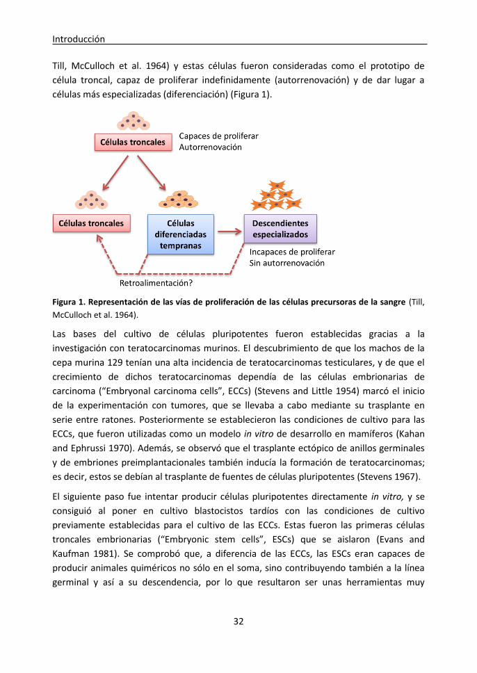
Introducción
32
Till, McCulloch et al. 1964) y estas células fueron consideradas como el prototipo de
célula troncal, capaz de proliferar indefinidamente (autorrenovación) y de dar lugar a
células más especializadas (diferenciación) (Figura 1).
Figura 1. Representación de las vías de proliferación de las células precursoras de la sangre (Till,
McCulloch et al. 1964).
Las bases del cultivo de células pluripotentes fueron establecidas gracias a la
investigación con teratocarcinomas murinos. El descubrimiento de que los machos de la
cepa murina 129 tenían una alta incidencia de teratocarcinomas testiculares, y de que el
crecimiento de dichos teratocarcinomas dependía de las células embrionarias de
carcinoma (“Embryonal carcinoma cells”, ECCs) (Stevens and Little 1954) marcó el inicio
de la experimentación con tumores, que se llevaba a cabo mediante su trasplante en
serie entre ratones. Posteriormente se establecieron las condiciones de cultivo para las
ECCs, que fueron utilizadas como un modelo in vitro de desarrollo en mamíferos (Kahan
and Ephrussi 1970). Además, se observó que el trasplante ectópico de anillos germinales
y de embriones preimplantacionales también inducía la formación de teratocarcinomas;
es decir, estos se debían al trasplante de fuentes de células pluripotentes (Stevens 1967).
El siguiente paso fue intentar producir células pluripotentes directamente in vitro, y se
consiguió al poner en cultivo blastocistos tardíos con las condiciones de cultivo
previamente establecidas para el cultivo de las ECCs. Estas fueron las primeras células
troncales embrionarias (“Embryonic stem cells”, ESCs) que se aislaron (Evans and
Kaufman 1981). Se comprobó que, a diferencia de las ECCs, las ESCs eran capaces de
producir animales quiméricos no sólo en el soma, sino contribuyendo también a la línea
germinal y así a su descendencia, por lo que resultaron ser unas herramientas muy

Introducción
33
eficaces para la manipulación genética (Bradley, Evans et al. 1984, Robertson, Bradley et
al. 1986) (revisado en (Solter 2006)).
En 1992 se aislaron células pluripotentes in vitro a partir de una fuente diferente, las
células germinales primordiales (“Primordial germ cells”, PGCs), mediante el uso de
factores de crecimiento como suplemento para el medio de cultivo convencional de ESCs
(Matsui, Zsebo et al. 1992). Estas células compartían importantes características con las
ESCs, incluyendo su morfología, pluripotencia y capacidad de transmisión a línea germinal
en animales quiméricos. Fueron llamadas células germinales embrionarias (“Embryonic
germ cells”, EGCs), para distinguirlas de las ESCs derivadas a partir de blastocistos.
Desde que las ESCs fueron aisladas por primera vez en el ratón, pasaron diecisiete años
hasta que se aislaron las primeras ESCs humanas (Thomson, Itskovitz-Eldor et al. 1998).
Esta demora probablemente fue debida a las diferencias existentes entre el ratón y el
humano a nivel de desarrollo embrionario. Además, la pluripotencia de estas células es
menor a la de sus equivalentes murinas, pues no son capaces de generar quimeras en sus
condiciones de cultivo estándar (recientemente se ha descubierto que utilizando ciertos
factores en el medio de cultivo se puede incrementar su pluripotencia, haciendo así que
se asemejen a las ESCs murinas y que sean capaces de generar quimeras (Gafni,
Weinberger et al. 2013)). Además, las ESCs humanas forman colonias de morfología
plana, asemejándose a las líneas celulares derivadas a partir de epiblastos tardíos
murinos, llamadas células troncales del epiblasto (EpiSCs) (Tesar, Chenoweth et al. 2007);
a diferencia de las ESCs murinas, que forman colonias tridimensionales. Por lo tanto, se
han propuesto dos tipos de pluripotencia: la naïve, donde se incluyen las ESCs murinas
que dependen de la vía LIF/Stat3, y la primed, donde se incluyen las ESCs de primates y
ungulados y las EpiSCs murinas que dependen de la señalización de Fgf/ERK (Nichols and
Smith 2009).
Aunque el aislamiento de ESCs humanas fue reconocido como un gran avance médico,
generó conflictos éticos respecto a la utilización de embriones humanos para
investigación. Estos inconvenientes fueron superados gracias al descubrimiento de los
factores de reprogramación (Oct4, Sox2, Klf4, c-Myc) por Yamanaka et al., que
permitieron la reprogramación de células somáticas a células pluripotentes, dando lugar a
las células de pluripotencia inducida (“induced pluripotent stem cells”, iPSCs) por primera
vez en ratón (Takahashi and Yamanaka 2006), y posteriormente en humanos omitiendo
cMyc y usando LIN28 en su lugar (Takahashi, Tanabe et al. 2007).
En resumen, en las últimas décadas se han obtenido diferentes tipos de células
pluripotentes en diferentes momentos del desarrollo que pueden ser cultivadas in vitro a
la vez que mantienen su autorrenovación y pluripotencia (Figura 2).
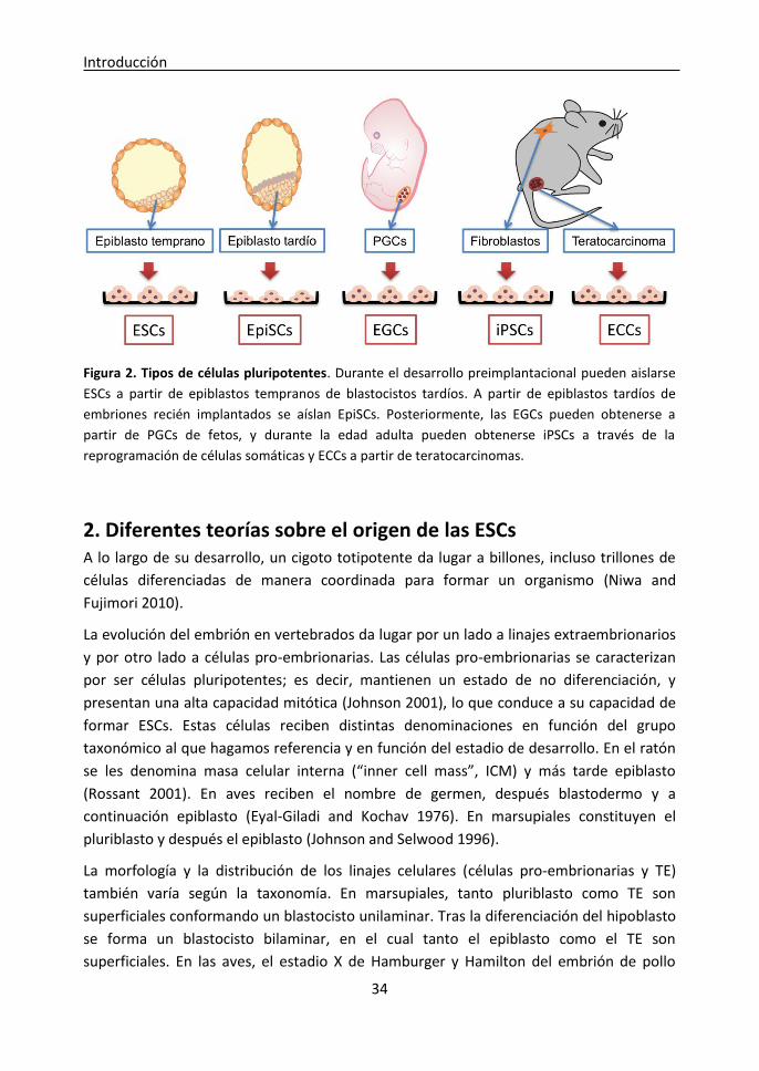
Introducción
34
Figura 2. Tipos de células pluripotentes. Durante el desarrollo preimplantacional pueden aislarse
ESCs a partir de epiblastos tempranos de blastocistos tardíos. A partir de epiblastos tardíos de
embriones recién implantados se aíslan EpiSCs. Posteriormente, las EGCs pueden obtenerse a
partir de PGCs de fetos, y durante la edad adulta pueden obtenerse iPSCs a través de la
reprogramación de células somáticas y ECCs a partir de teratocarcinomas.
2. Diferentes teorías sobre el origen de las ESCs A lo largo de su desarrollo, un cigoto totipotente da lugar a billones, incluso trillones de
células diferenciadas de manera coordinada para formar un organismo (Niwa and
Fujimori 2010).
La evolución del embrión en vertebrados da lugar por un lado a linajes extraembrionarios
y por otro lado a células pro-embrionarias. Las células pro-embrionarias se caracterizan
por ser células pluripotentes; es decir, mantienen un estado de no diferenciación, y
presentan una alta capacidad mitótica (Johnson 2001), lo que conduce a su capacidad de
formar ESCs. Estas células reciben distintas denominaciones en función del grupo
taxonómico al que hagamos referencia y en función del estadio de desarrollo. En el ratón
se les denomina masa celular interna (“inner cell mass”, ICM) y más tarde epiblasto
(Rossant 2001). En aves reciben el nombre de germen, después blastodermo y a
continuación epiblasto (Eyal-Giladi and Kochav 1976). En marsupiales constituyen el
pluriblasto y después el epiblasto (Johnson and Selwood 1996).
La morfología y la distribución de los linajes celulares (células pro-embrionarias y TE)
también varía según la taxonomía. En marsupiales, tanto pluriblasto como TE son
superficiales conformando un blastocisto unilaminar. Tras la diferenciación del hipoblasto
se forma un blastocisto bilaminar, en el cual tanto el epiblasto como el TE son
superficiales. En las aves, el estadio X de Hamburger y Hamilton del embrión de pollo

Introducción
35
consta de dos regiones visiblemente diferenciadas: el area pellucida, a partir del cual se
desarrolla el embrión, y la zona opaca circundante (area opaca) que da lugar al saco
vitelino extraembrionario (Eyal-Giladi and Kochav 1976). Es también en el estadio X,
donde comienza a formarse el hipoblasto, de modo que en el estadio XI, el area pellucida
comprende tanto epiblasto como hipoblasto. Por ello, el estadio X de desarrollo es el
empleado en aves para el aislamiento de ESCs y es también el momento para realizar
gene targeting y generar animales quiméricos. En el pez cebra, el pez medaka y la dorada,
tras la décima división (mid-blástula), el genoma del cigoto se activa dando lugar a los
primeros tres linajes celulares. Dos de ellos son linajes extra-embrionarios: la capa
sincitial de la yema, y la capa externa que la rodea, y la tercera capa es el pluriblasto, que
se encuentra en el interior (Fan, Crodian et al. 2004). Para el aislamiento de ESCs en estas
tres especies de peces se ha empleado el estadio de mid-blástula, y también el anillo
germinal en el caso del pez cebra.
En el ratón, a día 3,5 de desarrollo embrionario, el blastocisto comprende dos linajes
celulares, la ICM y el trofoectodermo (“trophectoderm”, TE). La ICM da lugar al
ectodermo primitivo o epiblasto, mientras que el trofoblasto sólo contribuye al tejido
placentario (Rossant 2001). Tras la diferenciación del hipoblasto (endodermo primitivo),
la ICM residual se convierte en el epiblasto temprano post-implantacional, y estas células
pluripotentes darán lugar rápidamente a las hojas germinales primarias durante la
gastrulación. Históricamente se ha asumido que las ESCs murinas eran equivalentes a las
células de la ICM, debido a que el aislamiento de las ESCs se consiguió gracias el cultivo de
blastocistos de día 3,5. Sin embargo, cada vez hay más indicios de que las ESCs tienen un
origen diferente. Hace unos años surgió una teoría que suponía la existencia de una
subpoblación celular del epiblasto que era seleccionada durante el proceso de
aislamiento y daba lugar a las ESCs; y un candidato interesante para esta subpoblación
eran las células predispuestas a diferenciarse hacia el linaje germinal (Zwaka and
Thomson 2005). Apoyando esta teoría, por una parte las PGCs son capaces de generar
líneas celulares pluripotentes que son idénticas a las ESCs (Matsui, Zsebo et al. 1992,
Resnick, Bixler et al. 1992), y por otra parte, de todos los linajes celulares que se
diferencian a partir del epiblasto, sólo las células germinales expresan genes relacionados
con la pluripotencia como Oct4, Nanog y Sox2 (Durcova-Hills and Surani 2008, Chu, Surani
et al. 2011). Además, se ha observado que el factor de especificación del linaje germinal
Blimp1 y otros marcadores de células germinales como Dppa3 (Stella) y Prdm14 se
activan cuando se ponen en cultivo blastocistos para aislar ESCs en medio suplementado
con suero fetal bovino (“fetal calf serum”; FCS) y LIF (Chu, Surani et al. 2011), lo que
sugiere que las células que van a dar lugar a las ESCs activan transitoriamente un
programa transcripcional específico de PGCs (Hochedlinger 2011). De hecho, cuando
estas células que sobreexpresan Blimp1 son aisladas y trasplantadas a embriones

Introducción
36
deficientes en células germinales a día 8,5, migran a los anillos germinales y comienzan a
sobreexpresar el marcador de desarrollo de linaje germinal Mvh. Además, aislando las
células que sobreexpresan Blimp1 se pudieron obtener ESCs con una eficiencia nueve
veces mayor que con todo el cúmulo de células de la ICM. Sin embargo, este estado
transitorio de célula germinal no parece ser estrictamente necesario para la obtención de
ESCs, ya que las ESCs pueden aislarse a partir de blastocistos knockout que no expresan
Blimp1 (Chu, Surani et al. 2011, Nichols and Smith 2011). Por otra parte, la obtención de
ESCs con el sistema 2i (explicado más adelante en la introducción) no provoca
sobreexpresión de Blimp1, sino que permite capturar directamente el potencial de auto-
renovación de las células del epiblasto sin pasar por un programa germinal transitorio
(Hochedlinger 2011).
Señales y pruebas de pluripotencia
Las células pluripotentes se caracterizan por su capacidad de dividirse indefinidamente a
la vez que mantienen un estado indiferenciado o auto-renovación, y por su potencial para
diferenciarse en cualquiera de las tres hojas embrionarias (endodermo, mesodermo y
ectodermo).
Existen diferentes técnicas in vivo e in vitro para analizar la pluripotencia de una
población celular. Las células pluripotentes “naïve” crecen in vitro como colonias
tridimensionales de bordes redondeados, mientras que la aparición de colonias planas es
característica de células pluripotentes “primed” o puede ser señal de diferenciación
(Nichols, Silva et al. 2009, Nichols and Smith 2009).
Uno de los protocolos más utilizados para detectar pluripotencia es la tinción de la
Fosfatasa Alcalina, ya que las células pluripotentes indiferenciadas tienen unos niveles
elevados de esta enzima. Por otra parte, existe un amplio panel de marcadores
moleculares y bioquímicos específicos de células pluripotentes, que son fundamentales
para mantener su estado indiferenciado. En el ratón, los principales marcadores de
pluripotencia son Oct4, Nanog, Sox2 y Fut4 (Ssea1) (Marti, Mulero et al. 2013).
Asimismo pueden llevarse a cabo estudios de diferenciación in vivo e in vitro. Las células
pluripotentes pueden diferenciarse in vitro formando cuerpos embrioides (Embryoid
bodies, EBs); agregados celulares redondos y compactos que crecen en suspensión
generando las tres hojas embrionarias (endodermo, mesodermo y ectodermo). De la
misma manera, cuando las células pluripotentes son inyectadas en lugares ectópicos en
animales receptores, producen teratomas que contienen diferentes tejidos diferenciados
representativos de las tres hojas embrionarias in vivo. Curiosamente, no sólo las células
Introducción
37
totalmente pluripotentes se diferencian formando las tres hojas embrionarias, ya que las
ESCs humanas y las EpiSCs murinas también son capaces de formar EBs y teratomas
(Alvarez, Garcia-Lavandeira et al. 2012).
La prueba más importante para asegurar que una población celular es pluripotente es la
generación de animales quiméricos combinando embriones receptores con células
pluripotentes, que sean capaces de transmitir la información genética procedente de las
células pluripotentes al linaje germinal. Uno de los métodos para generar estas quimeras
es el ensayo de complementación con tetraploides. Los embriones tetraploides pueden
generarse fusionando las células de un embrión en estadio de dos células mediante una
corriente eléctrica. Estos embriones pueden desarrollarse de manera normal hasta el
estadio de blastocisto, y las células tetraploides son capaces de formar los tejidos
extraembrionarios; sin embargo, raramente son capaces de formar el feto. En el ensayo
de complementación con tetraploides, un embrión tetraploide en estadio de mórula o de
blastocisto se combina con células pluripotentes diploides. De esta manera el embrión es
capaz de desarrollarse de manera normal porque procede exclusivamente de las células
pluripotentes, mientras que los tejidos extraembrionarios procederán de las células
tetraploides. Otro método para generar animales quiméricos es la microinyección de
células pluripotentes en un embrión receptor en estadio de mórula o de blastocisto
(Ramirez, Fernandez-Gonzalez et al. 2009) o la agregación de células pluripotentes con un
embrión en estadio de 8 blastómeras (Ramirez, Pericuesta et al. 2007). Sólo las células
verdaderamente pluripotentes serán capaces de integrarse en el blastocisto receptor y de
contribuir a la formación de todos los órganos del animal, y en especial del linaje germinal
para permitir la transmisión de la información genética a las siguientes generaciones, y
esto es característico de las células pluripotentes en estado naïve. Por el contrario, las
células pluripotentes en estado primed, como las EpiSCs murinas y las ESCs en especies
como el cerdo, el bovino y el humano, son incapaces de contribuir a la formación de
animales quiméricos, lo que demuestra que no son completamente pluripotentes
(Brevini, Antonini et al. 2008, Alvarez, Garcia-Lavandeira et al. 2012). Sin embargo, en
algunos casos se ha conseguido obtener animales quiméricos a partir de células
pluripotentes en estado naïve en el cerdo (Fujishiro, Nakano et al. 2013), en humanos
(Gafni, Weinberger et al. 2013) y en la oveja (Sartori, DiDomenico et al. 2012).

Introducción
38
Influencia de la fuente embrionaria o celular en la
epigenética y la pluripotencia de las ESCs
1. Modificaciones epigenéticas Las modificaciones epigenéticas son todos aquellos cambios que puede sufrir la
cromatina que modifican la expresión génica sin que se vea afectada la secuencia de
nucleótidos. Esto se consigue principalmente mediante dos mecanismos: la metilación del
ADN, que consiste en la adición de un grupo metilo en el carbono 5 de las citosinas, y las
modificaciones en las histonas. La metilación del ADN en la región promotora de un gen
supone una marca represiva sobre la cromatina que normalmente no permite que dicho
gen se exprese; mientras que la falta de metilación permite su expresión.
La metilación del ADN tiene lugar principalmente en los dinucleótidos CG, y está
catalizada por las ADN metil-transferasas (Dnmt), siendo Dnmt1 la responsable del
mantenimiento de la metilación, y Dnmt3a y Dnmt3b las encargadas de la metilación de
novo. Algunos de los mecanismos más importantes regulados epigenéticamente son la
impronta genómica y la inactivación del cromosoma X.
1.1. La impronta genómica La impronta genómica es un mecanismo que permite que determinados genes se
expresen sólo a partir del alelo heredado de la madre o del alelo heredado del padre, a
diferencia de la mayoría de los genes del genoma, que se expresan a partir de ambos
alelos. Este mecanismo está regulado por la metilación del ADN en las regiones
diferencialmente metiladas (“Differentially methylated regions”, DMRs) del genoma
(Arnaud 2010).
Las marcas de imprinting deben ser borradas y reestablecidas durante la gametogénesis
para que puedan ser transmitidas a la siguiente generación de manera específica en
función del sexo. Para ello, durante el desarrollo fetal tiene lugar una reprogramación
epigenética que permite borrar estas marcas de metilación en las PGCs (Surani 2001). El
borrado de las marcas de imprinting mediante desmetilación activa comienza en el feto
del ratón a día 10,5 y se completa a día 13,5 (Hajkova, Erhardt et al. 2002, Yamazaki,
Mann et al. 2003). Sin embargo, las marcas de imprinting deben ser restablecidas antes
del comienzo de la meiosis. En el linaje germinal femenino se reestablecen en los ovocitos
en desarrollo tras el nacimiento, durante la profase I de la meiosis (Hajkova, Erhardt et al.
2002, Allegrucci, Thurston et al. 2005), y en el masculino en los gonocitos durante el
desarrollo fetal (Lucifero, Mertineit et al. 2002) (Figura 3). Las alteraciones en la

Introducción
39
reprogramación de la impronta genómica pueden producir síndromes relacionados con el
crecimiento y el comportamiento (Clayton-Smith 2003, Chen, Robbins et al. 2013).
Figu
ra 3
. Re
pro
gram
ació
n d
e la
imp
ron
ta g
en
óm
ica
Introducción
40
1.2. La inactivación del cromosoma X La inactivación del cromosoma X es otro complejo mecanismo regulado
epigenéticamente que asegura que la mayoría de los genes localizados en el cromosoma
X se expresen de igual manera en ambos sexos (Lyon 1961). Este proceso compensa la
doble dosis de genes localizados en el cromosoma X mediante el silenciamiento de uno
de ellos en las hembras. Este silenciamiento tiene lugar en estadios tempranos de la
embriogénesis en el ratón; y en el embrión se produce al azar en el cromosoma X
heredado de la madre o en el heredado del padre, mientras que en los tejidos
extraembrionarios sólo se inactiva el cromosoma X paterno (Bermejo-Alvarez, Ramos-
Ibeas et al. 2012).
La inactivación del cromosoma X se inicia por la expresión del ARN Xist (X-inactive specific
transcript), que recluta diferentes proteínas que recubren el cromosoma X mediante
diferentes marcas epigenéticas. La más característica es la trimetilación de la histona H3
en la lisina 27 (H3K27me3), una marca represiva que impide la transcripción génica
(Bermejo-Alvarez, Ramos-Ibeas et al. 2012).
En el epiblasto temprano del blastocisto, ambos cromosomas X están activos en el caso
de las hembras. Por ello, cabría esperar que las ESCs obtenidas a partir de estos
epiblastos mostrasen una ausencia de inactivación del cromosoma X. Sin embargo, las
ESCs humanas son muy heterogéneas en su estado de inactivación del cromosoma X, por
lo que se ha propuesto que el hecho de que ambos cromosomas X se encuentren activos
es un indicador de calidad y de verdadera pluripotencia en las líneas de ESCs hembras. De
la misma manera, durante la reprogramación de células somáticas, la reactivación del
cromosoma X es un evento clave para conseguir un estado de pluripotencia naïve
(Bermejo-Alvarez, Ramos-Ibeas et al. 2012).
2. Influencia de las características del embrión en la epigenética y la pluripotencia de las ESCs Las ESCs pueden obtenerse a partir de embriones de diferentes características, y en
algunos casos dichas características, o incluso alteraciones, pueden transmitirse a sus
correspondientes ESCs.
El diagnóstico genético preimplantacional ha permitido el aislamiento de ESCs humanas
portadoras de enfermedades a partir de embriones afectados. De esta manera se han
obtenido ESCs portadoras de mutaciones responsables de distrofia miotónica tipo 1,
fibrosis quística, enfermedad de Huntington (Mateizel, De Temmerman et al. 2006),
adrenoleucodistrofia, distrofia muscular de Duchenne y de Becker y talasemia (Verlinsky,
Strelchenko et al. 2005). Estas líneas celulares suponen una poderosa herramienta para
estudiar la progresión de estas enfermedades, identificar mecanismos moleculares que
Introducción
41
puedan frenar esta progresión, y hacer estudios de toxicidad y de eficacia de fármacos in
vitro (Stephenson, Mason et al. 2009). De la misma manera, se han obtenido iPSC
humanas a partir de pacientes con diferentes enfermedades neurológicas, y se han
conseguido diferenciar para dar lugar a diferentes subtipos neuronales (Juopperi, Song et
al. 2011).
Sin embargo, en otras circunstancias, las características del embrión no se ven reflejadas
en las líneas de ESCs derivadas a partir de ellos. Antes del descubrimiento de las iPSCs, se
obtuvieron líneas de ESCs a partir de células somáticas de individuos portadores de
enfermedades gracias a la transferencia nuclear de células somáticas (“Somatic Cell
Nuclear Transfer”, SCNT), que podían ser diferenciadas hacia multitud de tipos celulares y
ser usadas para medicina regenerativa. Al principio, esta técnica fue rechazada para su
uso en humanos debido a las graves anormalidades observadas en animales clonados
(Brambrink, Hochedlinger et al. 2006). Sin embargo, posteriormente se demostró que
dichas anormalidades no se transmitían a las ESCs obtenidas a través de SCNT, ya que
varios estudios demostraron que estas ESCs eran funcional y transcripcionalmente
idénticas a las ESCs derivadas a partir de embriones fertilizados de manera natural. A
diferencia de lo que ocurre en los animales producidos mediante SCNT, el proceso de
obtención de las ESCs podría seleccionar sólo aquellas células que hubieran borrado la
“memoria epigenética” del núcleo de la célula donante (Brambrink, Hochedlinger et al.
2006, Ding, Guo et al. 2009).
La obtención de líneas de ESCs implica la manipulación y cultivo in vitro de blastocistos.
Estos embriones se encuentran en un estadio de desarrollo epigenéticamente dinámico y
muy sensible a los cambios ambientales, y al ser manipulados y sometidos a condiciones
de cultivo no fisiológicas, pueden producirse alteraciones epigenéticas que persistan
durante la edad adulta y que se traduzcan en problemas de salud como obesidad,
infertilidad o alteraciones del comportamiento y del crecimiento (Ramirez, Pericuesta et
al. 2006, Fernandez-Gonzalez, Moreira et al. 2008, Calle, Miranda et al. 2012).
Se ha demostrado que el cultivo in vitro de los embriones puede producir alteraciones en
la impronta genómica, resultando en la expresión bialélica de algunos genes como H19,
Igf2 e Igf2r. Estas alteraciones se transmiten a las líneas de ESCs, y en algunos casos el
cultivo continuado puede hacer que se corrijan (Ramirez, Pericuesta et al. 2007, Horii,
Yanagisawa et al. 2010); sin embargo, otros estudios indican que se mantienen tras la
diferenciación y dan lugar a patrones de expresión génica aberrantes (Feinberg, Ohlsson
et al. 2006). Muchos de los genes improntados se encargan de regular el crecimiento y la
proliferación celular, por lo que su alteración podría dar lugar a la aparición de tumores
(Morison, Ramsay et al. 2005). De hecho, se ha comprobado que los ratones quiméricos

Introducción
42
generados a partir de ESCs con pérdida global de impronta genómica desarrollan
múltiples tumores (Holm, Jackson-Grusby et al. 2005).
Esto es especialmente preocupante en humanos, donde debido a la legislación vigente y a
cuestiones éticas, todas las líneas de ESCs se generan a partir de embriones producidos in
vitro en las clínicas de infertilidad y donados al no ser apropiados para ser transferidos o
congelados. (Stephenson, Mason et al. 2009). En los últimos años se ha descrito un
incremento significativo en el riesgo de padecer ciertos síndromes en los niños
concebidos mediante técnicas de reproducción asistida (“Assisted reproductive
technologies”, ARTs), aunque es posible que este riesgo se deba a la infertilidad
subyacente de las parejas que se someten a ARTs, y no a las técnicas en sí (Wen, Jiang et
al. 2012, Hansen, Kurinczuk et al. 2013, Vermeiden and Bernardus 2013). Las
anormalidades observadas en los embriones producidos mediante ARTs podrían
transmitirse a las líneas de ESCs obtenidas a partir de ellos, como fue demostrado por
Horii et al., quienes obtuvieron líneas de ESCs a partir de embriones producidos por
fertilización in vitro (“in vitro fertilization”, IVF); y aunque la eficiencia de obtención fue
similar a la de embriones producidos in vivo, pudieron observar alteraciones en la
impronta genómica y en los patrones de expresión de genes relacionados con la
metilación en las líneas de ESCs de embriones producidos por IVF en pases tempranos.
Por el contrario, estas diferencias desaparecían en pases posteriores (Horii, Yanagisawa et
al. 2010).
La inyección intracitoplasmática de espermatozoides (“Intracytoplasmic sperm injection”,
ICSI) es la técnica más utilizada actualmente en los casos de infertilidad masculina; sin
embargo, algunos estudios sugieren que la ICSI se salta barreras de selección naturales,
permitiendo que los ovocitos sean fertilizados por espermatozoides con daños en el ADN
(Schultz and Williams 2002, Hourcade, Perez-Crespo et al. 2010). Además, el 40% de los
hombres infértiles que se someten a tratamiento con ICSI tienen una alta proporción de
roturas de hebras en el ADN u otros tipos de daño en el ADN de su esperma (Lopes,
Jurisicova et al. 1998, Sergerie, Laforest et al. 2005, Zini, Meriano et al. 2005). En el ratón
se ha descrito que los embriones producidos por ICSI utilizando esperma con ADN
fragmentado muestran alteraciones genéticas y epigenéticas. A pesar de ello, algunos son
capaces de implantarse y de dar lugar a animales que muestran crecimiento aberrante,
envejecimiento prematuro, comportamiento anormal y tumores mesenquimales
(Fernandez-Gonzalez, Moreira et al. 2008). Por ello, la ICSI utilizando esperma con ADN
fragmentado podría servir como un modelo para analizar si las líneas de ESCs obtenidas a
partir de estos embriones reflejan las alteraciones observadas en los animales, y si su
pluripotencia se ve afectada. De esta manera se evitaría tener que generar animales para
el estudio del impacto de las ARTs.
Introducción
43
3. Influencia de las características de la célula de origen en la epigenética y la pluripotencia de las iPSCs Tras el descubrimiento de la pluripotencia inducida, algunos estudios han sugerido que
las iPSCs retienen cierta memoria epigenética de la célula de la que proceden, ya que
mantienen la expresión de algunos transcritos de la célula original. Esto se ha asociado
con cierta variabilidad entre líneas en cuanto a su capacidad de diferenciación (Kim, Doi
et al. 2010, Polo, Liu et al. 2010). Kim et al., hicieron un estudio comparativo entre líneas
de ESCs derivadas a partir de embriones producidos mediante SCNT y fertilización in vivo,
y líneas de iPSCs producidas a partir de fibroblastos o células sanguíneas; todas ellas con
bajo número de pases. Observaron diferentes tendencias de diferenciación entre las
líneas celulares: mientras que todas las líneas de ESCs eran capaces de diferenciarse
fácilmente hacia cualquier linaje celular, las iPSCs mostraban una tendencia a
diferenciarse hacia ciertos linajes relacionados con su célula de origen; es decir, hacia los
linajes sanguíneo u óseo. Tras un análisis de las DMRs se vio que las líneas de ESCs
derivadas a partir de embriones producidos mediante SCNT y fertilización in vivo eran
muy similares, pero que las líneas de iPSCs eran diferentes a las ESCs, y a su vez unas de
otras (Kim, Doi et al. 2010).
Stadtfeld et al. también descubrieron que algunas líneas de iPSCs no tenían el mismo
potencial que las ESCs, y que esto estaba relacionado con silenciamiento del locus Dlk1-
Dio3. Demostraron que algunos transcritos codificados por el cluster improntado Dlk1-
Dio3 estaban aberrantemente silenciados en aquellos clones de iPSCs que contribuían
pobremente en la generación de quimeras, y que eran incapaces de mantener el
desarrollo de animales completamente generados a partir de iPSCs mediante el ensayo
de complementación con tetraploides. Sin embargo, este locus pudo ser reactivado
mediante el tratamiento con un inhibidor de deacetilasas de histonas, rescatando así la
capacidad de generar ratones íntegramente a partir de iPSCs que llegasen a término
(Stadtfeld, Apostolou et al. 2010).
Polo et al. señalaron que las diferencias entre líneas de iPSCs generadas a partir de
diferentes tipos celulares eran más obvias cuando los análisis epigenéticos se extendían a
las modificaciones de histonas. Además, su estudio demuestra que cuando las iPSCs son
mantenidas en cultivo, estas diferencias se borran a lo largo de repetidos pases; ya que
en un pase temprano (pase 4), las líneas de iPSCs mostraban diferencias a nivel
transcripcional, epigenético y en cuanto a potencial de diferenciación; pero en el pase 16
estas diferencias se eliminaban. Sin embargo, el silenciamiento del cluster Dlk1-Dio3 no
se veía modificado por el número de pases, lo que sugiere que no todas las
modificaciones epigenéticas pueden ser restauradas (Polo, Liu et al. 2010).

Introducción
44
Influencia de las condiciones de cultivo en la
epigenética y en la pluripotencia
1. Influencia de las condiciones de cultivo en la captura de la pluripotencia in vitro Aunque los detalles de las condiciones de cultivo mostrados en la literatura no siempre
son suficientes, y los patrones de expresión de marcadores de ESCs no son extensos o no
se han descrito para la mayoría de las especies, existen más similitudes que diferencias en
las condiciones de cultivo utilizadas para obtener ESCs en diferentes especies de
vertebrados.
Se han probado muchos tipos celulares homólogos y heterólogos como “feeder layers”
para el cultivo de ESCs, incluyendo fibroblastos embrionarios (“embryonic fibroblasts”,
EFs) murinos. Los EFs homólogos han sido empleados con éxito para obtener ESCs en
visón, marsupial, humano y porcino. En otros casos como la oveja, la vaca y el pollo, el
uso de EFs homólogos no ha sido adecuado (Familari and Selwood 2006). Esto puede
sugerir que los EFs de estas especies no secretan los factores necesarios para la auto-
renovación del epiblasto, o tal vez que la etapa gestacional en la que se tomó el tejido
para obtener tales “feeder layers” no era equivalente a la etapa funcional en la que se
obtienen los EF de ratón (mitad de la gestación). Entre las “feeder layers” heterólogas que
se han utilizado con éxito para derivar ESCs se incluyen líneas celulares de hígado de rata
Bufalo para ratón (Smith and Hooper 1987); cordón umbilical bovino para caballo (Saito,
Ugai et al. 2002); fibroblastos de pulmón fetal humano para vaca (Gjorret and Maddox-
Hyttel 2005); y fibroblastos de bazo de trucha arco iris para pez cebra (Fan, Crodian et al.
2004). Pero en la mayoría de los casos en que se requieren “feeder layers”, los EF de
ratón o bien STO (una línea de fibroblastos de ratón transformada) permiten la obtención
de la mayoría de las ESCs de mamíferos y todavía se usan en la actualidad.
Las ESCs murinas crecen como colonias compactas y redondas de células de pequeño
tamaño que dependen de la ruta de LIF/STAT3. Tradicionalmente las condiciones de
cultivo consistían en una monocapa de EF inactivados que hacía de soporte para las ESCs,
y medio de cultivo suplementado con FCS y “Leukemia inhibitory factor” (LIF) (Evans and
Kaufman 1981). LIF es una citoquina producida por el endometrio que permite la
implantación del blastocisto (Pera and Tam 2010). En ESCs, LIF se une a su receptor,
Gp130, y mantiene la auto-renovación y la pluripotencia al fosforilar STAT3 (Williams,
Hilton et al. 1988, Niwa, Burdon et al. 1998). El efecto de LIF en el desarrollo in vitro de
los embriones ha sido ampliamente estudiado, pero los resultados a veces son
contradictorios. Algunos trabajos sostienen que LIF incrementa la formación de
Introducción
45
blastocistos y reduce la fragmentación embrionaria en el ratón (Tsai, Chang et al. 1999,
Cheng, Huang et al. 2004), mientras que otros muestran resultados opuestos, como que
al suplementar con LIF el medio de cultivo no mejora el desarrollo embrionario en
humano y bovino (Jurisicova, Ben-Chetrit et al. 1995, Gutierrez-Adan, Perez-Crespo et al.
2006). Sin embargo, estos resultados contradictorios pueden deberse a que los
experimentos utilizaron diferentes especies y sistemas de cultivo. No obstante, LIF parece
tener protagonismo tanto en el mantenimiento de las ESCs como en la diapausa
embrionaria. Esta dependencia común de LIF ha constituido un argumento a favor de la
teoría de que las ESCs naïve podrían considerarse como un estado similar a la diapausa.
Además, la entrada en diapausa incrementa la eficiencia de obtención de ESCs a partir de
blastocistos (Brook and Gardner 1997, Welling and Geijsen 2013). Por lo tanto, sería
interesante investigar si los embriones cultivados in vitro en medio suplementado con LIF
son más aptos para la obtención de ESCs.
Posteriormente surgió un innovador sistema de cultivo para la obtención de ESCs en el
cual la señalización de la proteína quinasa activada por mitógenos (MAPK/MEK) era
eliminada, y la actividad de la quinasa sintetizadora de glucógeno (GSK3) era reducida
(Ying, Wray et al. 2008). Este sistema de cultivo combina el uso de LIF con dos inhibidores
de quinasas (2i): PD0325901, un inhibidor de las enzimas proteína quinasas activadas por
mitógenos MEK1 y MEK2, y CHIR99021, un inhibidor de GSK3β, en un medio de cultivo
definido que evita el uso de FCS. El sistema LIF-2i permite la eficiente obtención y
expansión de ESCs capaces de transmitir a linaje germinal en diferentes cepas de ratón de
las que previamente había sido imposible obtener ESCs (Ying, Wray et al. 2008, Nichols,
Jones et al. 2009), y por primera vez, de la rata (Buehr, Meek et al. 2008, Li, Tong et al.
2008, Ying, Wray et al. 2008, Leitch, Blair et al. 2010). Igualmente, al suplementar el
medio de cultivo embrionario con 2i, se suprime la formación del hipoblasto, lo que
supone que toda la ICM se convierte en epiblasto pluripotente, incrementando la
eficiencia de obtención de ESCs (Nichols, Silva et al. 2009). Además, la eficiencia de
reprogramación de células somáticas a iPSCs en el ratón se incrementa también al
suplementar el medio de cultivo con 2i (Silva, Barrandon et al. 2008).
Por otra parte, se han usado diversos factores de crecimiento para suplementar el medio
de cultivo y aislar células pluripotentes que son notablemente parecidas a las ESCs: las
EGCs, aisladas a partir de PGCs suplementando el medio de cultivo con LIF, factor de
células troncales (“stem cell factor”, SCF) y factor de crecimiento de fibroblastos básico
(“basic fibroblast growth factor”, bFGF) (Resnick, Bixler et al. 1992). De la misma manera
se han aislado GSCs tras el cultivo in vitro de gonocitos o células troncales
espermatogénicas (“spermatogonial stem cells”, SSCs) en presencia de LIF, bFGF, factor
de crecimiento epidérmico (“epidermal growth factor”, EGF) y factor neurotrópico
derivado de células gliales (“glial cell line-derived neurotrophic factor”, GDNF) (Kanatsu-
Introducción
46
Shinohara, Ogonuki et al. 2003, Kanatsu-Shinohara, Inoue et al. 2004). Todos estos
factores también afectan a la proliferación y mantenimiento de otras poblaciones de
células pluripotentes: EGF es un potente mitógeno que incrementa la regeneración tisular
en diferentes órganos como la piel, el hígado y el epitelio intestinal en la edad adulta
(Reynolds and Weiss 1992); y se ha observado que GDNF estimula la auto-renovación de
las células troncales espermatogénicas in vivo (Meng, Lindahl et al. 2000).
Curiosamente, estudios recientes han descrito que el transcriptoma de las células
pluripotentes depende más de las condiciones de cultivo que de su origen o
procedimiento de obtención. Cuando se comparó la expresión génica de células
cultivadas en medio con FCS vs. cultivadas en el sistema 2i obtenidas en las mismas
condiciones se observaron grandes diferencias, pero no al comparar ESCs obtenidas a
partir de blastocistos con EGCs obtenidas a partir de PGCs, ambas cultivadas en las
mismas condiciones (Marks, Kalkan et al. 2012, Leitch, McEwen et al. 2013).
Por lo tanto, sería de gran interés analizar el proceso de obtención de ESCs bajo
diferentes condiciones de cultivo; y en particular bajo condiciones optimizadas para el
cultivo de células germinales para así poder probar la teoría de que durante el proceso de
obtención de ESCs existe un intermediario similar a una célula germinal.
2. Influencia de las condiciones de cultivo en la estabilidad epigenética y el mantenimiento de la pluripotencia Una vez establecidas, las líneas de ESCs pueden ser mantenidas en cultivo durante
periodos indefinidos; no obstante, en ocasiones esto hace que pierdan su pluripotencia
(Nagy, Gocza et al. 1990) debido a la aparición de alteraciones epigenéticas como
modificaciones en la metilación de los genes improntados (Fujimoto, Mitalipov et al.
2005, Rugg-Gunn, Ferguson-Smith et al. 2005, Horii, Yanagisawa et al. 2010). En ESCs
humanas se ha descrito que el cultivo prolongado afecta a la metilación de la región
promotora de genes oncosupresores (Maitra, Arking et al. 2005). Otros estudios indican
que el ADN retroviral que se introduce en las ESCs y que es previamente silenciado,
puede volver a ser reactivado por una pérdida de metilación en función de las
condiciones de cultivo (Allegrucci, Wu et al. 2007, Minoguchi and Iba 2008). Estos
cambios son impredecibles y variables entre líneas celulares, y una vez establecidos
pueden ser estables y transmitidos a las células diferenciadas a partir de ellas (Lund,
Narva et al. 2012). Sin embargo, hasta ahora no se han descrito cuáles serían las
condiciones de cultivo óptimas para una mayor estabilidad epigenética.
En el epiblasto temprano del blastocisto, ambos cromosomas X se encuentran aún activos
en las hembras, por lo que cabría esperar la misma situación para las ESCs obtenidas a
Introducción
47
partir de estos embriones. Sin embargo, se ha encontrado gran variabilidad entre líneas
celulares, sobre todo en las ESCs humanas (Bermejo-Alvarez, Ramos-Ibeas et al. 2012).
Las condiciones de cultivo también pueden afectar a la inactivación del cromosoma X, ya
que cambios en la concentración de oxígeno pueden promover su inactivación (Lengner,
Gimelbrant et al. 2010), así como otros estímulos estresantes como la congelación y la
descongelación (Lund, Narva et al. 2012).
Por otra parte, algunas líneas XX muestran una pérdida global de metilación del ADN,
posiblemente asociada con el hecho de que sus dos cromosomas X se encuentran activos.
Todo esto hace que las líneas XX sean más inestables en cultivo que las XY, por lo que en
investigación son preferibles las líneas de ESCs XY (Zvetkova, Apedaile et al. 2005,
Pannetier and Feil 2007).
Pluripotencia en tejidos adultos
En los últimos años se ha descrito la presencia de células troncales adultas (“adult stem
cells”, ASCs) que dan soporte y mantienen a múltiples tejidos. Estas células son
multipotentes, lo que significa que están limitadas a diferenciarse hacia linajes celulares
específicos. Se han identificado ASCs en varios tejidos y órganos: músculo esquelético,
hueso, cartílago, piel, vasos sanguíneos, corazón, hígado, intestino, pulmón, tejido graso,
pulpa dentaria, fluido amniótico, sangre periférica, epitelio ovárico, testículo y médula
ósea (Macaluso and Myburgh 2012). Las ASCs se encuentran en áreas específicas dentro
de estos órganos llamadas “nichos”; microambientes especializados donde están
protegidas mientras mantienen un bajo índice de división. Estos nichos están compuestos
por una matriz extracelular específica y células de soporte, y tienen la capacidad de
modular la señalización para promover la auto-renovación o la diferenciación celular
(Greco and Guo 2010).
A lo largo de la vida, cierto número de ASCs serán activadas con el fin de mantener la
homeostasis del tejido, mientras que otras ASCs permanecerán en un estado quiescente,
todas coexistiendo en la misma zona (Li and Clevers 2010). Su localización y su bajo índice
de proliferación hace que estas células sean mucho más difíciles de estudiar que otras
células pluripotentes como las ESCs (Alvarez, Garcia-Lavandeira et al. 2012). A pesar de
ello, hoy en día se han descrito numerosos subtipos de ASCs: células troncales
hematopoyéticas y mesenquimales localizadas en la médula ósea, células troncales
intestinales en las criptas de Lieberhahn, células troncales del hígado, células troncales
del hueso y del cartílago, células troncales del pelo y de la piel, células troncales
neuronales, células troncales pancreáticas, células troncales de la retina, células troncales
cardiacas, células troncales de la pulpa dentaria y células troncales del músculo

Introducción
48
esquelético (Schabort, Myburgh et al. 2009). Todas estas células tienen un gran potencial
regenerativo, por lo que se han utilizado en modelos animales de daño tisular. Del mismo
modo se ha investigado su habilidad para trans-diferenciarse in vitro con el objetivo de
utilizarlas para trasplantes (Macaluso and Myburgh 2012). Tras ser purificadas, las ASCs
pueden ser cultivadas in vitro si se suplementa el medio con factores de crecimiento,
micronutrientes, antioxidantes y hormonas a concentraciones fisiológicas (Alvarez,
Garcia-Lavandeira et al. 2012).
Probablemente, uno de los subtipos de ASCs más estudiado sean las SSCs. Estas células
son capaces de dividirse para producir nuevas SSCs, o de diferenciarse hacia
espermatogonias para mantener la espermatogénesis (Kanatsu-Shinohara, Inoue et al.
2011). Aunque en el testículo del ratón las SSCs suponen sólo un 0,03% del total de
células (de Rooij and Mizrak 2008), se han desarrollado técnicas que permiten su
aislamiento y cultivo a largo plazo dando lugar a las GSCs. Además, se ha descrito la
aparición de colonias similares a las ESCs que han sido llamadas células troncales de linaje
germinal multipotentes (“multipotent germline stem cells” mGSCs) (Kanatsu-Shinohara,
Ogonuki et al. 2003, Kanatsu-Shinohara, Inoue et al. 2004, Guan, Nayernia et al. 2006).
A pesar de que los marcadores característicos de las ESCs como Oct4, Sox2, Nanog, Klf4,
Lin28 o Fut4 (Ssea1) se expresan en algunos tipos de ASCs como las HSCs (Wang, Zhou et
al. 2010), las células troncales mesenquimales (“mesenchymal stem cells”, MSCs)
(Jaramillo-Ferrada, Wolvetang et al. 2012) y las SSCs (Izadyar, Pau et al. 2008); las ASCs
también expresan marcadores específicos en función de la localización del nicho al que
pertenecen. Sin embargo, aún se desconocen buenos marcadores comunes a todas las
ASCs, o específicos de cada órgano, ya que algunos de ellos se expresan también en
células diferenciadas.

Introducción
49
Marcadores comunes de pluripotencia y de linaje
germinal
1. Especificación y desarrollo del linaje germinal La especificación del linaje germinal es uno de los primeros eventos de diferenciación
celular que se produce en el embrión en mamíferos. La inducción de las PGCs comienza
antes de la gastrulación en el epiblasto proximal a día 6,25 de desarrollo embrionario
gracias a la estimulación de las proteínas morfogenéticas del hueso (“bone
morphogenetic proteins”, BMPs); principalmente Bmp4 y en menor medida Bmp8b, que
son liberadas por el ectodermo extraembrionario (Lawson, Dunn et al. 1999, Ying, Qi et al.
2001). Estos factores activan el receptor serina-treonina quinasa ACVR1 (Alk2) (de Sousa
Lopes, Roelen et al. 2004), que induce la fosforilación y la translocación nuclear de los
factores de transcripción SMAD1, SMAD5 y SMAD8, permitiendo modular la expresión de
ciertos genes (de Sousa Lopes, Hayashi et al. 2007). Estos eventos hacen que un grupo de
4-8 células sobreexprese Blimp1 y Prdm14, los dos determinantes cruciales de la
especificación hacia el linaje germinal (Ohinata, Payer et al. 2005, Saitou and Yamaji
2012). Estas células proliferan hasta formar un grupo de 20 que sucesivamente expresa
otros marcadores de PGCs como Fragilis (Ifitm3) y Stella (Dppa3) a día 7,5. Los factores
asociados a la pluripotencia Pou5f1 (Oct4) y Nanog se encuentran inicialmente en la ICM,
pero su expresión se restringe al linaje germinal a partir del día 7,75-8,5 (Young, Dias et
al. 2010). Posteriormente, las PGCs comienzan su migración y la colonización de los
tubérculos germinales, y su número incrementa hasta 4000 a día 12,5 (Saitou, Barton et
al. 2002, Sato, Kimura et al. 2002). Una vez que las PGCs alcanzan las gónadas, otros
marcadores específicos del linaje germinal comienzan a expresarse, incluyendo Ddx4
(Toyooka, Tsunekawa et al. 2000) y Dazl (Lin and Page 2005).
Figura 4. Especificación y desarrollo de las PGCs
Introducción
50
2. Marcadores de linaje germinal expresados en células pluripotentes Existen diferentes tipos de células pluripotentes en función de la fuente celular empleada
para su establecimiento: ESCs obtenidas a partir de epiblastos tempranos, EGCs a partir
de PGCs, ECCs aisladas a partir de tumores de células germinales, GSCs a partir de células
troncales espermatogénicas, e iPSCs a partir de la reprogramación de células somáticas
(Xu, Pantakani et al. 2011). Muchas de estas células tienen un origen germinal, excepto
las ESCs y las iPSCs, pero todas ellas comparten marcadores de células germinales o
premeióticos, lo que podría indicar un origen germinal común (Zwaka and Thomson
2005).
Curiosamente, los principales genes de pluripotencia Oct4, Sox2 y Nanog, parecen ser
necesarios para la supervivencia de las células germinales (Kehler, Tolkunova et al. 2004,
Yamaguchi, Kimura et al. 2005, Chambers, Silva et al. 2007, Zhao, Ji et al. 2012). Además,
varios estudios han demostrado la expresión de marcadores de linaje germinal o
premeióticos como Blimp1, Dppa3, Ifitm3, Piwil2, Dazl, Ddx4, Stra8, Rnf17 y Rnh2 en las
ESCs (Geijsen, Horoschak et al. 2004, Qing, Shi et al. 2007, Mise, Fuchikami et al. 2008, Xu,
Pantakani et al. 2011). Asimismo un estudio reciente ha asociado Dazl con la
desmetilación en ESCs mediada por proteínas Tet (ten-eleven translocation) (Blaschke,
Ebata et al. 2013). También se ha descubierto otro marcador común de linaje germinal y
de pluripotencia, Prdm14, cuyo rol principal está relacionado con el establecimiento del
linaje germinal (Yamaji, Seki et al. 2008), y que además juega un papel fundamental en la
regulación epigenética de la pluripotencia naïve en las ESCs (Yamaji, Ueda et al. 2013).
De la misma manera, estos marcadores de linaje germinal, premeióticos e incluso los
marcadores meióticos Sycp3, Pgk2, y Creb3/4, han sido detectados en iPSC, GSCs y EGCs,
tanto en líneas celulares masculinas como en las femeninas. Al analizar el proceso de
reprogramación de células somáticas, se ha observado que los marcadores germinales
Blimp1, Dppa3 e Ifitm3 comienzan a expresarse entre los días 6 y 9 de reprogramación, y
que su expresión va incrementando hasta el día 22, cuando alcanzan los niveles
observados en ESCs. Por el contrario, la expresión de los marcadores de pluripotencia
Pou5f1 y Sox2 comienza más tarde, a día 12. Otros marcadores premeióticos como Stra8,
Dazl y Ddx4 se detectan sólo a partir del día 22 de reprogramación (Xu, Pantakani et al.
2011). Además, se ha descrito recientemente una nueva isoforma de splicing alternativo
de Dazl en el ratón que carece del exón 8, conocida como Dazl_Δ8, que se expresa
conjuntamente con la isoforma completa de Dazl en diferentes tipos de células
pluripotentes: ESCs, EGCs, iPSCs, GSCs y ECCs (Xu, Tan et al. 2013).
En embriones preimplantacionales de ratón se ha detectado la expresión de los
marcadores de linaje germinal Dppa3, Dazl y Ddx4 a través de todos los estadios de
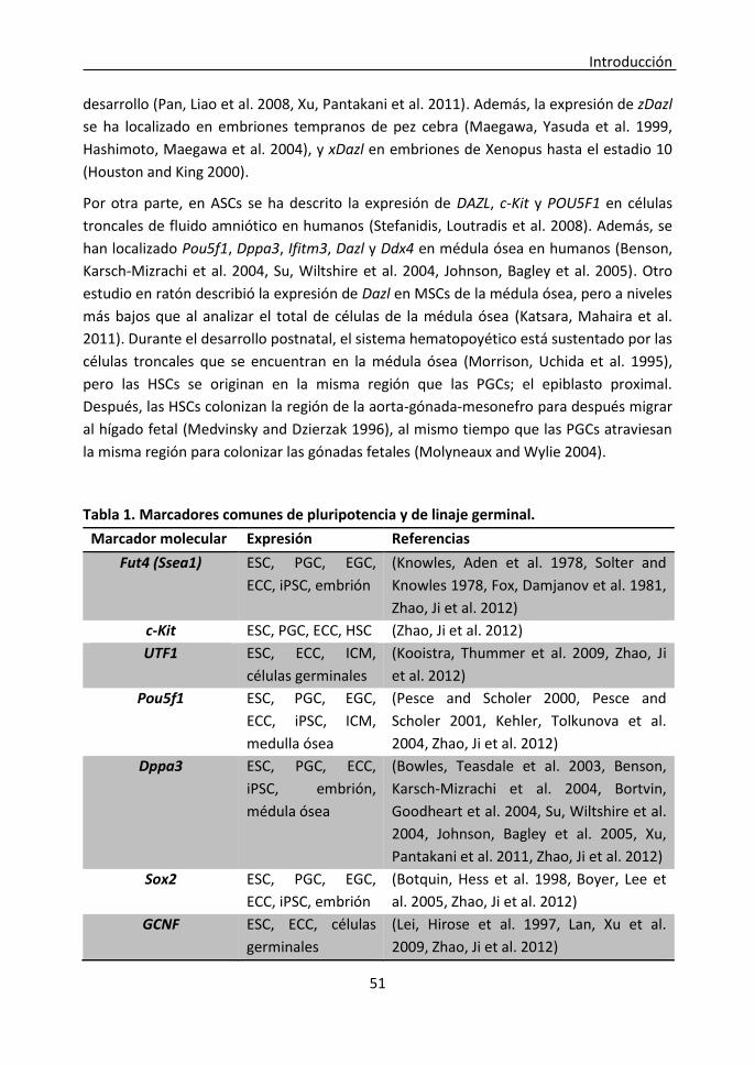
Introducción
51
desarrollo (Pan, Liao et al. 2008, Xu, Pantakani et al. 2011). Además, la expresión de zDazl
se ha localizado en embriones tempranos de pez cebra (Maegawa, Yasuda et al. 1999,
Hashimoto, Maegawa et al. 2004), y xDazl en embriones de Xenopus hasta el estadio 10
(Houston and King 2000).
Por otra parte, en ASCs se ha descrito la expresión de DAZL, c-Kit y POU5F1 en células
troncales de fluido amniótico en humanos (Stefanidis, Loutradis et al. 2008). Además, se
han localizado Pou5f1, Dppa3, Ifitm3, Dazl y Ddx4 en médula ósea en humanos (Benson,
Karsch-Mizrachi et al. 2004, Su, Wiltshire et al. 2004, Johnson, Bagley et al. 2005). Otro
estudio en ratón describió la expresión de Dazl en MSCs de la médula ósea, pero a niveles
más bajos que al analizar el total de células de la médula ósea (Katsara, Mahaira et al.
2011). Durante el desarrollo postnatal, el sistema hematopoyético está sustentado por las
células troncales que se encuentran en la médula ósea (Morrison, Uchida et al. 1995),
pero las HSCs se originan en la misma región que las PGCs; el epiblasto proximal.
Después, las HSCs colonizan la región de la aorta-gónada-mesonefro para después migrar
al hígado fetal (Medvinsky and Dzierzak 1996), al mismo tiempo que las PGCs atraviesan
la misma región para colonizar las gónadas fetales (Molyneaux and Wylie 2004).
Tabla 1. Marcadores comunes de pluripotencia y de linaje germinal.
Marcador molecular Expresión Referencias
Fut4 (Ssea1) ESC, PGC, EGC,
ECC, iPSC, embrión
(Knowles, Aden et al. 1978, Solter and
Knowles 1978, Fox, Damjanov et al. 1981,
Zhao, Ji et al. 2012)
c-Kit ESC, PGC, ECC, HSC (Zhao, Ji et al. 2012)
UTF1 ESC, ECC, ICM,
células germinales
(Kooistra, Thummer et al. 2009, Zhao, Ji
et al. 2012)
Pou5f1 ESC, PGC, EGC,
ECC, iPSC, ICM,
medulla ósea
(Pesce and Scholer 2000, Pesce and
Scholer 2001, Kehler, Tolkunova et al.
2004, Zhao, Ji et al. 2012)
Dppa3 ESC, PGC, ECC,
iPSC, embrión,
médula ósea
(Bowles, Teasdale et al. 2003, Benson,
Karsch-Mizrachi et al. 2004, Bortvin,
Goodheart et al. 2004, Su, Wiltshire et al.
2004, Johnson, Bagley et al. 2005, Xu,
Pantakani et al. 2011, Zhao, Ji et al. 2012)
Sox2 ESC, PGC, EGC,
ECC, iPSC, embrión
(Botquin, Hess et al. 1998, Boyer, Lee et
al. 2005, Zhao, Ji et al. 2012)
GCNF ESC, ECC, células
germinales
(Lei, Hirose et al. 1997, Lan, Xu et al.
2009, Zhao, Ji et al. 2012)
Introducción
52
Nanog ESC, PGC, EGC,
ECC, iPSC
(Chambers, Silva et al. 2007, Yamaguchi,
Kurimoto et al. 2009)
Prdm14 ESC, PGC (Yamaji, Seki et al. 2008, Leitch, McEwen
et al. 2013, Yamaji, Ueda et al. 2013)
Blimp1 ESC, PGC, iPSC (Xu, Pantakani et al. 2011)
Ifitm3 ESC, PGC, iPSC,
medulla ósea
(Benson, Karsch-Mizrachi et al. 2004, Su,
Wiltshire et al. 2004, Johnson, Bagley et
al. 2005, Xu, Pantakani et al. 2011)
Piwil2 ESC, PGC (Xu, Pantakani et al. 2011)
Dazl ESC, PGC, EGC,
GSC, ECC, iPSC,
embrión, médula
ósea
(Benson, Karsch-Mizrachi et al. 2004, Su,
Wiltshire et al. 2004, Johnson, Bagley et
al. 2005, Pan, Liao et al. 2008, Katsara,
Mahaira et al. 2011, Xu, Pantakani et al.
2011, Xu, Tan et al. 2013)
Ddx4 ESC, PGC, iPSC,
embrión, médula
ósea
(Benson, Karsch-Mizrachi et al. 2004, Su,
Wiltshire et al. 2004, Johnson, Bagley et
al. 2005, Xu, Pantakani et al. 2011)
Stra8 ESC, PGC, iPSC (Xu, Pantakani et al. 2011)
Rnf17 ESC, PGC (Xu, Pantakani et al. 2011)
Rnh2 ESC, PGC (Xu, Pantakani et al. 2011)
ESC: células troncales embrionarias; PGC: células primordiales germinales; EGC: células germinales embrionarias; ECC: células embrionarias de carcinoma; iPSC: células de pluripotencia inducida; EGC: células troncales de linaje germinal; CTH: células troncales hematopoyéticas; ICM: masa celular interna
En conclusión, se ha observado que numerosos marcadores de linaje germinal y
premeióticos se expresan en varios tipos de células pluripotentes, lo que podría indicar
un origen germinal común para todos ellos. Según esta teoría, el blastocisto (d3,5)
expresa los principales marcadores de pluripotencia Pou5f1 y Sox2. A lo largo del
desarrollo embrionario, la especificación de las PGCs comienza cuando se activa la
expresión de Blimp1, reprimiendo el programa somático y reactivando las vías de
pluripotencia antes de que comience la migración de las PGCs (Saitou 2009). De la misma
manera, la obtención de ESCs comienza con el cultivo de blastocistos a día 3,5, cuyas
células progresarían hacia el programa de especificación y desarrollo de las PGCs gracias a
la señalización de las BMPs, y comenzarían a expresar Blimp1, Ifitm3 y Dppa3 entre otros
marcadores, y a reactivar los genes de pluripotencia para adquirir propiedades de auto-
renovación. Del mismo modo, durante la reprogramación de células somáticas mediante
los factores Pou5f1, Sox2, c-Myc y Klf4, la señalización de las BMPs dispararía la expresión
de los marcadores de linaje germinal y finalmente se reactivarían las vías de la
pluripotencia (Xu, Pantakani et al. 2011) (Figura 5).

Introducción
53
Figura 5. Teoría del origen germinal de las células pluripotentes
La creación de un ratón transgénico en el que la expresión de un gen determinado venga
marcada por una proteína flurescente como “Enhanced Green fluorescent protein”
(eGFP) constituye un modelo ideal para estudiar la localización de dicho gen en diferentes
tejidos o poblaciones celulares en distintos momentos del desarrollo sin interferir con
ningún proceso biológico. Dazl es uno de los genes cuya principal función está
relacionada con el desarrollo del linaje germinal y la meiosis (Ruggiu, Speed et al. 1997),
aunque en los últimos años se han publicado estudios que lo relacionan con la
pluripotencia (Haston, Tung et al. 2009, Xu, Tan et al. 2013).

Introducción
54
Especificación del trofoectodermo
La primera diferenciación de linajes celulares tiene lugar en el blastocisto, cuando las
células se diferencian hacia masa celular interna (ICM), precursora del embrión, y
trofoectodermo (TE), que dará lugar a la placenta (Schiffmacher and Keefer 2013). Dicha
diferenciación está controlada por determinados genes reguladores, pero existen
diferencias interespecíficas.
1. Diferencias interespecíficas En el ratón existe una interacción antagonista entre los genes reguladores de la ICM y del
TE; ya que la ICM y su estado pluripotente están mantenidos por Pou5f1, mientras que la
especificación del TE depende de Cdx2, que es capaz de suprimir la expresión de Pou5f1 y
viceversa. Sin embargo, esta situación es muy diferente en los embriones de otras
especies de mamíferos. La expresión de Pou5f1 está restringida a la ICM en el ratón, pero
en embriones bovinos y porcinos Pou5f1 se expresa en el TE también, por lo que el
embrión del ratón no es un buen modelo para estudiar la embriogénesis de otras
especies como los ungulados (Wolf, Serup et al. 2011). Algunos autores han sugerido que
el rápido silenciamiento de Pou5f1 en roedores podría haber evolucionado para permitir
una veloz diferenciación del TE y un periodo preimplantacional más corto, mientras que
en otros mamíferos en los que el desarrollo preimplantacional es más largo no habría esta
necesidad (Berg, Smith et al. 2011).
Los precursores de las células diferenciadas de la placenta son las células troncales del
trofoblasto (“trophoblast stem cells”, TSCs). Su existencia se supone en todos los
mamíferos euterios, especialmente durante los estadios tempranos del desarrollo de la
placenta cuando el crecimiento del TE es máximo. Sin embargo, se sabe poco acerca de su
localización y fenotipo en otras especies diferentes al ratón y al macaco Rhesus (Roberts
and Fisher 2011). En el ratón, el nicho de TSCs parece estar localizado en el ectodermo
extraembrionario adyacente al epiblasto, del que depende para nutrirse de factores de
crecimiento esenciales. Este nicho tan especializado es transitorio y existe sólo durante 3
o 4 días durante el desarrollo postimplantacional. Las TSCs de ratón han sido aisladas a
partir del cultivo in vitro de explantes de TE polar, obtenidos a partir una región
adyacente al epiblasto, sobre una monocapa de fibroblastos mitóticamente inactivados y
con medio suplementado con FCS, factor de crecimiento fibrobástico 4 (“fibroblast
growth factor”, FGF4) y heparina (Tanaka, Kunath et al. 1998). Del mismo modo se han
obtenido TSCs a partir de blastocistos de macaco Rhesus (Vandevoort, Thirkill et al. 2007).
En algunas especies domésticas como el cerdo, la oveja y la vaca, se produce una enorme
elongación del TE antes de la implantación, con el objetivo de aprovechar al máximo las
Introducción
55
secreciones uterinas a lo largo de un gran área en el lumen uterino (Bindon 1971, Geisert,
Brookbank et al. 1982, Thatcher, Meyer et al. 1995). A diferencia del ratón, el TE polar
que recubre el disco embrionario en estas especies desaparece rápidamente y no
contribuye a la formación de la placenta, mientras que el TE mural es el responsable de la
elongación. Se desconoce si la elongación del TE mural depende de una población
pluripotente que se encontraría alrededor del disco embrionario, o de células
pluripotentes diseminadas a lo largo de todo el TE; o si por el contrario este gran
crecimiento se debe a la proliferación de células progenitoras que se encuentran
parcialmente diferenciadas (Roberts and Fisher 2011). En consecuencia, no han podido
aislarse TSCs de ninguna especie de ungulados; sin embargo se han conseguido aislar
líneas celulares de trofoectodermo en oveja y cabra (Miyazaki, Imai et al. 2002), cerdo
(Ramsoondar, Christopherson et al. 1993, Flechon, Laurie et al. 1995, La Bonnardiere,
Flechon et al. 2002) y vaca (Talbot, Caperna et al. 2000, Shimada, Nakano et al. 2001,
Suzuki, Koshi et al. 2011).
2. Aislamiento de líneas celulares de trofoblasto bovino Las células del trofoblasto crecen in vitro formando una monocapa en la que a veces
aparecen estructuras con forma de caverna que acumulan líquido en su interior hasta
transformarse en esferoides que son liberados al medio de cultivo.
Las primeras líneas de trofoblasto bovino fueron producidas por Talbot et al., a partir de
blastocistos de día 7-8 producidos in vitro y cultivados sobre una monocapa de
fibroblastos STO (Talbot, Caperna et al. 2000). Sin embargo, el co-cultivo con fibroblastos
murinos tiene el riesgo de contaminar las células de trofoblasto bovino, y esto podría ser
perjudicial para el uso de estas líneas celulares en estudios posteriores. Más tarde,
Shimada et al. (Shimada, Nakano et al. 2001) establecieron una línea de células de
trofoblasto bovino en ausencia de fibroblastos, utilizando medio condicionado por
fibroblastos endometriales, pero su eficiencia de obtención fue muy baja; plaquearon
más de 50 blastocistos obteniendo sólo una línea celular. El mayor problema parecía
radicar en la adherencia del blastocisto a la placa de cultivo y en la proliferación inicial.
Desde entonces, la mayoría de los estudios de señalización y diferenciación del
trofoectodermo se han llevado a cabo con estas dos únicas líneas celulares producidas
por Talbot y Shimada (Nakano, Shimada et al. 2002, Hirata, Sato et al. 2003, Ushizawa,
Takahashi et al. 2005, Michael, Wagner et al. 2006, Das, Ezashi et al. 2008, Nakaya, Kizaki
et al. 2009, Bai, Sakurai et al. 2011, Schiffmacher and Keefer 2012, Schiffmacher and
Keefer 2013). Por lo tanto, sería muy útil conseguir un número mayor de líneas celulares
de trofoblasto apropiadas para estudios in vitro, que carezcan de posibles
contaminaciones por otros tipos celulares.

Introducción
56
Se han obtenido líneas celulares de trofoblasto a partir de embriones producidos
mediante SCNT o por partenogénesis, y se han observado diferencias significativas en
cuanto a su eficiencia de obtención, ya que los embriones partenogenéticos mostraban
un menor índice de formación de colonias que los embriones producidos por IVF o por
SCNT. De la misma manera, la cantidad de interferón tau (IFN-t), proteína necesaria para
el establecimiento de la gestación, producida por las líneas de células del trofoblasto
procedentes de embriones partenogenéticos y de SCNT, era menor que la de aquellos
producidos por IVF. Además, las líneas procedentes de SCNT contaban con menor
cantidad de proteínas anexinas, relacionadas con el mantenimiento de la placentación
(Talbot, Powell et al. 2007, Talbot, Powell et al. 2008, Talbot, Powell et al. 2010). Por lo
tanto, las líneas celulares trofoblásticas podrían reflejar las posibles alteraciones
embrionarias y ser utilizadas como un modelo para su estudio.
En un estudio más reciente, basándose en que el aporte exógeno de BMP4 estimula a las
ESCs para convertirse en células trofoblásticas (Murohashi, Nakamura et al. 2010), Suzuki
et al. utilizaron BMP4 para producir líneas de células trofoblásticas de bovino, pero no
encontraron diferencias significativas en la eficiencia de obtención, y sólo pudieron
obtener 12 líneas celulares a partir de 172 embriones. Al analizar la expresión de los
genes necesarios para el establecimiento de la gestación: interferón tau (IFNt), lactógeno
placentario (CSH1), proteína relacionada con la prolactina 1 (PRP1) y glicoproteína
asociada a la gestación (PAG1), encontraron patrones de expresión muy variables entre
las diferentes líneas celulares (Suzuki, Koshi et al. 2011). Por lo tanto, existe una alta
variabilidad entre las líneas de células trofoblásticas obtenidas bajo condiciones idénticas.
Muchas de las líneas de células trofoblasticas obtenidas en diferentes estudios crecen
continuamente sin mostrar signos de envejecimiento y expresan POU5F1 (Suzuki, Koshi et
al. 2011, Schiffmacher and Keefer 2013), por lo que es posible que estas células
representen la población celular pluripotente a partir de la cual se diferencia la placenta
en el bovino, o quizás un equivalente a las TSCs de ratón algo más diferenciado.
Las líneas de células trofoblásticas constituyen un modelo muy útil para los estudios de
implantación y placentogénesis, y podrían reflejar las características genéticas y
epigenéticas y las alteraciones presentes en los embriones de los que proceden. Sin
embargo, tan sólo un reducido número de líneas celulares se utiliza para investigación
debido a la dificultad de establecer nuevas líneas. Por ello, sería interesante establecer un
sistema para la obtención de líneas de células trofoblásticas de manera eficaz a partir de
una biopsia de TE que permitiera al embrión seguir siendo viable, mientras sus
características o alteraciones son analizadas en su correspondiente línea de células
trofoblásticas.

Bibliography / Bibliografía
57
Bibliography/Bibliografía

Bibliography / Bibliografía
58

Bibliography / Bibliografía
59
Allegrucci, C., A. Thurston, E. Lucas and L. Young (2005). "Epigenetics and the germline."
Reproduction 129(2): 137-149. Allegrucci, C., Y. Z. Wu, A. Thurston, C. N. Denning, H. Priddle, C. L. Mummery, D. Ward-van
Oostwaard, P. W. Andrews, M. Stojkovic, N. Smith, T. Parkin, M. E. Jones, G. Warren, L. Yu, R. M. Brena, C. Plass and L. E. Young (2007). "Restriction landmark genome scanning identifies culture-induced DNA methylation instability in the human embryonic stem cell epigenome." Hum Mol Genet 16(10): 1253-1268.
Alvarez, C. V., M. Garcia-Lavandeira, M. E. Garcia-Rendueles, E. Diaz-Rodriguez, A. R. Garcia-Rendueles, S. Perez-Romero, T. V. Vila, J. S. Rodrigues, P. V. Lear and S. B. Bravo (2012). "Defining stem cell types: understanding the therapeutic potential of ESCs, ASCs, and iPS cells." J Mol Endocrinol 49(2): R89-111.
Arnaud, P. (2010). "Genomic imprinting in germ cells: imprints are under control." Reproduction 140(3): 411-423.
Bai, H., T. Sakurai, Y. Someya, T. Konno, A. Ideta, Y. Aoyagi and K. Imakawa (2011). "Regulation of trophoblast-specific factors by GATA2 and GATA3 in bovine trophoblast CT-1 cells." J Reprod Dev 57(4): 518-525.
Becker, A. J., C. E. Mc and J. E. Till (1963). "Cytological demonstration of the clonal nature of spleen colonies derived from transplanted mouse marrow cells." Nature 197: 452-454.
Benson, D. A., I. Karsch-Mizrachi, D. J. Lipman, J. Ostell and D. L. Wheeler (2004). "GenBank: update." Nucleic Acids Res 32(Database issue): D23-26.
Berg, D. K., C. S. Smith, D. J. Pearton, D. N. Wells, R. Broadhurst, M. Donnison and P. L. Pfeffer (2011). "Trophectoderm lineage determination in cattle." Dev Cell 20(2): 244-255.
Bermejo-Alvarez, P., P. Ramos-Ibeas and A. Gutierrez-Adan (2012). "Solving the "X" in embryos and stem cells." Stem Cells Dev 21(8): 1215-1224.
Bindon, B. M. (1971). "Systematic study of preimplantation stages of pregnancy in the sheep." Aust J Biol Sci 24(1): 131-147.
Bird, A. (2002). "DNA methylation patterns and epigenetic memory." Genes Dev 16(1): 6-21. Blaschke, K., K. T. Ebata, M. M. Karimi, J. A. Zepeda-Martínez, P. Goyal, S. Mahapatra, A. Tam, D. J.
Laird, M. Hirst, A. Rao, M. C. Lorincz and M. Ramalho-Santos (2013). "Vitamin[thinsp]C induces Tet-dependent DNA demethylation and a blastocyst-like state in ES cells." Nature 500: 222-226.
Bortvin, A., M. Goodheart, M. Liao and D. C. Page (2004). "Dppa3 / Pgc7 / stella is a maternal factor and is not required for germ cell specification in mice." BMC Dev Biol 4: 2.
Botquin, V., H. Hess, G. Fuhrmann, C. Anastassiadis, M. K. Gross, G. Vriend and H. R. Scholer (1998). "New POU dimer configuration mediates antagonistic control of an osteopontin preimplantation enhancer by Oct-4 and Sox-2." Genes Dev 12(13): 2073-2090.
Boveri, T. (1892). d. Gesellschaft f. Morphologie und Physiologie, Sitzungsber: 114-225. Bowles, J., R. P. Teasdale, K. James and P. Koopman (2003). "Dppa3 is a marker of pluripotency and
has a human homologue that is expressed in germ cell tumours." Cytogenet Genome Res 101(3-4): 261-265.
Boyer, L. A., T. I. Lee, M. F. Cole, S. E. Johnstone, S. S. Levine, J. P. Zucker, M. G. Guenther, R. M. Kumar, H. L. Murray, R. G. Jenner, D. K. Gifford, D. A. Melton, R. Jaenisch and R. A. Young (2005). "Core transcriptional regulatory circuitry in human embryonic stem cells." Cell 122(6): 947-956.
Bradley, A., M. Evans, M. H. Kaufman and E. Robertson (1984). "Formation of germ-line chimaeras from embryo-derived teratocarcinoma cell lines." Nature 309(5965): 255-256.
Brambrink, T., K. Hochedlinger, G. Bell and R. Jaenisch (2006). "ES cells derived from cloned and fertilized blastocysts are transcriptionally and functionally indistinguishable." Proc Natl Acad Sci U S A 103(4): 933-938.

Bibliography / Bibliografía
60
Brevini, T. A., S. Antonini, G. Pennarossa and F. Gandolfi (2008). "Recent progress in embryonic stem cell research and its application in domestic species." Reprod Domest Anim 43 Suppl 2: 193-199.
Brook, F. A. and R. L. Gardner (1997). "The origin and efficient derivation of embryonic stem cells in the mouse." Proc Natl Acad Sci U S A 94(11): 5709-5712.
Buehr, M., S. Meek, K. Blair, J. Yang, J. Ure, J. Silva, R. McLay, J. Hall, Q. L. Ying and A. Smith (2008). "Capture of authentic embryonic stem cells from rat blastocysts." Cell 135(7): 1287-1298.
Calle, A., A. Miranda, R. Fernandez-Gonzalez, E. Pericuesta, R. Laguna and A. Gutierrez-Adan (2012). "Male mice produced by in vitro culture have reduced fertility and transmit organomegaly and glucose intolerance to their male offspring." Biol Reprod 87(2): 34.
Chambers, I., J. Silva, D. Colby, J. Nichols, B. Nijmeijer, M. Robertson, J. Vrana, K. Jones, L. Grotewold and A. Smith (2007). "Nanog safeguards pluripotency and mediates germline development." Nature 450(7173): 1230-1234.
Chen, Z., K. M. Robbins, K. D. Wells and R. M. Rivera (2013). "Large offspring syndrome: a bovine model for the human loss-of-imprinting overgrowth syndrome Beckwith-Wiedemann." Epigenetics 8(6): 591-601.
Cheng, T. C., C. C. Huang, C. I. Chen, C. H. Liu, Y. S. Hsieh, C. Y. Huang, M. S. Lee and J. Y. Liu (2004). "Leukemia inhibitory factor antisense oligonucleotide inhibits the development of murine embryos at preimplantation stages." Biol Reprod 70(5): 1270-1276.
Chu, L. F., M. A. Surani, R. Jaenisch and T. P. Zwaka (2011). "Blimp1 expression predicts embryonic stem cell development in vitro." Curr Biol 21(20): 1759-1765.
Clayton-Smith, J. (2003). "Genomic imprinting as a cause of disease: Is increasingly recognised, especially after assisted reproduction." BMJ 327(7424): 1121-1122.
Das, P., T. Ezashi, R. Gupta and R. M. Roberts (2008). "Combinatorial roles of protein kinase A, Ets2, and 3',5'-cyclic-adenosine monophosphate response element-binding protein-binding protein/p300 in the transcriptional control of interferon-tau expression in a trophoblast cell line." Mol Endocrinol 22(2): 331-343.
de Rooij, D. G. and S. C. Mizrak (2008). "Deriving multipotent stem cells from mouse spermatogonial stem cells: a new tool for developmental and clinical research." Development 135(13): 2207-2213.
de Sousa Lopes, S. M., K. Hayashi and M. A. Surani (2007). "Proximal visceral endoderm and extraembryonic ectoderm regulate the formation of primordial germ cell precursors." BMC Dev Biol 7: 140.
de Sousa Lopes, S. M., B. A. Roelen, R. M. Monteiro, R. Emmens, H. Y. Lin, E. Li, K. A. Lawson and C. L. Mummery (2004). "BMP signaling mediated by ALK2 in the visceral endoderm is necessary for the generation of primordial germ cells in the mouse embryo." Genes Dev 18(15): 1838-1849.
Ding, J., Y. Guo, S. Liu, Y. Yan, G. Chang, Z. Kou, Y. Zhang, Y. Jiang, F. He, S. Gao and J. Sang (2009). "Embryonic stem cells derived from somatic cloned and fertilized blastocysts are post-transcriptionally indistinguishable: a MicroRNA and protein profile comparison." Proteomics 9(10): 2711-2721.
Durcova-Hills, G. and A. Surani (2008). "Reprogramming primordial germ cells (PGC) to embryonic germ (EG) cells." Curr Protoc Stem Cell Biol Chapter 1: Unit1A.3.
Evans, M. J. and M. H. Kaufman (1981). "Establishment in culture of pluripotential cells from mouse embryos." Nature 292(5819): 154-156.
Eyal-Giladi, H. and S. Kochav (1976). "From cleavage to primitive streak formation: a complementary normal table and a new look at the first stages of the development of the chick. I. General morphology." Dev Biol 49(2): 321-337.

Bibliography / Bibliografía
61
Familari, M. and L. Selwood (2006). "The potential for derivation of embryonic stem cells in vertebrates." Mol Reprod Dev 73(1): 123-131.
Fan, L., J. Crodian, X. Liu, A. Alestrom, P. Alestrom and P. Collodi (2004). "Zebrafish embryo cells remain pluripotent and germ-line competent for multiple passages in culture." Zebrafish 1(1): 21-26.
Feinberg, A. P., R. Ohlsson and S. Henikoff (2006). "The epigenetic progenitor origin of human cancer." Nat Rev Genet 7(1): 21-33.
Fernandez-Gonzalez, R., P. N. Moreira, M. Perez-Crespo, M. Sanchez-Martin, M. A. Ramirez, E. Pericuesta, A. Bilbao, P. Bermejo-Alvarez, J. de Dios Hourcade, F. R. de Fonseca and A. Gutierrez-Adan (2008). "Long-term effects of mouse intracytoplasmic sperm injection with DNA-fragmented sperm on health and behavior of adult offspring." Biol Reprod 78(4): 761-772.
Flechon, J. E., S. Laurie and E. Notarianni (1995). "Isolation and characterization of a feeder-dependent, porcine trophectoderm cell line obtained from a 9-day blastocyst." Placenta 16(7): 643-658.
Fox, N., I. Damjanov, A. Martinez-Hernandez, B. B. Knowles and D. Solter (1981). "Immunohistochemical localization of the early embryonic antigen (SSEA-1) in postimplantation mouse embryos and fetal and adult tissues." Dev Biol 83(2): 391-398.
Fujimoto, A., S. M. Mitalipov, L. L. Clepper and D. P. Wolf (2005). "Development of a monkey model for the study of primate genomic imprinting." Mol Hum Reprod 11(6): 413-422.
Fujishiro, S. H., K. Nakano, Y. Mizukami, T. Azami, Y. Arai, H. Matsunari, R. Ishino, T. Nishimura, M. Watanabe, T. Abe, Y. Furukawa, K. Umeyama, S. Yamanaka, M. Ema, H. Nagashima and Y. Hanazono (2013). "Generation of naive-like porcine-induced pluripotent stem cells capable of contributing to embryonic and fetal development." Stem Cells Dev 22(3): 473-482.
Gafni, O., L. Weinberger, A. A. Mansour, Y. S. Manor, E. Chomsky, D. Ben-Yosef, Y. Kalma, S. Viukov, I. Maza, A. Zviran, Y. Rais, Z. Shipony, Z. Mukamel, V. Krupalnik, M. Zerbib, S. Geula, I. Caspi, D. Schneir, T. Shwartz, S. Gilad, D. Amann-Zalcenstein, S. Benjamin, I. Amit, A. Tanay, R. Massarwa, N. Novershtern and J. H. Hanna (2013). "Derivation of novel human ground state naive pluripotent stem cells." Nature.
Geijsen, N., M. Horoschak, K. Kim, J. Gribnau, K. Eggan and G. Q. Daley (2004). "Derivation of embryonic germ cells and male gametes from embryonic stem cells." Nature 427(6970): 148-154.
Geisert, R. D., J. W. Brookbank, R. M. Roberts and F. W. Bazer (1982). "Establishment of pregnancy in the pig: II. Cellular remodeling of the porcine blastocyst during elongation on day 12 of pregnancy." Biol Reprod 27(4): 941-955.
Gjorret, J. O. and P. Maddox-Hyttel (2005). "Attempts towards derivation and establishment of bovine embryonic stem cell-like cultures." Reprod Fertil Dev 17(1-2): 113-124.
Greco, V. and S. Guo (2010). "Compartmentalized organization: a common and required feature of stem cell niches?" Development 137(10): 1586-1594.
Guan, K., K. Nayernia, L. S. Maier, S. Wagner, R. Dressel, J. H. Lee, J. Nolte, F. Wolf, M. Li, W. Engel and G. Hasenfuss (2006). "Pluripotency of spermatogonial stem cells from adult mouse testis." Nature 440(7088): 1199-1203.
Gutierrez-Adan, A., M. Perez-Crespo, R. Fernandez-Gonzalez, M. A. Ramirez, P. Moreira, B. Pintado, P. Lonergan and D. Rizos (2006). "Developmental consequences of sexual dimorphism during pre-implantation embryonic development." Reprod Domest Anim 41 Suppl 2: 54-62.
Haeckel, E. (1868). Natürliche Schöpfungsgeschichte. Berlin, Georg Reimer. Haeckel, E. (1877). Anthropogenie. Lepizig, Wilhelm Engelmann. Hajkova, P., S. Erhardt, N. Lane, T. Haaf, O. El-Maarri, W. Reik, J. Walter and M. A. Surani (2002).
"Epigenetic reprogramming in mouse primordial germ cells." Mech Dev 117(1-2): 15-23.

Bibliography / Bibliografía
62
Hansen, M., J. J. Kurinczuk, E. Milne, N. de Klerk and C. Bower (2013). "Assisted reproductive technology and birth defects: a systematic review and meta-analysis." Hum Reprod Update 19(4): 330-353.
Hashimoto, Y., S. Maegawa, T. Nagai, E. Yamaha, H. Suzuki, K. Yasuda and K. Inoue (2004). "Localized maternal factors are required for zebrafish germ cell formation." Dev Biol 268(1): 152-161.
Haston, K. M., J. Y. Tung and R. A. Reijo Pera (2009). "Dazl Functions in Maintenance of Pluripotency and Genetic and Epigenetic Programs of Differentiation in Mouse Primordial Germ Cells In Vivo and In Vitro." PLoS ONE 4(5).
Hirata, M., T. Sato, M. Tsumagari, A. Shimada, H. Nakano, K. Hashizume and A. Ito (2003). "Differential regulation of the expression of matrix metalloproteinases and tissue inhibitors of metalloproteinases by cytokines and growth factors in bovine endometrial stromal cells and trophoblast cell line BT-1 in vitro." Biol Reprod 68(4): 1276-1281.
Hochedlinger, K. (2011). "Embryonic stem cells: testing the germ-cell theory." Curr Biol 21(20): R850-852.
Holm, T. M., L. Jackson-Grusby, T. Brambrink, Y. Yamada, W. M. Rideout, 3rd and R. Jaenisch (2005). "Global loss of imprinting leads to widespread tumorigenesis in adult mice." Cancer Cell 8(4): 275-285.
Horii, T., E. Yanagisawa, M. Kimura, S. Morita and I. Hatada (2010). "Epigenetic differences between embryonic stem cells generated from blastocysts developed in vitro and in vivo." Cell Reprogram 12(5): 551-563.
Hourcade, J. D., M. Perez-Crespo, R. Fernandez-Gonzalez, B. Pintado and A. Gutierrez-Adan (2010). "Selection against spermatozoa with fragmented DNA after postovulatory mating depends on the type of damage." Reprod Biol Endocrinol 8: 9.
Houston, D. W. and M. L. King (2000). "A critical role for Xdazl, a germ plasm-localized RNA, in the differentiation of primordial germ cells in Xenopus." Development 127(3): 447-456.
Izadyar, F., F. Pau, J. Marh, N. Slepko, T. Wang, R. Gonzalez, T. Ramos, K. Howerton, C. Sayre and F. Silva (2008). "Generation of multipotent cell lines from a distinct population of male germ line stem cells." Reproduction 135(6): 771-784.
Jaramillo-Ferrada, P. A., E. J. Wolvetang and J. J. Cooper-White (2012). "Differential mesengenic potential and expression of stem cell-fate modulators in mesenchymal stromal cells from human-term placenta and bone marrow." J Cell Physiol 227(9): 3234-3242.
Johnson, J., J. Bagley, M. Skaznik-Wikiel, H. J. Lee, G. B. Adams, Y. Niikura, K. S. Tschudy, J. C. Tilly, M. L. Cortes, R. Forkert, T. Spitzer, J. Iacomini, D. T. Scadden and J. L. Tilly (2005). "Oocyte generation in adult mammalian ovaries by putative germ cells in bone marrow and peripheral blood." Cell 122(2): 303-315.
Johnson, M. H. (2001). "Mammalian development: axes in the egg?" Curr Biol 11(7): R281-284. Johnson, M. H. and L. Selwood (1996). "Nomenclature of early development in mammals." Reprod
Fertil Dev 8(4): 759-764. Juopperi, T. A., H. Song and G. L. Ming (2011). "Modeling neurological diseases using patient-
derived induced pluripotent stem cells." Future Neurol 6(3): 363-373. Jurisicova, A., A. Ben-Chetrit, S. L. Varmuza and R. F. Casper (1995). "Recombinant human leukemia
inhibitory factor does not enhance in vitro human blastocyst formation." Fertil Steril 64(5): 999-1002.
Kahan, B. W. and B. Ephrussi (1970). "Developmental potentialities of clonal in vitro cultures of mouse testicular teratoma." J Natl Cancer Inst 44(5): 1015-1036.
Kanatsu-Shinohara, M., K. Inoue, J. Lee, M. Yoshimoto, N. Ogonuki, H. Miki, S. Baba, T. Kato, Y. Kazuki, S. Toyokuni, M. Toyoshima, O. Niwa, M. Oshimura, T. Heike, T. Nakahata, F. Ishino, A.

Bibliography / Bibliografía
63
Ogura and T. Shinohara (2004). "Generation of pluripotent stem cells from neonatal mouse testis." Cell 119(7): 1001-1012.
Kanatsu-Shinohara, M., K. Inoue, N. Ogonuki, H. Morimoto, A. Ogura and T. Shinohara (2011). "Serum- and feeder-free culture of mouse germline stem cells." Biol Reprod 84(1): 97-105.
Kanatsu-Shinohara, M., N. Ogonuki, K. Inoue, H. Miki, A. Ogura, S. Toyokuni and T. Shinohara (2003). "Long-term proliferation in culture and germline transmission of mouse male germline stem cells." Biol Reprod 69(2): 612-616.
Katsara, O., L. G. Mahaira, E. G. Iliopoulou, A. Moustaki, A. Antsaklis, D. Loutradis, K. Stefanidis, C. N. Baxevanis, M. Papamichail and S. A. Perez (2011). "Effects of donor age, gender, and in vitro cellular aging on the phenotypic, functional, and molecular characteristics of mouse bone marrow-derived mesenchymal stem cells." Stem Cells Dev 20(9): 1549-1561.
Kehler, J., E. Tolkunova, B. Koschorz, M. Pesce, L. Gentile, M. Boiani, H. Lomeli, A. Nagy, K. J. McLaughlin, H. R. Scholer and A. Tomilin (2004). "Oct4 is required for primordial germ cell survival." EMBO Rep 5(11): 1078-1083.
Kim, K., A. Doi, B. Wen, K. Ng, R. Zhao, P. Cahan, J. Kim, M. J. Aryee, H. Ji, L. I. Ehrlich, A. Yabuuchi, A. Takeuchi, K. C. Cunniff, H. Hongguang, S. McKinney-Freeman, O. Naveiras, T. J. Yoon, R. A. Irizarry, N. Jung, J. Seita, J. Hanna, P. Murakami, R. Jaenisch, R. Weissleder, S. H. Orkin, I. L. Weissman, A. P. Feinberg and G. Q. Daley (2010). "Epigenetic memory in induced pluripotent stem cells." Nature 467(7313): 285-290.
Knowles, B. B., D. P. Aden and D. Solter (1978). "Monoclonal antibody detecting a stage-specific embryonic antigen (SSEA-1) on preimplantation mouse embryos and teratocarcinoma cells." Curr Top Microbiol Immunol 81: 51-53.
Kooistra, S. M., R. P. Thummer and B. J. Eggen (2009). "Characterization of human UTF1, a chromatin-associated protein with repressor activity expressed in pluripotent cells." Stem Cell Res 2(3): 211-218.
La Bonnardiere, C., J. E. Flechon, S. Battegay, B. Flechon, J. Degrouard and F. Lefevre (2002). "Polarized porcine trophoblastic cell lines spontaneously secrete interferon-gamma." Placenta 23(10): 716-726.
Lan, Z. J., X. Xu, A. C. K. Chung and A. J. Cooney (2009). "Extra-Germ Cell Expression of Mouse Nuclear Receptor Subfamily 6, Group A, Member 1 (NR6A1)1." Biol Reprod 80(5): 905-912.
Lawson, K. A., N. R. Dunn, B. A. Roelen, L. M. Zeinstra, A. M. Davis, C. V. Wright, J. P. Korving and B. L. Hogan (1999). "Bmp4 is required for the generation of primordial germ cells in the mouse embryo." Genes Dev 13(4): 424-436.
Lei, W., T. Hirose, L. X. Zhang, H. Adachi, M. J. Spinella, E. Dmitrovsky and A. M. Jetten (1997). "Cloning of the human orphan receptor germ cell nuclear factor/retinoid receptor-related testis-associated receptor and its differential regulation during embryonal carcinoma cell differentiation." J Mol Endocrinol 18(2): 167-176.
Leitch, H. G., K. Blair, W. Mansfield, H. Ayetey, P. Humphreys, J. Nichols, M. A. Surani and A. Smith (2010). "Embryonic germ cells from mice and rats exhibit properties consistent with a generic pluripotent ground state." Development 137(14): 2279-2287.
Leitch, H. G., K. R. McEwen, A. Turp, V. Encheva, T. Carroll, N. Grabole, W. Mansfield, B. Nashun, J. G. Knezovich, A. Smith, M. A. Surani and P. Hajkova (2013). "Naive pluripotency is associated with global DNA hypomethylation." Nat Struct Mol Biol 20(3): 311-316.
Lengner, C. J., A. A. Gimelbrant, J. A. Erwin, A. W. Cheng, M. G. Guenther, G. G. Welstead, R. Alagappan, G. M. Frampton, P. Xu, J. Muffat, S. Santagata, D. Powers, C. B. Barrett, R. A. Young, J. T. Lee, R. Jaenisch and M. Mitalipova (2010). "Derivation of pre-X inactivation human embryonic stem cells under physiological oxygen concentrations." Cell 141(5): 872-883.
Li, L. and H. Clevers (2010). "Coexistence of quiescent and active adult stem cells in mammals." Science 327(5965): 542-545.

Bibliography / Bibliografía
64
Li, P., C. Tong, R. Mehrian-Shai, L. Jia, N. Wu, Y. Yan, R. E. Maxson, E. N. Schulze, H. Song, C. L. Hsieh, M. F. Pera and Q. L. Ying (2008). "Germline competent embryonic stem cells derived from rat blastocysts." Cell 135(7): 1299-1310.
Lin, Y. and D. C. Page (2005). "Dazl deficiency leads to embryonic arrest of germ cell development in XY C57BL/6 mice." Dev Biol 288(2): 309-316.
Lopes, S., A. Jurisicova, J. G. Sun and R. F. Casper (1998). "Reactive oxygen species: potential cause for DNA fragmentation in human spermatozoa." Hum Reprod 13(4): 896-900.
Lucifero, D., C. Mertineit, H. J. Clarke, T. H. Bestor and J. M. Trasler (2002). "Methylation dynamics of imprinted genes in mouse germ cells." Genomics 79(4): 530-538.
Lund, R. J., E. Narva and R. Lahesmaa (2012). "Genetic and epigenetic stability of human pluripotent stem cells." Nat Rev Genet 13(10): 732-744.
Lyon, M. F. (1961). "Gene action in the X-chromosome of the mouse (Mus musculus L.)." Nature 190: 372-373.
Macaluso, F. and K. H. Myburgh (2012). "Current evidence that exercise can increase the number of adult stem cells." J Muscle Res Cell Motil 33(3-4): 187-198.
Maegawa, S., K. Yasuda and K. Inoue (1999). "Maternal mRNA localization of zebrafish DAZ-like gene." Mech Dev 81(1-2): 223-226.
Maitra, A., D. E. Arking, N. Shivapurkar, M. Ikeda, V. Stastny, K. Kassauei, G. Sui, D. J. Cutler, Y. Liu, S. N. Brimble, K. Noaksson, J. Hyllner, T. C. Schulz, X. Zeng, W. J. Freed, J. Crook, S. Abraham, A. Colman, P. Sartipy, S. Matsui, M. Carpenter, A. F. Gazdar, M. Rao and A. Chakravarti (2005). "Genomic alterations in cultured human embryonic stem cells." Nat Genet 37(10): 1099-1103.
Marks, H., T. Kalkan, R. Menafra, S. Denissov, K. Jones, H. Hofemeister, J. Nichols, A. Kranz, A. F. Stewart, A. Smith and H. G. Stunnenberg (2012). "The transcriptional and epigenomic foundations of ground state pluripotency." Cell 149(3): 590-604.
Marti, M., L. Mulero, C. Pardo, C. Morera, M. Carrio, L. Laricchia-Robbio, C. R. Esteban and J. C. Izpisua Belmonte (2013). "Characterization of pluripotent stem cells." Nat Protoc 8(2): 223-253.
Mateizel, I., N. De Temmerman, U. Ullmann, G. Cauffman, K. Sermon, H. Van de Velde, M. De Rycke, E. Degreef, P. Devroey, I. Liebaers and A. Van Steirteghem (2006). "Derivation of human embryonic stem cell lines from embryos obtained after IVF and after PGD for monogenic disorders." Hum Reprod 21(2): 503-511.
Matsui, Y., K. Zsebo and B. L. Hogan (1992). "Derivation of pluripotential embryonic stem cells from murine primordial germ cells in culture." Cell 70(5): 841-847.
Medvinsky, A. and E. Dzierzak (1996). "Definitive hematopoiesis is autonomously initiated by the AGM region." Cell 86(6): 897-906.
Meng, X., M. Lindahl, M. E. Hyvonen, M. Parvinen, D. G. de Rooij, M. W. Hess, A. Raatikainen-Ahokas, K. Sainio, H. Rauvala, M. Lakso, J. G. Pichel, H. Westphal, M. Saarma and H. Sariola (2000). "Regulation of cell fate decision of undifferentiated spermatogonia by GDNF." Science 287(5457): 1489-1493.
Michael, D. D., S. K. Wagner, O. M. Ocon, N. C. Talbot, J. A. Rooke and A. D. Ealy (2006). "Granulocyte-macrophage colony-stimulating-factor increases interferon-tau protein secretion in bovine trophectoderm cells." Am J Reprod Immunol 56(1): 63-67.
Minoguchi, S. and H. Iba (2008). "Instability of retroviral DNA methylation in embryonic stem cells." Stem Cells 26(5): 1166-1173.
Mise, N., T. Fuchikami, M. Sugimoto, S. Kobayakawa, F. Ike, T. Ogawa, T. Tada, S. Kanaya, T. Noce and K. Abe (2008). "Differences and similarities in the developmental status of embryo-derived stem cells and primordial germ cells revealed by global expression profiling." Genes Cells 13(8): 863-877.
Miyazaki, H., M. Imai, T. Hirayama, S. Saburi, M. Tanaka, M. Maruyama, C. Matsuo, H. Meguro, K. Nishibashi, F. Inoue, J. Djiane, A. Gertler, S. Tachi, K. Imakawa and C. Tachi (2002).

Bibliography / Bibliografía
65
"Establishment of feeder-independent cloned caprine trophoblast cell line which expresses placental lactogen and interferon tau." Placenta 23(8-9): 613-630.
Molyneaux, K. and C. Wylie (2004). "Primordial germ cell migration." Int J Dev Biol 48(5-6): 537-544.
Morison, I. M., J. P. Ramsay and H. G. Spencer (2005). "A census of mammalian imprinting." Trends Genet 21(8): 457-465.
Morrison, S. J., N. Uchida and I. L. Weissman (1995). "The biology of hematopoietic stem cells." Annu Rev Cell Dev Biol 11: 35-71.
Murohashi, M., T. Nakamura, S. Tanaka, T. Ichise, N. Yoshida, T. Yamamoto, M. Shibuya, J. Schlessinger and N. Gotoh (2010). "An FGF4-FRS2alpha-Cdx2 axis in trophoblast stem cells induces Bmp4 to regulate proper growth of early mouse embryos." Stem Cells 28(1): 113-121.
Nagy, A., E. Gocza, E. M. Diaz, V. R. Prideaux, E. Ivanyi, M. Markkula and J. Rossant (1990). "Embryonic stem cells alone are able to support fetal development in the mouse." Development 110(3): 815-821.
Nakano, H., A. Shimada, K. Imai, T. Takezawa, T. Takahashi and K. Hashizume (2002). "Bovine trophoblastic cell differentiation on collagen substrata: formation of binucleate cells expressing placental lactogen." Cell Tissue Res 307(2): 225-235.
Nakaya, Y., K. Kizaki, T. Takahashi, O. V. Patel and K. Hashizume (2009). "The characterization of DNA methylation-mediated regulation of bovine placental lactogen and bovine prolactin-related protein-1 genes." BMC Mol Biol 10: 19.
Nichols, J., K. Jones, J. M. Phillips, S. A. Newland, M. Roode, W. Mansfield, A. Smith and A. Cooke (2009). "Validated germline-competent embryonic stem cell lines from nonobese diabetic mice." Nat Med 15(7): 814-818.
Nichols, J., J. Silva, M. Roode and A. Smith (2009). "Suppression of Erk signalling promotes ground state pluripotency in the mouse embryo." Development 136(19): 3215-3222.
Nichols, J. and A. Smith (2009). "Naive and primed pluripotent states." Cell Stem Cell 4(6): 487-492. Nichols, J. and A. Smith (2011). "The origin and identity of embryonic stem cells." Development
138(1): 3-8. Niwa, H., T. Burdon, I. Chambers and A. Smith (1998). "Self-renewal of pluripotent embryonic stem
cells is mediated via activation of STAT3." Genes Dev 12(13): 2048-2060. Niwa, H. and T. Fujimori (2010). "Stem cell systems in development of mammals." Dev Growth
Differ 52(3): 251. Ohinata, Y., B. Payer, D. O'Carroll, K. Ancelin, Y. Ono, M. Sano, S. C. Barton, T. Obukhanych, M.
Nussenzweig, A. Tarakhovsky, M. Saitou and M. A. Surani (2005). "Blimp1 is a critical determinant of the germ cell lineage in mice." Nature 436(7048): 207-213.
Pan, H. A., R. W. Liao, C. L. Chung, Y. N. Teng, Y. M. Lin and P. L. Kuo (2008). "DAZL protein expression in mouse preimplantation embryo." Fertil Steril 89(5 Suppl): 1324-1327.
Pannetier, M. and R. Feil (2007). "Epigenetic stability of embryonic stem cells and developmental potential." Trends Biotechnol 25(12): 556-562.
Pappenheim, A. (1896). Ueber entwickelung und ausbildung der erythroblasten, Virchows Arch Pathol Anat: 587-643.
Pera, M. F. and P. P. Tam (2010). "Extrinsic regulation of pluripotent stem cells." Nature 465(7299): 713-720.
Pesce, M. and H. R. Scholer (2000). "Oct-4: control of totipotency and germline determination." Mol Reprod Dev 55(4): 452-457.
Pesce, M. and H. R. Scholer (2001). "Oct-4: gatekeeper in the beginnings of mammalian development." Stem Cells 19(4): 271-278.
Polo, J. M., S. Liu, M. E. Figueroa, W. Kulalert, S. Eminli, K. Y. Tan, E. Apostolou, M. Stadtfeld, Y. Li, T. Shioda, S. Natesan, A. J. Wagers, A. Melnick, T. Evans and K. Hochedlinger (2010). "Cell type
Bibliography / Bibliografía
66
of origin influences the molecular and functional properties of mouse induced pluripotent stem cells." Nat Biotechnol 28(8): 848-855.
Qing, T., Y. Shi, H. Qin, X. Ye, W. Wei, H. Liu, M. Ding and H. Deng (2007). "Induction of oocyte-like cells from mouse embryonic stem cells by co-culture with ovarian granulosa cells." Differentiation 75(10): 902-911.
Ramalho-Santos, M. and H. Willenbring (2007). "On the origin of the term "stem cell"." Cell Stem Cell 1(1): 35-38.
Ramirez, M. A., R. Fernandez-Gonzalez, M. Perez-Crespo, E. Pericuesta and A. Gutierrez-Adan (2009). "Effect of stem cell activation, culture media of manipulated embryos, and site of embryo transfer in the production of F0 embryonic stem cell mice." Biol Reprod 80(6): 1216-1222.
Ramirez, M. A., E. Pericuesta, R. Fernandez-Gonzalez, P. Moreira, B. Pintado and A. Gutierrez-Adan (2006). "Transcriptional and post-transcriptional regulation of retrotransposons IAP and MuERV-L affect pluripotency of mice ES cells." Reprod Biol Endocrinol 4: 55.
Ramirez, M. A., E. Pericuesta, R. Fernandez-Gonzalez, B. Pintado and A. Gutierrez-Adan (2007). "Inadvertent presence of pluripotent cells in monolayers derived from differentiated embryoid bodies." Int J Dev Biol 51(5): 397-407.
Ramsoondar, J., R. J. Christopherson, L. J. Guilbert and T. G. Wegmann (1993). "A porcine trophoblast cell line that secretes growth factors which stimulate porcine macrophages." Biol Reprod 49(4): 681-694.
Resnick, J. L., L. S. Bixler, L. Cheng and P. J. Donovan (1992). "Long-term proliferation of mouse primordial germ cells in culture." Nature 359(6395): 550-551.
Reynolds, B. A. and S. Weiss (1992). "Generation of neurons and astrocytes from isolated cells of the adult mammalian central nervous system." Science 255(5052): 1707-1710.
Roberts, R. M. and S. J. Fisher (2011). "Trophoblast stem cells." Biol Reprod 84(3): 412-421. Robertson, E., A. Bradley, M. Kuehn and M. Evans (1986). "Germ-line transmission of genes
introduced into cultured pluripotential cells by retroviral vector." 323(6087): 445-448. Rossant, J. (2001). "Stem cells from the Mammalian blastocyst." Stem Cells 19(6): 477-482. Rugg-Gunn, P. J., A. C. Ferguson-Smith and R. A. Pedersen (2005). "Epigenetic status of human
embryonic stem cells." Nat Genet 37(6): 585-587. Ruggiu, M., R. Speed, M. Taggart, S. J. McKay, F. Kilanowski, P. Saunders, J. Dorin and H. J. Cooke
(1997). "The mouse Dazla gene encodes a cytoplasmic protein essential for gametogenesis." Nature 389(6646): 73-77.
Saito, S., H. Ugai, K. Sawai, Y. Yamamoto, A. Minamihashi, K. Kurosaka, Y. Kobayashi, T. Murata, Y. Obata and K. Yokoyama (2002). "Isolation of embryonic stem-like cells from equine blastocysts and their differentiation in vitro." FEBS Lett 531(3): 389-396.
Saitou, M. (2009). "Germ cell specification in mice." Curr Opin Genet Dev 19(4): 386-395. Saitou, M., S. C. Barton and M. A. Surani (2002). "A molecular programme for the specification of
germ cell fate in mice." Nature 418(6895): 293-300. Saitou, M. and M. Yamaji (2012). "Primordial germ cells in mice." Cold Spring Harb Perspect Biol
4(11). Sartori, C., A. I. DiDomenico, A. J. Thomson, E. Milne, S. G. Lillico, T. G. Burdon and C. B. Whitelaw
(2012). "Ovine-induced pluripotent stem cells can contribute to chimeric lambs." Cell Reprogram 14(1): 8-19.
Sato, M., T. Kimura, K. Kurokawa, Y. Fujita, K. Abe, M. Masuhara, T. Yasunaga, A. Ryo, M. Yamamoto and T. Nakano (2002). "Identification of PGC7, a new gene expressed specifically in preimplantation embryos and germ cells." Mech Dev 113(1): 91-94.

Bibliography / Bibliografía
67
Schabort, E. J., K. H. Myburgh, J. M. Wiehe, J. Torzewski and C. U. Niesler (2009). "Potential myogenic stem cell populations: sources, plasticity, and application for cardiac repair." Stem Cells Dev 18(6): 813-830.
Schiffmacher, A. T. and C. L. Keefer (2012). "Optimization of a lipitoid-based plasmid DNA transfection protocol for bovine trophectoderm CT-1 cells." In Vitro Cell Dev Biol Anim 48(7): 403-406.
Schiffmacher, A. T. and C. L. Keefer (2013). "CDX2 regulates multiple trophoblast genes in bovine trophectoderm CT-1 cells." Mol Reprod Dev.
Schultz, R. M. and C. J. Williams (2002). "The science of ART." Science 296(5576): 2188-2190. Sergerie, M., G. Laforest, L. Bujan, F. Bissonnette and G. Bleau (2005). "Sperm DNA fragmentation:
threshold value in male fertility." Hum Reprod 20(12): 3446-3451. Shimada, A., H. Nakano, T. Takahashi, K. Imai and K. Hashizume (2001). "Isolation and
characterization of a bovine blastocyst-derived trophoblastic cell line, BT-1: development of a culture system in the absence of feeder cell." Placenta 22(7): 652-662.
Silva, J., O. Barrandon, J. Nichols, J. Kawaguchi, T. W. Theunissen and A. Smith (2008). "Promotion of reprogramming to ground state pluripotency by signal inhibition." PLoS Biol 6(10): e253.
Smith, A. G. and M. L. Hooper (1987). "Buffalo rat liver cells produce a diffusible activity which inhibits the differentiation of murine embryonal carcinoma and embryonic stem cells." Dev Biol 121(1): 1-9.
Solter, D. (2006). "From teratocarcinomas to embryonic stem cells and beyond: a history of embryonic stem cell research." Nat Rev Genet 7(4): 319-327.
Solter, D. and B. B. Knowles (1978). "Monoclonal antibody defining a stage-specific mouse embryonic antigen (SSEA-1)." Proc Natl Acad Sci U S A 75(11): 5565-5569.
Stadtfeld, M., E. Apostolou, H. Akutsu, A. Fukuda, P. Follett, S. Natesan, T. Kono, T. Shioda and K. Hochedlinger (2010). "Aberrant silencing of imprinted genes on chromosome 12qF1 in mouse induced pluripotent stem cells." Nature 465(7295): 175-181.
Stefanidis, K., D. Loutradis, L. Koumbi, V. Anastasiadou, V. Dinopoulou, E. Kiapekou, A. A. Lavdas, S. Mesogitis and A. Antsaklis (2008). "Deleted in Azoospermia-Like (DAZL) gene-expressing cells in human amniotic fluid: a new source for germ cells research?" Fertil Steril 90(3): 798-804.
Stephenson, E. L., C. Mason and P. R. Braude (2009). "Preimplantation genetic diagnosis as a source of human embryonic stem cells for disease research and drug discovery." Bjog 116(2): 158-165.
Stevens, L. C. (1967). "The biology of teratomas." Adv Morphog 6: 1-31. Stevens, L. C. and C. C. Little (1954). "Spontaneous Testicular Teratomas in an Inbred Strain of
Mice*." Proc Natl Acad Sci U S A 40(11): 1080-1087. Su, A. I., T. Wiltshire, S. Batalov, H. Lapp, K. A. Ching, D. Block, J. Zhang, R. Soden, M. Hayakawa, G.
Kreiman, M. P. Cooke, J. R. Walker and J. B. Hogenesch (2004). "A gene atlas of the mouse and human protein-encoding transcriptomes." Proc Natl Acad Sci U S A 101(16): 6062-6067.
Surani, M. A. (2001). "Reprogramming of genome function through epigenetic inheritance." Nature 414(6859): 122-128.
Suzuki, Y., K. Koshi, K. Imai, T. Takahashi, K. Kizaki and K. Hashizume (2011). "Bone morphogenetic protein 4 accelerates the establishment of bovine trophoblastic cell lines." Reproduction 142(5): 733-743.
Takahashi, K., K. Tanabe, M. Ohnuki, M. Narita, T. Ichisaka, K. Tomoda and S. Yamanaka (2007). "Induction of pluripotent stem cells from adult human fibroblasts by defined factors." Cell 131(5): 861-872.
Takahashi, K. and S. Yamanaka (2006). "Induction of pluripotent stem cells from mouse embryonic and adult fibroblast cultures by defined factors." Cell 126(4): 663-676.

Bibliography / Bibliografía
68
Talbot, N. C., T. J. Caperna, J. L. Edwards, W. Garrett, K. D. Wells and A. D. Ealy (2000). "Bovine blastocyst-derived trophectoderm and endoderm cell cultures: interferon tau and transferrin expression as respective in vitro markers." Biol Reprod 62(2): 235-247.
Talbot, N. C., A. M. Powell, M. Camp and A. D. Ealy (2007). "Establishment of a bovine blastocyst-derived cell line collection for the comparative analysis of embryos created in vivo and by in vitro fertilization, somatic cell nuclear transfer, or parthenogenetic activation." In Vitro Cell Dev Biol Anim 43(2): 59-71.
Talbot, N. C., A. M. Powell, T. J. Caperna and W. M. Garrett (2010). "Proteomic analysis of the major cellular proteins of bovine trophectoderm cell lines derived from IVP, parthenogenetic and nuclear transfer embryos: Reduced expression of annexins I and II in nuclear transfer-derived cell lines." Anim Reprod Sci 120(1-4): 187-202.
Talbot, N. C., A. M. Powell, O. M. Ocon, T. J. Caperna, M. Camp, W. M. Garrett and A. D. Ealy (2008). "Comparison of the interferon-tau expression from primary trophectoderm outgrowths derived from IVP, NT, and parthenogenote bovine blastocysts." Mol Reprod Dev 75(2): 299-308.
Tanaka, S., T. Kunath, A. K. Hadjantonakis, A. Nagy and J. Rossant (1998). "Promotion of trophoblast stem cell proliferation by FGF4." Science 282(5396): 2072-2075.
Tesar, P. J., J. G. Chenoweth, F. A. Brook, T. J. Davies, E. P. Evans, D. L. Mack, R. L. Gardner and R. D. McKay (2007). "New cell lines from mouse epiblast share defining features with human embryonic stem cells." Nature 448(7150): 196-199.
Thatcher, W. W., M. D. Meyer and G. Danet-Desnoyers (1995). "Maternal recognition of pregnancy." J Reprod Fertil Suppl 49: 15-28.
Thomson, J. A., J. Itskovitz-Eldor, S. S. Shapiro, M. A. Waknitz, J. J. Swiergiel, V. S. Marshall and J. M. Jones (1998). "Embryonic stem cell lines derived from human blastocysts." Science 282(5391): 1145-1147.
Till, J. E. and C. E. Mc (1961). "A direct measurement of the radiation sensitivity of normal mouse bone marrow cells." Radiat Res 14: 213-222.
Till, J. E., E. A. McCulloch and L. Siminovitch (1964). "A STOCHASTIC MODEL OF STEM CELL PROLIFERATION, BASED ON THE GROWTH OF SPLEEN COLONY-FORMING CELLS*." Proc Natl Acad Sci U S A 51(1): 29-36.
Toyooka, Y., N. Tsunekawa, Y. Takahashi, Y. Matsui, M. Satoh and T. Noce (2000). "Expression and intracellular localization of mouse Vasa-homologue protein during germ cell development." Mech Dev 93(1-2): 139-149.
Tsai, H. D., C. C. Chang, Y. Y. Hsieh, H. Y. Lo, L. W. Hsu and S. C. Chang (1999). "Recombinant human leukemia inhibitory factor enhances the development of preimplantation mouse embryo in vitro." Fertil Steril 71(4): 722-725.
Ushizawa, K., T. Takahashi, K. Kaneyama, T. Tokunaga, Y. Tsunoda and K. Hashizume (2005). "Gene expression profiles of bovine trophoblastic cell line (BT-1) analyzed by a custom cDNA microarray." J Reprod Dev 51(2): 211-220.
Vandevoort, C. A., T. L. Thirkill and G. C. Douglas (2007). "Blastocyst-derived trophoblast stem cells from the rhesus monkey." Stem Cells Dev 16(5): 779-788.
Verlinsky, Y., N. Strelchenko, V. Kukharenko, S. Rechitsky, O. Verlinsky, V. Galat and A. Kuliev (2005). "Human embryonic stem cell lines with genetic disorders." Reprod Biomed Online 10(1): 105-110.
Vermeiden, J. P. and R. E. Bernardus (2013). "Are imprinting disorders more prevalent after human in vitro fertilization or intracytoplasmic sperm injection?" Fertil Steril 99(3): 642-651.
Wang, J., X. Zhou, L. Cui, L. Yan, J. Liang, X. Cheng, L. Qiao, Y. Shi, Z. Han, Y. Cao, Y. Han and D. Fan (2010). "The significance of CD14+ monocytes in peripheral blood stem cells for the treatment of rat liver cirrhosis." Cytotherapy 12(8): 1022-1034.
Bibliography / Bibliografía
69
Welling, M. and N. Geijsen (2013). "Uncovering the true identity of naive pluripotent stem cells." Trends Cell Biol 23(9): 442-448.
Wen, J., J. Jiang, C. Ding, J. Dai, Y. Liu, Y. Xia, J. Liu and Z. Hu (2012). "Birth defects in children conceived by in vitro fertilization and intracytoplasmic sperm injection: a meta-analysis." Fertil Steril 97(6): 1331-1337.e1331-1334.
Wen, J., J. Jiang, C. Ding, J. Dai, Y. Liu, Y. Xia, J. Liu and Z. Hu (2012). "Birth defects in children conceived by in vitro fertilization and intracytoplasmic sperm injection: a meta-analysis." Fertil Steril 97(6): 1331-1337 e1331-1334.
Williams, R. L., D. J. Hilton, S. Pease, T. A. Willson, C. L. Stewart, D. P. Gearing, E. F. Wagner, D. Metcalf, N. A. Nicola and N. M. Gough (1988). "Myeloid leukaemia inhibitory factor maintains the developmental potential of embryonic stem cells." Nature 336(6200): 684-687.
Wilson, E. (1896). The Cell in Development and Inheritance. New York, Macmillan. Wolf, X. A., P. Serup and P. Hyttel (2011). "Three-dimensional localisation of NANOG, OCT4, and E-
CADHERIN in porcine pre- and peri-implantation embryos." Dev Dyn 240(1): 204-210. Xu, X., D. V. Pantakani, S. Luhrig, X. Tan, T. Khromov, J. Nolte, R. Dressel, U. Zechner and W. Engel
(2011). "Stage-specific germ-cell marker genes are expressed in all mouse pluripotent cell types and emerge early during induced pluripotency." PLoS One 6(7): e22413.
Xu, X., X. Tan, Q. Lin, B. Schmidt, W. Engel and D. V. Pantakani (2013). "Mouse Dazl and its novel splice variant functions in translational repression of target mRNAs in embryonic stem cells." Biochim Biophys Acta 1829(5): 425-435.
Yamaguchi, S., H. Kimura, M. Tada, N. Nakatsuji and T. Tada (2005). "Nanog expression in mouse germ cell development." Gene Expr Patterns 5(5): 639-646.
Yamaguchi, S., K. Kurimoto, Y. Yabuta, H. Sasaki, N. Nakatsuji, M. Saitou and T. Tada (2009). "Conditional knockdown of Nanog induces apoptotic cell death in mouse migrating primordial germ cells." Development 136(23): 4011-4020.
Yamaji, M., Y. Seki, K. Kurimoto, Y. Yabuta, M. Yuasa, M. Shigeta, K. Yamanaka, Y. Ohinata and M. Saitou (2008). "Critical function of Prdm14 for the establishment of the germ cell lineage in mice." Nat Genet 40(8): 1016-1022.
Yamaji, M., J. Ueda, K. Hayashi, H. Ohta, Y. Yabuta, K. Kurimoto, R. Nakato, Y. Yamada, K. Shirahige and M. Saitou (2013). "PRDM14 ensures naive pluripotency through dual regulation of signaling and epigenetic pathways in mouse embryonic stem cells." Cell Stem Cell 12(3): 368-382.
Yamazaki, Y., M. R. Mann, S. S. Lee, J. Marh, J. R. McCarrey, R. Yanagimachi and M. S. Bartolomei (2003). "Reprogramming of primordial germ cells begins before migration into the genital ridge, making these cells inadequate donors for reproductive cloning." Proc Natl Acad Sci U S A 100(21): 12207-12212.
Ying, Q. L., J. Wray, J. Nichols, L. Batlle-Morera, B. Doble, J. Woodgett, P. Cohen and A. Smith (2008). "The ground state of embryonic stem cell self-renewal." Nature 453(7194): 519-523.
Ying, Y., X. Qi and G. Q. Zhao (2001). "Induction of primordial germ cells from murine epiblasts by synergistic action of BMP4 and BMP8B signaling pathways." Proc Natl Acad Sci U S A 98(14): 7858-7862.
Young, J. C., V. L. Dias and K. L. Loveland (2010). "Defining the window of germline genesis in vitro from murine embryonic stem cells." Biol Reprod 82(2): 390-401.
Zhao, W., X. Ji, F. Zhang, L. Li and L. Ma (2012). "Embryonic stem cell markers." Molecules 17(6): 6196-6236.
Zini, A., J. Meriano, K. Kader, K. Jarvi, C. A. Laskin and K. Cadesky (2005). "Potential adverse effect of sperm DNA damage on embryo quality after ICSI." Hum Reprod 20(12): 3476-3480.
Zvetkova, I., A. Apedaile, B. Ramsahoye, J. E. Mermoud, L. A. Crompton, R. John, R. Feil and N. Brockdorff (2005). "Global hypomethylation of the genome in XX embryonic stem cells." Nat Genet 37(11): 1274-1279.

Bibliography / Bibliografía
70
Zwaka, T. P. and J. A. Thomson (2005). "A germ cell origin of embryonic stem cells?" Development 132(2): 227-233.

Objectives
71
Objectives

Objectives
72
Objectives
73
General objective
Although over the last few decades much research has been done on pluripotent cells in
mammals due to their numerous applications, most critical aspects remain unknowk,
such as the optimal conditions for their identification, isolation and in vitro culture. Thus,
in this thesis, several determinant aspects for cell lines derivation, as the effect of
embryonic source or culture conditions, have been analyzed. Furthermore, the theory
that all pluripotent cells may share a common germinal origin has been corroborated, and
the ability of the germ cell-specific marker Dazl to identify pluripotent cells in adult
tissues has been analyzed. Finally, an efficient system to derive bovine trophoblastic cell
lines from embryo biopsies has been developed in order to perform trophectoderm and
placentation studies, and for genomic and epigenetic diagnosis before embryo transfer.
Specific objectives
1. To analyze if ESCs derivation is affected by embryonic source, by examining ESCs
derivation efficiency and by analyzing the characteristics of ESCs derived from
bad quality blastocysts produced by ICSI with DNA fragmented sperm.
2. To analyze if ESCs lines can be used for embryonic quality testing and as a safety
indicator of the methodology used for embryo production.
3. To determine the effect of culture medium on ESCs derivation and to investigate
the existence of an intermediate state during the process that shows germline
markers expression.
4. To analyze the efficiency of the germline-specific gene Dazl to track the existence
of pluripotent cells during in vivo development, in adult tissues and during in vitro
differentiation.
5. To develop a system to efficiently derive bovine trophoblastic cell lines from
embryo biopsies as a model for trophectoderm and placentation studies, and for
embryonic genomic and epigenetic diagnosis.

74

Objetivos
75
Objetivos

Objetivos
76
Objetivos
77
Objetivo general
A pesar de que durante las últimas décadas se ha producido un enorme avance en el
conocimiento sobre las células pluripotentes en mamíferos, aún se desconocen con
precisión muchos aspectos esenciales, como cuáles son las condiciones óptimas para su
identificación, aislamiento y cultivo in vitro. Por ello, en esta tesis se han analizado
algunos aspectos determinantes para la obtención de líneas celulares, como son el efecto
de la calidad de la fuente embrionaria o de las condiciones de cultivo. Además se ha
contrastado la teoría de que las células pluripotentes podrían compartir un origen
germinal común, y se ha analizado la utilidad del marcador de células germinales Dazl
para identificarcélulas pluripotentes en tejidos adultos. Finalmente, se ha examinado si es
posible desarrollar una metodología eficiente para obtener líneas celulares de trofoblasto
bovino a partir de biopsias embrionarias que pudieran ser utilizadas tanto para estudios
de trofectodermo y placentación, como para el diagnóstico genómico y epigenético del
embrión antes de su transferencia.
Objetivos específicos
1. Analizar si la obtención de ESCs se ve afectada por la calidad de la fuente
embrionaria mediante el análisis de la eficiencia de obtención de ESCs y las
características de las ESCs obtenidas a partir de blastocistos de “mala calidad”
producidos mediante ICSI con espermatozoides con ADN fragmentado.
2. Analizar si las líneas de ESCs pueden ser utilizadas para el diagnóstico de la
calidad embrionaria y como indicador de la seguridad de la metodología utilizada
para la producción de los embriones a partir de los cuales se generan las ESCs.
3. Determinar el efecto del medio de cultivo en la obtención de ESCs e investigar si
la transición de embrión a ESCs es mediada por un estadio intermedio con
expresion de marcadores de células germinales.
4. Analizar la utilidad del marcador de células germinales Dazl para identificar
células pluripotentes durante el desarrollo in vivo, en tejidos adultos y en
diferenciación in vitro.
5. Desarrollar un sistema para obtener de manera eficiente líneas de células
trofoblásticas bovinas a partir de biopsias embrionarias como modelo para
estudios de trofectodermo y diagnóstico génomico y epigenético del embrión.

78
Chapter I / Capítulo I
79
Intracytoplasmic sperm injection using DNA-fragmented sperm
in mice negatively affects embryo-derived ES cells, reduces the
fertility of male offspring and induces heritable changes in epialleles
Priscila Ramos-Ibeas, Alexandra Calle, Raúl Fernández-González, Ricardo Laguna-Barraza,
Eva Pericuesta, Antonia Calero, Miguel Ángel Ramírez and Alfonso Gutiérrez-Adán
PlosONE; 9 (4): e95625
Chapter I / Capítulo I

Chapter I / Capítulo I
80
Chapter I / Capítulo I
81
Summary
The in vitro fertilization procedure intracytoplasmic sperm injection (ICSI) is currently one
of the most commonly used methods to overcome male infertility. However, concern has
been recently raised that ICSI bypasses the natural selection process of the fertilizing
sperm, allowing sperm with fragmented DNA to fertilize an oocyte, and generating
embryos with genetic and epigenetic abnormalities. Furthermore, the sperm of infertile
men frequently shows DNA strand breaks or other types of DNA damage. Therefore, ICSI
using DNA-fragmented sperm (DFS-ICSI) in mice was used as a model to generate bad
quality-embryos.
This chapter examines whether embryonic stem cells (ESCs) derived from DFS-ICSI
embryos reflect the abnormalities that would be observed in the DFS-ICSI progeny. DFS-
ICSI embryos showed a lower ESCs derivation efficiency, although ESCs lines had normal
karyotype. Gene expression alterations were observed at early passages, though these
differences were lost after several passages.
Furthermore, it was observed that DFS-ICSI reduces fertility in the male progeny and
affects the postnatal expression of a defined epigenetically sensitive allele, and this
modification may be inherited across generations.
In conclusion, ESCs derivation is affected by embryo features, and embryo abnormalities
are transmitted to their corresponding ESCs lines. However, ESCs cannot be used as a
model because these alterations are amended along passages.

Chapter I / Capítulo I
82
Resumen
El procedimiento de fertilización in vitro conocido como ICSI (inyección
intracitoplasmática de espermatozoides) es actualmente uno de los métodos más
utilizados para tratar la infertilidad masculina. Sin embargo, recientemente se ha
cuestionado la seguridad de la ICSI ya que evita el proceso de selección natural del
esperma, permitiendo que cualquier espermatozoide con ADN fragmentado (“DNA
fragmented sperm”, DFS) pueda fertilizar un ovocito y generar embriones con
anormalidades genéticas y epigenéticas. Además, el esperma de los hombres infértiles
con frecuencia muestra roturas u otros daños del ADN. Por lo tanto, se ha utilizado un
modelo murino de ICSI con espermatozoides con DFS (ICSI-DFS) para generar embriones
de “mala calidad”.
Este capítulo analiza si las ESCs obtenidas a partir de embriones ICSI-DFS reflejan las
anormalidades que se observarían en los animales generados mediante ICSI-DFS. Los
embriones DFS-ICSI mostraron una menor eficiencia de obtención de ESCs, aunque las
líneas de ESCs generadas exhibieron cariotipos normales. En pases celulares tempranos
pudieron observarse alteraciones en la expresión génica que fueron corregidas durante el
cultivo a largo plazo.
Por otra parte se observó que la ICSI-DFS reduce la fertilidad en los machos de la
descendencia, y que afecta a la expresión postnatal de un alelo definido
epigenéticamente cuya modificación puede ser heredada transgeneracionalmente.
En conclusión, la obtención de ESCs se ve afectada por las características del embrión, y
las anormalidades de los embriones son transmitidas a sus correspondientes líneas de
ESCs. Sin embargo, las ESCs no pueden ser usadas como un modelo porque las
alteraciones producidas se corrigen a través de sucesivos pases celulares.
Chapter I / Capítulo I
83
Intracytoplasmic sperm injection using DNA-fragmented sperm in
mice negatively affects embryo-derived embryonic stem cells,
reduces the fertility of male offspring and induces heritable changes
in epialleles
Priscila Ramos-Ibeas, Alexandra Calle, Raúl Fernández-González, Ricardo Laguna-Barraza,
Eva Pericuesta, Antonia Calero, Miguel Ángel Ramírez and Alfonso Gutiérrez-Adán
Departamento de Reproduccion Animal, INIA, Av. Puerta de Hierro n 12, Local 10 Madrid
28040, Spain
Published in PlosONE; 2014 Apr 17; 9 (4): e95625.
Abstract
Intracytoplasmic sperm injection (ICSI) in mice using DNA-fragmented sperm (DFS) has
been linked to an increased risk of genetic and epigenetic abnormalities both in embryos
and offspring. This study examines: whether embryonic stem cells (ESCs) derived from
DFS-ICSI embryos reflect the abnormalities observed in the DFS-ICSI progeny; the effect of
DFS-ICSI on male fertility; and whether DFS-ICSI induces epigenetic changes that lead to a
modified heritable phenotype. DFS-ICSI-produced embryos showed a low potential to
generate ESC lines. However, these lines had normal karyotype accompanied by early
gene expression alterations, though a normal expression pattern was observed after
several passages. The fertility of males in the DFS-ICSI and control groups was compared
by mating test. Sperm quantity, vaginal plug and pregnancy rates were significantly lower
for the DFS-ICSI-produced males compared to in vivo-produced mice, while the number of
females showing resorptions was higher. The epigenetic effects of DFS-ICSI were assessed
by analyzing the phenotype rendered by the Axin1Fu allele, a locus that is highly sensitive
to epigenetic perturbations. Oocytes were injected with spermatozoa from Axin1Fu/+ mice
and the DFS-ICSI-generated embryos were transferred to females. A significantly higher
proportion of pups expressed the active kinky-tail epiallele in the DFS-ICSI group than the
controls. In conclusion: 1) ESCs cannot be used as a model of DFS-ICSI; 2) DFS-ICSI reduces
sperm production and fertility in the male progeny; and 3) DFS-ICSI affects the postnatal
expression of a defined epigenetically sensitive allele and this modification may be
inherited across generations.

Chapter I / Capítulo I
84
Introduction
The in vitro fertilization procedure intracytoplasmic sperm injection (ICSI) is currently the
most commonly used method to overcome male infertility. However, concern has been
recently raised that ICSI bypasses the natural selection process of the fertilizing sperm [1],
allowing sperm with fragmented or otherwise damaged DNA to fertilize an oocyte [2].
Sperm DNA integrity is crucial for paternal reproductive potential and many studies have
shown that the sperm of infertile men have more DNA strand breaks or other types of
DNA damage than the sperm of fertile donors [3,4]. We recently reported that ICSI using
DNA-fragmented sperm (DFS) gives rise to genetic and epigenetic alterations in
preimplantation embryos. These modifications include delayed male pronucleus
demethylation, different sized telomeres, altered gene expression at the blastocyst stage,
and modified expression of imprinting genes [5]. However, some of these suboptimal
blastocysts are capable of implantation and our data suggest that the use of DFS for ICSI
can produce effects later on in life such as aberrant growth, premature ageing, abnormal
behavior, and mesenchymal tumors [5]. However, it is not yet known if ICSI may affect
the fertility of the adult male offspring.
Embryonic stem cells (ESC) are clonal populations of cultured cells derived from the
blastocyst-stage embryo that can give rise to all of the cell types that constitute the adult
organism, and offer an in vitro model for early development and diseases, thus enabling
teratogenicity testing in a cell culture system and enabling the generation of disease-
specific cell lines [6]. Human ESC are generally generated form blastocysts produced by in
vitro manipulations such as IVF and ICSI; however, in vitro manipulated embryos may
already possess abnormalities that can be maintained in the ESCs lines generated from
these embryos. On the other hand, if ESCs reflects the abnormalities of the embryo, they
could be used as a method of testing quality of the embryos produced by assisted
reproduction techniques. Moreover, these ESCs could enable us to analyze possible
causes of the anomalies observed in adults generated by assisted reproductive
technologies without the need to generate new animals.
Recently, it has been reported that ICSI procedures produce primary epimutations in mice
that are, nevertheless, corrected in the germ line by epigenetic reprogramming and thus
not propagated to subsequent generations [7]. However, ICSI using DFS can cause
secondary epimutations or affect metastable epialleles and these abnormalities are
transmitted to following generations. Many of these epialleles are comprised of
transposable elements, and half of each mammalian genome is made up of these mobile,
repetitive elements [8]. In the present study, we examined the mouse metastable
epiallele Axin 1 fused (Axin1Fu). This epiallele has a well-characterized locus, whose
methylation pattern determines dramatic phenotypic outcomes. The Axin1Fu allele seems
Chapter I / Capítulo I
85
to be particularly vulnerable to environmental factors and its modifications may persist
across several generations [9] through a process known as transgenerational epigenetic
inheritance. Axin1Fu is a dominant gain-of-function allele that has a 5.1-kb intracisternal-A
particle (IAP) retrotransposon (subtype I1) inserted in an antisense direction in intron 6 of
Axin1[10]. The Axin 1 gene regulates embryonic axis formation in vertebrates by
inhibiting the Wnt signaling pathway [11]. The characteristic Axin1Fu phenotype consists
of kinks in the tail caused by axial duplications during embryogenesis [11]. This phenotype
is variably expressed among Axin1Fu individuals, and this variable expressivity correlates
with differential DNA methylation at a cryptic promoter within the long terminal repeat
(LTR) sequence of the IAP inserted in intron 6 of Axin1[12]. Rakyan et al. [12] observed
that the methylation state of Axin1Fu in mature sperm reflects the methylation state of
the allele in the somatic tissue of the animal, suggesting that it is not epigenetically
reprogrammed during gametogenesis. In prior studies, we detected that ICSI causes
epigenetic defects in preimplantation mouse embryos [13,14,15]. However, we are
unaware if some of these epigenetic effects of ICSI will persist in subsequent generations
when mice produced by ICSI are naturally mated.
This study was designed to determine whether the epigenetically inherited Axin1Fu allele
is also sensitive to preimplantation development alterations induced by DFS-ICSI by
examining whether DFS-ICSI causes a shift in Axin1Fu epiallele expression in the resulting
progeny that is inherited by the next generation. In addition, we assessed the fertility of
male mice generated by DFS-ICSI and determined whether ESC derived from blastocysts
generated by DFS-ICSI differed from ESC derived from in vivo produced blastocysts as a
possible method of testing assisted reproduction techniques without the need to
generate new animals.

Chapter I / Capítulo I
86
Materials and methods
Ethics statement
All experimental procedures using mice were approved by our Institutional Review Board
(INIA), permit number CEEA2012/021, and performed according to the Guide for Care and
Use of Laboratory Animals endorsed by the Society for the Study of Reproduction and
European legislation.
Animals and Embryo Production
Mice were fed a standard diet (Harland Ibérica) ad libitum and kept in a temperature- and
light controlled room (22-24ºC, 14L:10D). B6D2F1 (C57BL/6 x DBA/2) female mice (8–10
weeks old) were superovulated by intraperitoneal injection of 7.5 IU of equine chorionic
gonadotropin (eCG; Foligon 5000 Intervet), followed 48 h later by 7.5 IU of human
chorionic gonadotropin (hCG; Veterin Corion, Equinvest) [16]. Oocytes in the
superovulated B6D2F1 females were obtained from the ampulla of the oviduct and
fertilized by ICSI using the DFS of B6D2F1 males [5] to generate ICSI embryos and then
transferred at 2-cell stage to generate animals to study fertility. Embryos produced by
B6D2F1 females naturally mated with B6D2F1 males and in vitro cultured from zygote to
2-cell, were used as controls. In the experiment to analyze postnatal expression of an
epigenetically sensitive allele, oocytes obtained from B6D2F1 superovulated females
were fertilized with the sperm of 129/Rr Axin1Fu /+ males to generate an ICSI Axin1Fu group
[9]. Animals produced by natural mating of B6D2F1 females with 129/Rr Axin1Fu /+ males
formed the Axin1Fu control in vivo group. In addition, a 2-cell embryo transfer Axin1Fu
group was set up using zygotes obtained 0.5 day p.c. from the uterus of superovulated
B6D2F1 females naturally mated with 129/Rr Axin1Fu /+ males. All embryos obtained were
cultured in KSOMaa + BSA for 24 h and those reaching the 2-cell stage were transferred to
CD1 pseudopregnant females [17]. The tail phenotypes of the ICSI Axin1Fu group, 2-cell
embryo transfer Axin1Fu group, and natural mating offspring group were analyzed and
genotyped [9] to establish kinkiness categories. The tail phenotypes of the obtained pups
were classified as no visible kink or slight kinking (1) (a small kink forming an angle <45º to
the main tail axis), medium kinking (2) (one kink >30º but <45º), and kinky or very kinky
(3) (several kinks >45º). The pups were genotyped by multiplex PCR of genomic DNA [12].
ICSI using Frozen-Thawed Sperm
ICSI was performed as previously described [18]. Epididymal sperm cells collected in a
minimal volume for freezing-thawing were placed in the bottom of a 1.5-ml
polypropylene centrifuge tube and overlaid with the volume of fresh medium necessary
Chapter I / Capítulo I
87
to obtain a final concentration of 2.5 million cells per ml. The sperm extender used did
not contain cryoprotectants such as EDTA or EGTA to induce DNA fragmentation [2].
Sperm samples were frozen in liquid nitrogen and stored for periods ranging from 1 day
to 4 weeks at -80˚C. Asepsis was maintained throughout the procedure. A volume of
frozen-thawed sperm cells was mixed with 5 volumes of a 10% solution of polyvinyl-
pyrrolidone (PVP; Mw 360,000) in M2 to give a final volume of 40-50 µl and placed in a
culture dish for microinjection. ICSI was performed in M2 medium at room temperature.
Sperm were mixed with M2 medium containing 10% PVP to reduce stickiness. Individual
sperm heads, either mechanically obtained by decapitation using the piezo unit (for fresh
sperm) or by freezing/thawing, were injected into oocytes as groups of ten oocytes. After
a 15 min recovery period at room temperature in M2 medium, surviving oocytes were
returned to mineral oil-covered KSOM and cultured at 37ºC in a 5% CO2 air atmosphere
for up to 24 h. Embryos that reached the 2-cell stage were transferred to the oviduct of
Day 0.5 pseudopregnant females.
Male Fertility Tests
Three virgin female B6D2F1 mice of 8-12 weeks of age were partnered with each male
produced by ICSI or by natural mating on 5 consecutive days. The males in each group
were classified according to age as young (4-6 months), adult (10-12 months) or old (16-
18 months age) (control group: N=16, N=10 and N=14; ICSI group: N=16, N=23, and N=14
respectively). Every day during cohabitation, females were examined for plugs as
evidence of mating. On gestation Day 14, females were euthanized using CO2 and the
variables percentage of pregnant females, number of vaginal plugs, resorptions per litter
and litter size recorded. Live fetuses were euthanized after examination. This fertility
study was repeated 2-3 times.
Sperm Motility
Adult 9-month-old ICSI males were sacrificed by cervical dislocation. The testis,
epididymis, and vas deferens were immediately removed, and fat and veins dissected
away to avoid contamination. For the motility test, the sperm were harvested into a 35
mm-well containing 500 µl of M2 medium (Sigma-Aldrich) by exerting soft pressure from
the cauda epididymis to the end of the vas deferens with the help of watchmaker’s
tweezers. The sperm sample was incubated at 37ºC for 15 min until the sperm were
homogeneously distributed in the M2 drop. A sample of 25 µl from the surface of the
drop (swim-up) was placed on a microscope slide to obtain quantitative sperm motility
variables. Sperm motility and progressive motility measurements were analyzed using an
Integrated Semen Analysis System (ISAS). The parameters used for this analysis were SPV
(Smoothed Path Velocity), TV (Track Velocity), STR (Straightness: ratio of VSL/VAP) and

Chapter I / Capítulo I
88
ALH (Amplitude of Lateral Head displacement), based on total motility, progressive
motility and speed (static, medium and slow sperm cells) [19]. For sperm counts, a sample
of sperm was diluted 1/10 in milli-Q water and 10 µl were placed in a Bürker chamber to
obtain sperm cells concentrations (million spermatozoa/ml) using a standard procedure.
Histological and TUNEL Assessment of the Testes
Both testes were fixed in Bouin’s solution for 24 h. The immersion-fixed testes were
processed for paraffin embedding and posterior sectioning. Sections (5-μm thick) across
the seminiferous tubules were deparaffinated, hydrated and stained with hematoxylin for
histological examination. The TUNEL assay for apoptotic cell detection was performed
using the In Situ Cell Death Detection Kit (Boehringer Mannheim GmbH, Mannheim,
Germany) according to the manufacturer’s instructions. Apoptosis was visualized using
anti-fluorescein antibody Fab fragments conjugated with alkaline phosphatase (AP) and
converter-AP. The number of TUNEL positive cells in approximately 250 seminiferous
tubules of each mouse was counted, and apoptotic indices then determined by
calculating the ratio of the total number of TUNEL positive cells/number of counted
seminiferous tubules. For histological examination, seminiferous tubule cross sections
were randomly chosen in three non-serial sections per animal, totaling more than 100
tubules/animal and the percentage of tubules showing abnormal spermatogenesis
(irregularly outlined seminiferous tubules showing disarranged cell layers and loss of
germ cells, or premature release of germ cells into the seminiferous tubule lumen, or an
empty tubular lumen) and abnormal tubule morphology (empty tubules and seminiferous
tubules containing only Sertoli cells) compared in the control (N=5) and ICSI (N=8) groups.
Since germ cell numbers vary in tubule sections between stages I-VIII and stages IX-XIV,
the same number of tubules at each of these two stages was considered per animal.
Embryonic Stem Cell Production and Karyotyping
Female mice (8–10 weeks old) were superovulated as described above [16], and in vivo-
produced blastocysts were collected 3.5 days after the hCG injection and used as
controls. ICSI-produced blastocysts were obtained as described above. The in vivo- and
ICSI-produced blastocysts were plated individually onto 96-well plates containing
mitomycin-C treated (Sigma-Aldrich corporation St. Louis, MO, USA) mouse embryonic
fibroblast (MEF) cells on 0.1% gelatin-coated tissue plates containing Dulbecco’s modified
Eagle medium (DMEM plus 4500 mg/l glucose, glutaMAX, and pyruvate; Invitrogen,
Carlsbad, CA, USA) supplemented with 20% FBS (PAA Laboratories Cölbe Germany), 2 mM
glutamine, 1 mM MEM nonessential amino acids solution, 1 mM β-mercaptoethanol,
1000 U/ml LIF, an antibiotic mixture containing 100 U/ml penicillin and 100 μg/ml
streptomycin, 3 μM GSK3Beta inhibitor (Stemolecule CHIR99021, Stemgent, San Diego,

Chapter I / Capítulo I
89
CA, USA) and 0.5 μM MEK inhibitor (Stemolecule PD0325901, Stemgent, San Diego, CA,
USA). Blastocysts were allowed to attach to supportive MEFs and to expand for four days.
After this, all cell clumps were disaggregated by incubation in 0.05% Trypsin / 0.02% EDTA
in Ca2+-free and Mg2+-free Dulbecco’s phosphate-buffered saline (PBS) at 37ºC for 3 min
and transferred into 96-well plates containing MEFs and ES medium. Approximately 4
days after trypsinization, compact ESC colonies could be detected and these were then
trypsinized into 24- well plates containing MEFs and ES medium lacking GSK3Beta and
MEK inhibitors. For cell line expansion, cells were trypsinized at 80% confluence, and
clones not reaching confluence plated onto the same plate size. When ESCs were
transferred to a 35-mm dish, this was considered the first passage. The culture medium
was changed daily.
For karyotyping, ESC were arrested in metaphase by supplementing the culture medium
with 0.1 µg/ml Karyomax Colcemid Solution (Gibco, Paisley, Scotland, UK) for 2 h at 37°C
in a 5% CO2 air atmosphere. Cells were then disaggregated by incubation in 0.05%
Trypsin / 0.02% EDTA in Ca2+-free and Mg2+-free PBS at 37ºC for 2 min. After pipetting, a
single cell suspension was washed twice in PBS by centrifugation at 200 G for 5 min. The
pellet obtained was subjected to hypotonic shock by resuspending in 0.075 M KCl for 15
min at 37°C. After a second centrifugation step, the hypotonic solution was removed and
the pellet was fixed in a methanol/acetic acid solution (3:1; vol/vol) by gently pipetting.
Ten minutes later, cells were re-pelleted and fixed for a second time. Before slide
mounting, cells were washed twice with PBS. The slides were dried overnight at 55°C,
stained in freshly made 10% Giemsa solution for 30 min, and rinsed with distilled water.
Finally, chromosome spreads were observed using an Optishot II microscope (Nikon,
Tokyo, Japan) at a magnification of 1000x [20].
RNA Isolation, cDNA Synthesis, and qPCR
Poly (A) RNA was extracted from 7 ICSI-derived and 7 in vivo-derived ESC lines at passage
0 and passage 10 using the Dynabeads mRNA Purification Kit (Life Technologies, Oslo,
Norway) following the manufacturer’s instructions with minor modifications. Briefly, 100
µl of lysis buffer were added to the sample and incubated at RT for 10 min with gently
shaking. Then, 20 µl of beads were added and incubated at RT for 5 min with gentle
shaking, allowing beads/mRNA complexes formation. Finally, beads/mRNA complexes
were washed twice in washing buffer A and twice in washing buffer B, and resuspended
in 10 mM Tris-HCl pH 7.5. Immediately after extraction, the RT reaction was carried out
following the manufacturer's instructions to produce cDNA. Tubes were heated to 70°C
for 5 min to denature the secondary RNA structure, allowing Random Primer and Oligo dT
annealing, and the RT mix was then completed with the addition of 0.375 mM dNTPs
(Biotools, Madrid, Spain), 6.25 U RNAsin RNAse inhibitor (Promega, Madison, WI, USA),

Chapter I / Capítulo I
90
MMLV HP RT 10X reaction buffer, 5 mM DTT and 5 U MMLV high performance reverse
transcriptase (Epicentre, Madison, WI, USA). Tubes were first incubated at room
temperature for 10 min and then at 42°C for 60 min to allow the reverse transcription of
RNA, followed by 70°C for 10 min to denature the RT enzyme. To detect each transcript,
we used 2 µl of the cDNA sample in the RT-PCR. mRNA transcripts were quantified by
real-time qRT-PCR [21]. Two replicate PCR experiments were conducted for all genes of
interest. Experiments were designed to compare the relative levels of each transcript and
those of glyceraldehyde-3-phosphate dehydrogenase (Gapdh) in each sample. PCR was
performed by adding a 2-µl aliquot of each sample to the PCR mix (GoTaq qPCR Master
Mix, Promega, Madison, WI, USA) containing the specific primers. Primer sequences are
provided in Table S1.
Table S1. Primers used for RT-PCR
Gene Primer sequences 5´-> 3´
H2az AGGACGACTAGCCATGGACGTGTG / CCACCACCAGCAATTGTAGCCTTG
Sox2 GCACATGAACGGCTGGAGCAACG / TGCTGCGAGTAGGACATGCTGTAGG
Kap1 GGAATGGTTGTTCATTGGTG / ACCTTGGCCCATTTATTGATAAAG
Mecp2 ATATTTGATCAATCCCCAGGG / CTTAGGTGGTTTCTGCTCTC
Setdb1 CTTCTGGCTCTGACGGTGAT / GGAAGCCATGTTGGTTGATT
Hdac10 GTGCCTGCTTAGGAGCTCTG / CCTCCACCCTACAGAATTGG
Dnmt1 GCTTCTACTTCCTCGAGGCCTA / GTTGCAGTCCTCTGTGAACACTGTG
Dnmt3a CACAGAAGCATATCCAGGAGTG / GTCCTCACTTTGCTGAACTTGGC
(/
Dnmt3b ACGTCAATCCTGCCCGCAAAGGT / ACTGGGTTACATGCCAGGAATCTT
Osgin2 TCCGGCCTTACTGCCGCTGA / TGGCTGGCTTGAGTTACGGCC
Xrcc1 AGAATGGCGAGGACCCGTAT / CTCTGGGATTGGCAGGTCAG
Ercc1 GTGCTGCTGGTTCAAGTGGA / GCAGTCAGCCAAGATGCACAT
Xpa AATGCGGGAAAGAGTTCATGG / CATCAGCATCTCTGCAGCTGT
Ddit4 CTCTTGTCCGCAATCTTCGCT / GGACACCCCATCCAGGTATGA
Gadd45b CTTCTGGTCGCACGGGAAGG / GCTCCACCGCGGCAGTCACC
Alkbh3 GTGGACTGGCACAGCGACGA / CCAGTCGGCTTGTGTGGCTCC
Alkbh8 AGGAAGGCCACACCTCCATCCC / CAGCCAGCGCATCGCAGACTA
Chapter I / Capítulo I
91
The comparative cycle threshold (CT) method was used to quantify expression levels [22].
Quantification was normalized to the endogenous control Gapdh. Fluorescence was
acquired in each cycle to determine the threshold cycle, or the cycle during the log-linear
phase of the reaction wherein fluorescence increased above background for each sample.
Within this region of the amplification curve, a difference of one cycle is equivalent to
doubling of the amplified PCR product. According to the comparative CT method, the CT
value was determined by subtracting the Gapdh CT value for each sample from the CT
value of each gene in the sample. CT was calculated using the highest sample CT value
(i.e., the sample with the lowest target expression) as an arbitrary constant to be
subtracted from all other CT sample values. Fold changes in the relative gene expression
of the target were determined using the formula 2–CT.
Statistical Analysis
Pregnancy rates, number of implantations per litter, numbers and percentages of live
births, resorptions, and dead fetuses per litter were compared between the different
groups by one-way ANOVA and mean comparisons made using Holm-Sidak post hoc tests.
The effect of treatment on tail phenotype was analyzed by multinomial logistic regression
(SigmaStat, Jandel Scientific, San Rafael, CA). The number of pups showing the penetrant
or silent phenotype in each litter was entered in the statistical model as covariates. mRNA
expression data were also analyzed by one-way repeated-measures ANOVA with arcsine
data transformation when necessary. When main effects were detected, Holm-Sidak post
hoc tests were used to make comparisons with the control group. All statistical tests were
performed using the SigmaStat (Jandel Scientific, San Rafael, CA) package.
Chapter I / Capítulo I
92
Results
ICSI using DNA-fragmented Sperm Reduces Embryonic Stem Cell Derivation Efficiency
without affecting the Karyotype
To examine the effect of DFS-ICSI on ESC derivation, 31 DFS-ICSI-derived blastocysts and
30 control in vivo-derived blastocysts were plated onto feeder layers of MEFs in the
presence of GSK3β and MEK-inhibitors until the establishment of ESC lines. On Day 4 after
seeding on MEFs, we observed the attachment and proliferation up to Day 7 of 10 DFS-
ICSI-derived cell clumps and 27 in vivo-derived cell clumps, that were trypsinized and
passed onto new MEF-coated plates to establish ESC lines. Three days after passaging, 7
DFS-ICSI-derived ESC lines and 25 in vivo-derived ESC lines were obtained indicating an
efficiency of 83% for the in vivo-derived ESC lines and 23% for the DFS-ICSI-derived ESC
lines. Both groups of ESC lines were then subject to chromosome analysis. No karyotype
abnormalities were detected and appropriate chromosome numbers were observed in
81% (170/209 metaphase spreads) of metaphase spreads prepared from DFS-ICSI-derived
ESC lines and 78% (117/150 metaphase spreads) from in vivo-derived ESC lines.
Gene Expression in DFS-ICSI-Produced Embryonic Stem Cells
To compare the DFS-ICSI-derived and in vivo-derived ESC lines, gene expression profiles
were examined upon early passage (passage 0) and after several passages (passage 10).
The genes selected were Sox2, Kap1, Mecp2 and Setdb1 due to their known roles in
pluripotency and epigenetic repression. The profiles obtained indicated that Sox2 and
Kap1 were significantly downregulated in the early passage DFS-ICSI-derived ESC lines
compared to the in vivo-derived ESC lines (Figure 1A).
The expression of genes involved in DNA methylation and histone acetylation, Hdac10,
Dnmt3a and Dnmt3b, was also significantly lower in the early passage DFS-ICSI-derived
than the in vivo-derived ESC lines (Figure 1B). In contrast, no significant differences were
detected between the two ESC groups in their expression patterns of Osgin2, Ercc1, Xrcc1
and Xpa as markers of oxidative stress, base excision repair (BES) and nucleotide excision
repair (NES) (Figure 1C). Finally, we assessed DNA damage and repair by analyzing Ddit4,
Gadd45b, Alkbh3 and Alkbh8 expression. Significant upregulation was observed of
Gadd45b and downregulation of Alkbh3 in the DFS-ICSI-derived ESC lines upon early
passage (Figure 1D). No differences were detected in the expression profiles of the late
passage cell lines (Figure S1).
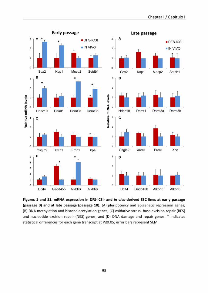
Chapter I / Capítulo I
93
Figures 1 and S1. mRNA expression in DFS-ICSI- and in vivo-derived ESC lines at early passage
(passage 0) and at late passage (passage 10). (A) pluripotency and epigenetic repression genes;
(B) DNA methylation and histone acetylation genes; (C) oxidative stress, base excision repair (BES)
and nucleotide excision repair (NES) genes; and (D) DNA damage and repair genes. * indicates
statistical differences for each gene transcript at P≤0.05; error bars represent SEM.
Early passage Late passage

Chapter I / Capítulo I
94
Effect of ICSI using DNA-fragmented Sperm on the Sperm Count and Motility
No significant differences were recorded in the average weight of either testes or
epididymis in the DFS-ICSI-produced and control male mice. However, four DFS-ICSI-
produced males (20%) (N=20 mice analyzed) showed a very low testes weight (<0.07 g vs
0.20 g in both WT and rest of ICSI mice), and morphological abnormalities (atrophied
testes without presence of spermatozoa). In addition, in three of the DFS-ICSI animals
(15%), testes weight was high (≥0.26 g; above the 90th percentile for the colony). In
contrast, the control males all showed a normal testes weight and normal testicular
morphology (N=20). Significant differences (P=0.002) were detected in the mean number
of sperm collected from the cauda epididymis and deferent conduct (6.77± 1.2x 10⁶
spz/ml for N=8 DFS-ICSI males versus 15.61± 2.2 x 10⁶ spz/ml for N=7 control males). No
differences were observed between the DFS-ICSI and control groups in overall sperm
motility (64.25% and 72.79% respectively) and progressive sperm motility (32.61% and
32.75% respectively).
Effect of ICSI using DNA-fragmented Sperm on the Copulation and Fertility Rates of the
Male Progeny
Pregnancy rates recorded in the B6D2F1 female mice partnered with DFS-ICSI-produced
males were significantly reduced compared to the rates observed in females mated with
control males (49.96% ±5.24, N = 237) vs. 86.52% ±3.13, 138), Table 1). Moreover, 100%
of the females with vaginal plugs mated with control mice became pregnant yet
pregnancy was only observed in 68.48% of the females with vaginal plugs mated with
DFS-ICSI males. In DFS-ICSI males, a statistically significant decrease in pregnancy rates
related to the age was observed. Thus, the ageing phenotype previously observed in
these animals [5] could affect fertility as well. No differences were detected in litter size
between the DFS-ICSI and control groups. However, significantly higher percentages of
pregnant females with resorptions or mummified fetuses were recorded for the DFS-ICSI
males. Furthermore, a statistically significant increase in the percentage of pregnant
females with resorptions was observed in old males from the control group (Table 1).
Some authors have described a decreased reproductive potential (in natural conception,
in vitro blastocyst development, and implantation potential) during ageing [23] . Although
old males from the control group showed high pregnancy rates, it could be possible that
aged sperm that should not fertilize yields a high number of resorptions. On the contrary,
this difference could not be found in old males from the DFS-ICSI group, as resorption
rates were high at all ages. In addition, more than 15% (8/53) of the males in the DFS-ICSI
group could be considered infertile since no pregnancies were recorded in response to
their partnering with at least 6 virgin females. In a subsequent examination of the testes
of these animals, we observed reduced testis sizes and low amounts of sperm.
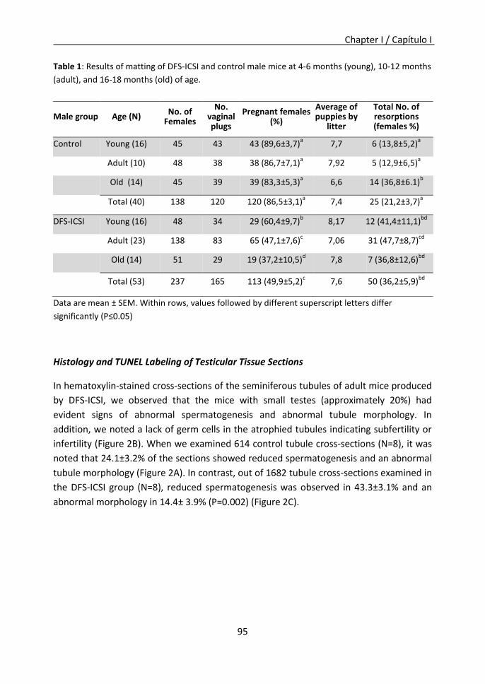
Chapter I / Capítulo I
95
Table 1: Results of matting of DFS-ICSI and control male mice at 4-6 months (young), 10-12 months
(adult), and 16-18 months (old) of age.
Data are mean ± SEM. Within rows, values followed by different superscript letters differ
significantly (P≤0.05)
Histology and TUNEL Labeling of Testicular Tissue Sections
In hematoxylin-stained cross-sections of the seminiferous tubules of adult mice produced
by DFS-ICSI, we observed that the mice with small testes (approximately 20%) had
evident signs of abnormal spermatogenesis and abnormal tubule morphology. In
addition, we noted a lack of germ cells in the atrophied tubules indicating subfertility or
infertility (Figure 2B). When we examined 614 control tubule cross-sections (N=8), it was
noted that 24.1±3.2% of the sections showed reduced spermatogenesis and an abnormal
tubule morphology (Figure 2A). In contrast, out of 1682 tubule cross-sections examined in
the DFS-ICSI group (N=8), reduced spermatogenesis was observed in 43.3±3.1% and an
abnormal morphology in 14.4± 3.9% (P=0.002) (Figure 2C).
Male group Age (N) No. of
Females
No. vaginal plugs
Pregnant females (%)
Average of puppies by
litter
Total No. of resorptions (females %)
Control Young (16) 45 43 43 (89,6±3,7)a 7,7 6 (13,8±5,2)
a
Adult (10) 48 38 38 (86,7±7,1)
a 7,92 5 (12,9±6,5)
a
Old (14) 45 39 39 (83,3±5,3)
a 6,6 14 (36,8±6.1)
b
Total (40) 138 120 120 (86,5±3,1)
a 7,4 25 (21,2±3,7)
a
DFS-ICSI Young (16) 48 34 29 (60,4±9,7)b 8,17 12 (41,4±11,1)
bd
Adult (23) 138 83 65 (47,1±7,6)
c 7,06 31 (47,7±8,7)
cd
Old (14) 51 29 19 (37,2±10,5)
d 7,8 7 (36,8±12,6)
bd
Total (53) 237 165 113 (49,9±5,2)
c 7,6 50 (36,2±5,9)
bd

Chapter I / Capítulo I
96
Figure 2. Histological comparisons between seminiferous tubules in adult offspring produced by DFS-ICSI or natural mating. (A) Seminiferous tubules in control mice show a normal shape with germ cells organized in concentric layers and exhibit ongoing germ cell production. (B) Abnormal seminiferous tubules observed in the testes of infertile DFS-ICSI-produced males (20% of the DFS-ICSI male mice with small testes); note their irregular shape and loss of germ cells in many atrophied seminiferous tubules. (C) The testis of an DFS-ICSI male showing reduced fertility (80% of DFS-ICSI males) containing both tubules showing a normal appearance and ongoing spermatogenesis as well as severely degenerated tubules, which are either empty or have only a small germ cell population (arrows).
The TUNEL assay was performed on paraffin-embedded cross-sections of testicular tissue
obtained from DFS-ICSI (N=7) (Figure S2C, D) and control (N=5) (Figure S2A, B) adult male
mice. At least 200 seminiferous tubules were examined per animal. Our results indicate
that compared to control mice, the testes of DFS-ICSI males showed more TUNEL-positive
cells/cross-sectioned tubule (apoptosis index) and more spermatogenic cells undergoing
apoptosis (Figure S2E).
Figure S2. TUNEL analysis in testes from DFS-ICSI- and in vivo-produced mice. TUNEL labeling of a cross-section of testes from in vivo- (A,B) and DFS-ICSI-produced (C,D) mice. (E) The mean number of TUNEL-positive cells/tubule cross section was higher in the testes of DFS-ICSI-produced mice than control testes. *P<0.05; error bars represent SEM.
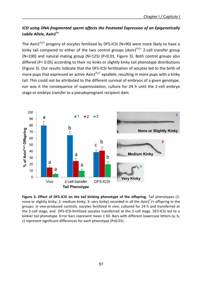
Chapter I / Capítulo I
97
ICSI using DNA-fragmented sperm affects the Postnatal Expression of an Epigenetically
Labile Allele, Axin1Fu
The Axin1Fu/+ progeny of oocytes fertilized by DFS-ICSI (N=90) were more likely to have a
kinky tail compared to either of the two control groups (Axin1Fu/+ 2-cell transfer group
(N=100) and natural mating group (N=125) (P<0.01, Figure 3). Both control groups also
differed (P< 0.05) according to their no kinks or slightly kinky tail phenotype distributions
(Figure 3). Our results indicate that the DFS-ICSI fertilization of oocytes led to the birth of
more pups that expressed an active Axin1Fu/+ epiallele, resulting in more pups with a kinky
tail. This could not be attributed to the different survival of embryos of a given genotype,
nor was it the consequence of superovulation, culture for 24 h until the 2-cell embryo
stage or embryo transfer to a pseudopregnant recipient dam.
Figure 3. Effect of DFS-ICSI on the tail kinking phenotype of the offspring. Tail phenotypes (1: none or slightly kinky; 2: medium kinky; 3: very kinky) recorded in all the Axin1
F/+ offspring in the
groups: in vivo-produced controls, oocytes fertilized in vivo, cultured for 24 h and transferred at the 2-cell stage, and DFS-ICSI-fertilized oocytes transferred at the 2-cell stage. DFS-ICSI led to a kinkier tail phenotype. Error bars represent mean ± SD. Bars with different lowercase letters (a, b, c) represent significant differences for each phenotype (P≤0.01).
Chapter I / Capítulo I
98
Discussion
In our model of ICSI we used frozen-thawed sperm because we have previously observed
that freezing in the absence of a cryoprotectant gives rise to DNA-fragmented
spermatozoa (DFS) with double-strand DNA breaks [5]. In effect, when ICSI is used in
human assisted reproduction to overcome male infertility, sperm with damaged DNA
incapable of oocyte fertilization in vivo are used for fertilization in vitro. ICSI with DNA
fragmented sperm produces genetic and epigenetic alterations in preimplantation
embryos that affect the phenotype of the offspring, and lead to the long term
manifestation of a variety of deleterious phenotypes in mice [5]. Here, we used ESCs
derived from embryos produced by DFS-ICSI to examine the effects of DFS-ICSI without
the need to produce animals. Our data indicate that DFS-ICSI-produced embryos show a
reduced potential to generate ESC lines compared to in vivo-produced embryos, and that
during early passages these ESCs differ in their expression of certain genes. Once
passaged 10 times, however, such gene expression differences were lost, confirming the
idea that mouse ESCs of different origins will eventually adopt a similar gene and protein
expression profiles after several passages [24,25]. The reduced derivation efficiency of
ESC lines produced by DFS-ICSI could be a consequence of the low quality of these
embryos both in terms of genetics and epigenetics, confirming the low rates of successful
implantation and fetal development observed for these embryos [18]. Several studies
have tried to correlate sperm DNA integrity with embryo quality and long term effects.
Thus, 40% of embryos generated by ICSI using DFS showed abnormal chromosome
segregation and chromosome fragmentation; and half of these embryos with abnormal
chromosomes developed into normal-looking blastocysts and were capable of
implantation. However, almost all of them aborted spontaneously before embryonic Day
7.5 [26]. The reduced number of ESC lines derived here from our DFS-ICSI embryos and
the fact that all the lines showed a normal karyotype suggests that embryos with an
abnormal karyotype are unable to produce ESC lines.
Our observation of gene expression differences between early passaged DFS-ICSI-derived
and in vivo-produced ESC lines including the down-regulation of both Hdac10 and the de
novo DNA methyltransferases Dnmt3a and Dnmt3b in the DFS-ICSI-derived ESC lines may
confirm epigenetic differences among DFS-ICSI-generated embryos [5]. Hypomethylation
in ESCs has been also linked to the down-regulation of Dnmt3a and Dnmt3b and to
differentiation dissimilarities [27]. Moreover, the lower expression of Sox2 observed here
could be related to the reduced pluripotency of these ESCs. Also, the transcriptional
repression of Kap1 along with Sox2, Hdac10, Dnmt3a, and Dnmt3b, as modifiers of
epigenetic gene silencing through the transcription of specific genes, involves changes in
chromatin state. Interestingly, these genes are retained in the small fraction of sperm

Chapter I / Capítulo I
99
DNA bound by nucleo-histones [28,29] suggesting that the mechanism whereby DFS-ICSI
modifies phenotype could be related to mechanisms of epigenetic gene silencing. The
higher expression of Gadd45 in our DFS-ICSI-derived ESC lines may support some sort of
DNA damage in these cells. In addition, Gadd45 family proteins have been attributed a
role in senescence and aging, and this phenotype is typical of DFS-ICSI-generated mice
[5]. The reduced expression of the repair enzyme-coding Alkbh3 [30] detected in our DFS-
ICSI-derived ESC lines is in agreement with the premature aging of DFS-ICSI-generated
mice [5].
When we examined the effects of DFS-ICSI on male fertility, we identified a group of mice
showing small testes and infertility that represented 20% of the male animals produced.
Remaining DFS-ICSI males with normal-sized testes showed reduced average fertility
during all stages of life. Other studies have analyzed the effect of ICSI with normal sperm
on fertility. When gene expression in the testes of both ICSI-produced and naturally
conceived mice by micro-array analysis was examined, 474 (150 up-regulated and 324
down-regulated in ICSI mice) differentially expressed genes were identified representing
several functional pathways, including those implicated in spermatogenesis, male meiosis
I, spermatid development, gonad development and male genitalia development. It has
been observed that such differential gene expression patterns are transmitted to the next
generation [31]. It has been also reported that ICSI-derived mice exhibit a high level of
spermatogenic cell apoptosis, suggesting a risk of the compromised fertility of male
progeny [32]. Our observation of the reduced in vivo fertility of DFS-ICSI male mice is
consistent with such findings and indicates that DFS-ICSI could compromise the fertility of
the male offspring in other mammals.
In this study, we also examined the effects of DFS-ICSI on the postnatal expression of an
epigenetically labile allele, Axin1Fu. Our results indicate that DFS-ICSI perturbs the
epigenetic reprogramming of Axin1Fu causing a shift towards the active state of the
epiallele. The manifestation of this was the birth of more pups expressing the active
epiallele rendering a kinky tail phenotype. Similar results have been reported for the
effect of 4 days of IVC on the Axin1Fu and Avy alleles [33]. Collectively, our results suggest
that such effects of ICSI and IVC are likely to affect metastable epialleles in general and
reveal that while primary epimutations produced by ICSI in mice can be properly
corrected in the germ line by epigenetic reprogramming [7], the alterations produced by
ICSI in some metastable epialleles like Axin1Fu are propagated to subsequent generations.
With regard to the question of whether the rest of the genome may have mechanisms
similar to the regulation of the Axin1F uallele, we know that transposable elements
represent up to 45-50% of mouse and human genomes [8]. Many of these new alleles
produced by the insertion of a transposable element are expressed during
Chapter I / Capítulo I
100
preimplantation development [34]. Given the conservation of epigenetic mechanisms
during evolution in mice and humans, it is likely that similar mechanisms of metastable
epialleles will be active in humans. Our results point to the notion that changes in the
epigenetic state of the genome can be induced early in development by environmental
conditions, and that these changes can have consequences for both gene expression in
adulthood [35,36] and the inheritance of epigenetic phenotypes. We are unaware of the
reason why DFS-ICSI renders a kinkier tail phenotype, though a kinked tail has been
described as an embryopathy produced by oxidative DNA damage due to ROS [37]. One
of the targets for oxidative DNA damage is the methylated base m5C found in mammalian
DNA. Thus, if DFS-ICSI increases ROS levels in the embryo, this may produce oxidative
DNA damage and preferentially affect m5C.
In humans, ICSI is currently used as a successful infertility treatment. Although a
significantly increased risk of birth defects in infants conceived by assisted reproductive
technology (ART) has been described in the last years, it has been reported that there is
no risk difference between children conceived by IVF and/or ICSI [38]. Some studies have
suggested that this increased risk may be due to the underlying infertility of the couples
pursuing ART, and not to ARTs themselves [38,39,40]. Furthermore, significant limitations
of human studies are the lack of a good comparison group for IVF or ICSI, which would be
babies naturally conceived by infertile couples rather than babies conceived by overall
population, and the low power of the studies due to the rarity of the diseases [39].
However, a potential risk of ICSI is the use of spermatozoa with apparently normal
morphology but with DNA fragmentation. A significant proportion of infertile men have
elevated levels of DNA damage in their ejaculated spermatozoa [41]. Sperm DNA damage
is a useful biomarker for male infertility diagnosis and it is associated with reduced
fertilization rates, embryo quality and pregnancy rates, and higher rates of spontaneous
miscarriage and childhood diseases [42]. It remains unclear whether assisted
reproductive techniques can compensate for DNA damage. Hence, studies conducted
with animal models are particularly important. Some of the effects described in our
manuscript regarding male infertility in DFS-ICSI mice have been previously described in
mice produced by ICSI with intact fresh sperm. Yu et al. reported decreased testis weight,
abnormal tubule morphology and increased apoptosis in testis of adult mice produced by
ICSI with fresh sperm [32]. Other studies with ICSI using fresh sperm described diverse
alterations as transcriptome perturbations that remained at the neonatal stage [43], or
alterations in glucose parameters in adult mice [44]. Studies that induce sperm DNA
damage and evaluate its biologic effects on the offspring and on next generations are of
vital importance. To date, few animal studies have assessed the effects of induced sperm
DNA damage on fertilization and embryo development. Yamagata et al. described
abnormal chromosome segregation in embryos generated by ICSI with fresh sperm or
Chapter I / Capítulo I
101
with DFS. We have previously described that more severe abnormalities appear when ICSI
is performed with DFS compared to ICSI performed with fresh sperm. Although there
were no differences in fertilization and embryo developmental competence, pregnancy
rates, live offspring rates and survival after 25 weeks were significantly lower.
Furthermore, ageing phenotype and tumor development were observed in DFS-ICSI
animals but not in animals generated by ICSI with fresh sperm [5]. Thus, from these data
we could speculate that although ICSI per se (performed with fresh sperm) can produce
several alterations, abnormal phenotypes are more severe when ICSI is performed with
DFS. However, comparative analyses of ICSI with fresh sperm vs. DFS-ICSI are very
important.
Our findings offer new motives for current concerns over the safety of ICSI with DFS.
Especially worrying is the frequent use of ICSI in cases of severe male factor infertility,
since a significant proportion of the spermatozoa used for ICSI are likely to have
fragmented DNA. The subacute nature of some of the aberrant embryo modifications
induced by ICSI with DFS means that many of these changes will be undetected in the
short term. Moreover, embryo development to the blastocyst stage, considered a
hallmark of ART system efficiency, is often possible despite detrimental environmental
effects and longer term consequences of this procedure [5]. Our first conclusion is that
we should only use early passage DFS-ICSI-derived ESC lines to assess specific alterations
associated with the DFS-ICSI technique. Secondly, we demonstrate here that DFS-ICSI in
the mouse modifies reprogramming in a manner that favors the active state of the
Axin1Fu epiallele, and that this epigenetic alteration is transmitted to the following
generations. Thirdly, our findings also reveal a dramatic influence of DFS-ICSI on the
reproductive lifespan of male progeny. This could have significant implications for the
reproductive management of livestock, endangered species and also humans.
Chapter I / Capítulo I
102
References
1. Schultz RM, Williams CJ (2002) The science of ART. Science 296: 2188-2190. 2. Hourcade JD, Perez-Crespo M, Fernandez-Gonzalez R, Pintado B, Gutierrez-Adan A (2010)
Selection against spermatozoa with fragmented DNA after postovulatory mating depends on the type of damage. Reprod Biol Endocrinol 8: 9.
3. Zini A, Meriano J, Kader K, Jarvi K, Laskin CA, et al. (2005) Potential adverse effect of sperm DNA damage on embryo quality after ICSI. Hum Reprod 20: 3476-3480.
4. Sergerie M, Laforest G, Bujan L, Bissonnette F, Bleau G (2005) Sperm DNA fragmentation: threshold value in male fertility. Hum Reprod 20: 3446-3451.
5. Fernandez-Gonzalez R, Moreira PN, Perez-Crespo M, Sanchez-Martin M, Ramirez MA, et al. (2008) Long-term effects of mouse intracytoplasmic sperm injection with DNA-fragmented sperm on health and behavior of adult offspring. Biol Reprod 78: 761-772.
6. Winkler J, Sotiriadou I, Chen S, Hescheler J, Sachinidis A (2009) The potential of embryonic stem cells combined with -omics technologies as model systems for toxicology. Curr Med Chem 16: 4814-4827.
7. Hollis RP, Stoll SM, Sclimenti CR, Lin J, Chen-Tsai Y, et al. (2003) Phage integrases for the construction and manipulation of transgenic mammals. Reprod Biol Endocrinol 1: 79.
8. van de Lagemaat LN, Landry JR, Mager DL, Medstrand P (2003) Transposable elements in mammals promote regulatory variation and diversification of genes with specialized functions. Trends Genet 19: 530-536.
9. Aviles M, Gutierrez-Adan A, Coy P (2010) Oviductal secretions: will they be key factors for the future ARTs? Mol Hum Reprod 16: 896-906.
10. Vasicek TJ, Zeng L, Guan XJ, Zhang T, Costantini F, et al. (1997) Two dominant mutations in the mouse fused gene are the result of transposon insertions. Genetics 147: 777-786.
11. Zeng L, Fagotto F, Zhang T, Hsu W, Vasicek TJ, et al. (1997) The mouse Fused locus encodes Axin, an inhibitor of the Wnt signaling pathway that regulates embryonic axis formation. Cell 90: 181-192.
12. Rakyan VK, Chong S, Champ ME, Cuthbert PC, Morgan HD, et al. (2003) Transgenerational inheritance of epigenetic states at the murine Axin(Fu) allele occurs after maternal and paternal transmission. Proc Natl Acad Sci U S A 100: 2538-2543.
13. Fernandez-Gonzalez R, Moreira P, Bilbao A, Jimenez A, Perez-Crespo M, et al. (2004) Long-term effect of in vitro culture of mouse embryos with serum on mRNA expression of imprinting genes, development, and behavior. Proc Natl Acad Sci U S A 101: 5880-5885.
14. Fernandez-Gonzalez R, Ramirez MA, Bilbao A, De Fonseca FR, Gutierrez-Adan A (2007) Suboptimal in vitro culture conditions: an epigenetic origin of long-term health effects. Mol Reprod Dev 74: 1149-1156.
15. Fernandez-Gonzalez R, de Dios Hourcade J, Lopez-Vidriero I, Benguria A, De Fonseca FR, et al. (2009) Analysis of gene transcription alterations at the blastocyst stage related to the long-term consequences of in vitro culture in mice. Reproduction 137: 271-283.
16. Gutierrez-Adan A, Pintado B (2000) Effect of flanking matrix attachment regions on the expression of microinjected transgenes during preimplantation development of mouse embryos. Transgenic Res 9: 81-89.
17. Ramirez MA, Fernandez-Gonzalez R, Perez-Crespo M, Pericuesta E, Gutierrez-Adan A (2009) Effect of stem cell activation, culture media of manipulated embryos, and site of embryo transfer in the production of F0 embryonic stem cell mice. Biol Reprod 80: 1216-1222.
18. Moreira PN, Perez-Crespo M, Ramirez MA, Pozueta J, Montoliu L, et al. (2007) Effect of Transgene Concentration, Flanking Matrix Attachment Regions, and RecA-Coating on the

Chapter I / Capítulo I
103
Efficiency of Mouse Transgenesis Mediated by Intracytoplasmic Sperm Injection. Biol Reprod 76: 336-343.
19. Perez-Crespo M, Pintado B, Gutierrez-Adan A (2008) Scrotal heat stress effects on sperm viability, sperm DNA integrity, and the offspring sex ratio in mice. Mol Reprod Dev 75: 40-47.
20. Gutierrez-Adan A, Perez-Crespo M, Fernandez-Gonzalez R, Ramirez MA, Moreira P, et al. (2006) Developmental consequences of sexual dimorphism during pre-implantation embryonic development. Reprod Domest Anim 41 Suppl 2: 54-62.
21. Bermejo-Alvarez P, Rizos D, Rath D, Lonergan P, Gutierrez-Adan A (2008) Epigenetic differences between male and female bovine blastocysts produced in vitro. Physiol Genomics 32: 264-272.
22. Bermejo-Alvarez P, Rizos D, Rath D, Lonergan P, Gutierrez-Adan A (2010) Sex determines the expression level of one third of the actively expressed genes in bovine blastocysts. Proc Natl Acad Sci U S A 107: 3394-3399.
23. Katz-Jaffe MG, Parks J, McCallie B, Schoolcraft WB (2013) Aging sperm negatively impacts in vivo and in vitro reproduction: a longitudinal murine study. Fertil Steril 100: 262-268 e261-262.
24. Brambrink T, Hochedlinger K, Bell G, Jaenisch R (2006) ES cells derived from cloned and fertilized blastocysts are transcriptionally and functionally indistinguishable. Proc Natl Acad Sci U S A 103: 933-938.
25. Ding J, Guo Y, Liu S, Yan Y, Chang G, et al. (2009) Embryonic stem cells derived from somatic cloned and fertilized blastocysts are post-transcriptionally indistinguishable: a MicroRNA and protein profile comparison. Proteomics 9: 2711-2721.
26. Yamagata K, Suetsugu R, Wakayama T (2009) Assessment of chromosomal integrity using a novel live-cell imaging technique in mouse embryos produced by intracytoplasmic sperm injection. Hum Reprod 24: 2490-2499.
27. Petazzi P, Sandoval J, Szczesna K, Jorge OC, Roa L, et al. (2013) Dysregulation of the long non-coding RNA transcriptome in a Rett syndrome mouse model. RNA Biol 10: 1197-1203.
28. Hammoud SS, Nix DA, Zhang H, Purwar J, Carrell DT, et al. (2009) Distinctive chromatin in human sperm packages genes for embryo development. Nature 460: 473-478.
29. Calle A, Miranda A, Fernandez-Gonzalez R, Pericuesta E, Laguna R, et al. (2012) Male mice produced by in vitro culture have reduced fertility and transmit organomegaly and glucose intolerance to their male offspring. Biol Reprod 87: 34.
30. Morita R, Nakane S, Shimada A, Inoue M, Iino H, et al. (2010) Molecular mechanisms of the whole DNA repair system: a comparison of bacterial and eukaryotic systems. J Nucleic Acids 2010: 179594.
31. Wang LY, Wang N, Le F, Li L, Li LJ, et al. (2013) Persistence and intergenerational transmission of differentially expressed genes in the testes of intracytoplasmic sperm injection conceived mice. J Zhejiang Univ Sci B 14: 372-381.
32. Yu Y, Zhao C, Lv Z, Chen W, Tong M, et al. (2011) Microinjection manipulation resulted in the increased apoptosis of spermatocytes in testes from intracytoplasmic sperm injection (ICSI) derived mice. PLoS One 6: e22172.
33. Morgan HD, Jin XL, Li A, Whitelaw E, O'Neill C (2008) The culture of zygotes to the blastocyst stage changes the postnatal expression of an epigentically labile allele, agouti viable yellow, in mice. Biol Reprod 79: 618-623.
34. Peaston AE, Evsikov AV, Graber JH, de Vries WN, Holbrook AE, et al. (2004) Retrotransposons regulate host genes in mouse oocytes and preimplantation embryos. Dev Cell 7: 597-606.
35. Gaudet F, Rideout WM, 3rd, Meissner A, Dausman J, Leonhardt H, et al. (2004) Dnmt1 expression in pre- and postimplantation embryogenesis and the maintenance of IAP silencing. Mol Cell Biol 24: 1640-1648.

Chapter I / Capítulo I
104
36. Reik W, Romer I, Barton SC, Surani MA, Howlett SK, et al. (1993) Adult phenotype in the mouse can be affected by epigenetic events in the early embryo. Development 119: 933-942.
37. Laposa RR, Henderson JT, Xu E, Wells PG (2004) Atm-null mice exhibit enhanced radiation-induced birth defects and a hybrid form of embryonic programmed cell death indicating a teratological suppressor function for ATM. FASEB J 18: 896-898.
38. Wen J, Jiang J, Ding C, Dai J, Liu Y, et al. (2012) Birth defects in children conceived by in vitro fertilization and intracytoplasmic sperm injection: a meta-analysis. Fertil Steril 97: 1331-1337 e1331-1334.
39. Vermeiden JP, Bernardus RE (2013) Are imprinting disorders more prevalent after human in vitro fertilization or intracytoplasmic sperm injection? Fertil Steril 99: 642-651.
40. Hansen M, Kurinczuk JJ, Milne E, de Klerk N, Bower C (2013) Assisted reproductive technology and birth defects: a systematic review and meta-analysis. Hum Reprod Update 19: 330-353.
41. Irvine DS, Twigg JP, Gordon EL, Fulton N, Milne PA, et al. (2000) DNA integrity in human spermatozoa: relationships with semen quality. J Androl 21: 33-44.
42. Lewis SE, John Aitken R, Conner SJ, Iuliis GD, Evenson DP, et al. (2013) The impact of sperm DNA damage in assisted conception and beyond: recent advances in diagnosis and treatment. Reprod Biomed Online 27: 325-337.
43. Kohda T, Ogonuki N, Inoue K, Furuse T, Kaneda H, et al. (2011) Intracytoplasmic sperm injection induces transcriptome perturbation without any transgenerational effect. Biochem Biophys Res Commun 410: 282-288.
44. Scott KA, Yamazaki Y, Yamamoto M, Lin Y, Melhorn SJ, et al. (2010) Glucose parameters are altered in mouse offspring produced by assisted reproductive technologies and somatic cell nuclear transfer. Biol Reprod 83: 220-227.

Chapter II / Capítulo II
105
Germ Cell culture conditions facilitate the reprogramming to
produce mouse ESCs
Priscila Ramos-Ibeas, Eva Pericuesta, Raúl Fernández-González, Alfonso Gutiérrez-Adán
and Miguel Ángel Ramírez
Chapter II / Capítulo II

Chapter II / Capítulo II
106

Chapter II / Capítulo II
107
Summary
Different cell types have been suggested to be the origin of ESCs since they were first
isolated. One of the solidest theories proposed that ESCs could arise from epiblast cells
that are already predisposed to a primordial germ cell (PGC) fate. In the same way, many
culture conditions for ESCs derivation have been developed that show different results.
The objective of this chapter was to determine the effect of a germ cells culture (GS)
medium during ESCs derivation. To do so, differential cell counting in blastocysts
produced in different culture conditions, ESCs derivation efficiency, karyotype, chimaera
generation ability and expression of genes related to pluripotency, germline and
epigenetics were analyzed. It was observed that LIF supplementation during embryo
culture increased the proportion of ICM cells. Thereafter, blastocysts cultured in GS
medium increased ESCs derivation efficiency, showing normal karyotype and higher
chimera formation capacity by blastocyst ESCs microinjection. ESCs lines derived in GS
medium showed an appropriate expression of pluripotency-related genes, and a
correlation was found between derivation efficiency and the expression of some
imprinting genes and retrotransposons. Although germ cell-specific genes were expressed
in all culture conditions, GS medium did not induce an upregulation of germline-specific
markers during the transition from blastocyst to ESCs.
In conclusion, ESCs derivation is conditioned by culture medium, and the combination of
LIF supplementation followed by culture in GS medium constitutes a high efficiency
method for ESCs derivation.
Chapter II / Capítulo II
108
Resumen
Desde las ESCs que fueron aisladas por vez primera, se han propuesto diferentes orígenes
para estas células. Una de las teorías más sólidas plantea que las ESCs podrían proceder
de células del epiblasto que estuvieran predestinadas a convertirse en células
primordiales germinales (PGCs). Del mismo modo se han desarrollado múltiples
condiciones de cultivo para la obtención de ESCs. El objetivo de este capítulo ha sido
determinar el efecto de diferentes condiciones de cultivo, y en concreto de un medio de
cultivo para células germinales (GS), en la obtención de ESCs. Para ello se hizo recuento
celular diferencial en los blastocistos producidos en las distintas condiciones de cultivo y
se analizó la eficiencia de obtención de ESCs, el cariotipo, la capacidad de formación de
animales quiméricos y la expresión de diferentes genes relacionados con pluripotencia,
linaje germinal y epigenética. Se observó que la proporción de células de la ICM aumentó
en los blastocistos producidos en medio suplementado con LIF. Posteriormente, el cultivo
de los blastocistos en medio GS aumentó la producción de líneas de ESCs, que además
mostraban un cariotipo normal y una capacidad superior de formación de animales
quiméricos mediante microinyección de ESCs en blastocistos. Las ESCs obtenidas en
medio GS mostraron una expresión correcta de genes relacionados con la pluripotencia, y
se encontró una correlación entre su eficiencia de obtención y la expresión de algunos
genes improntados y algunos retrotransposones. Aunque se detectó expresión de genes
relacionados con el linaje germinal en todas las condiciones de cultivo, el medio GS no
produjo una mayor expresión de marcadores de linaje germinal durante la transición de
blastocisto a ESCs.
En resumen, la obtención de ESCs está condicionada por la utilización de diferentes
medios de cultivo, y la combinación de LIF y de medio de cultivo GS constituye un método
eficiente para la obtención de ESCs.

Chapter II / Capítulo II
109
Germ Cell culture conditions facilitate the reprogramming to
produce mouse ESCs
Priscila Ramos-Ibeas, Eva Pericuesta, Raúl Fernández-González, Alfonso Gutiérrez-Adán
and Miguel Ángel Ramírez
Departamento de Reproduccion Animal, INIA, Av. Puerta de Hierro n 12, Local 10 Madrid
28040, Spain
Submitted to Molecular Reproduction and Development (second revision)
Abstract
The derivation of embryonic stem (ES) cell lines from blastocysts is a very inefficient
process. It has been proposed that murine ES cells could arise from epiblast cells that are
already predisposed to a primordial germ cell (PGC) fate. We have demonstrated that if
during the process of ES cell derivation in B6D2 F1 hybrid mice, we first culture the
embryo in medium supplemented with LIF we improve the quality of the blastocyst.
Thereafter, when blastocyst is cultured in a germ cells culture (GS) media, we are able to
obtain good quality ES cell lines with a higher efficiency, showing normal karyotype,
increasing the degree of chimerism and germline transmission by ES cells microinjection
in blastocysts. Although germ cell-specific genes were expressed in all culture medium
conditions, GS medium did not induce a more pronounced shift towards germ cell
specification. Furthermore, a correlation was observed between ES cell derivation
efficiency and some imprinted genes and retrotransposable elements expression. In
conclusion, the combination of LIF supplementation followed by culture in GS medium
constitutes a high efficiency method for ES cell derivation.
Chapter II / Capítulo II
110
Introduction
It is remarkable that pluripotent stem cell lines can be derived from preimplantation
embryos, since in vivo pluripotent cells of the early mammalian embryo only briefly
proliferate before differentiating to a more developmentaly restricted cell type. However,
certain cells from the blastocyst can be isolated and, under particular culture conditions,
give raise to embryonic stem (ES) cells, which can indefinitely self-renew in vitro while
maintaining an undifferentiated pluripotent state.
Mouse embryonic stem (mES) cells grow as round compact colonies of small cells, which
depend on leukemia inhibitory factor (LIF). LIF is a cytokine produced by the
endometrium to allow blastocyst implantation (Pera and Tam 2010). The effect of LIF on
the in vitro development of murine embryos has been widely studied with oftenly
contradictory results. In some studies LIF has been demonstrated to enhance mouse
blastocyst formation and decrease embryo fragmentation (Tsai et al. 1999; Cheng et al.
2004b), while in other studies LIF in standard medium did not enhance the development
of human or bovine early embryos (Jurisicova et al. 1995; Vejlsted et al. 2005). However,
these contradictory results may be attributable to the different species and culture
systems used for different experiments.
Although early studies indicated that murine ES cell lines were derived from the inner cell
mass (ICM), some later experiments suggested that ES cells more closely resemble cells
from the epiblast or primitive ectoderm (Brook and Gardner 1997). Thus, using
conventional methods, isolated mouse primitive ectoderm gives raise to ES cell lines at a
higher frequency than does isolated ICM. Moreover, the culture of primitive ectoderm
allows the isolation of ES cell lines from mouse strains that have been previously
refractory to ES cell isolation (Brook and Gardner 1997) and naïve ES cell lines can be
derived from single, isolated, mouse primitive ectoderm cells, which is not possible with
ICM cells (Nichols et al. 2009). However, although these data suggest that ES cells are
more closely related to primitive ectoderm than to ICM, they still cannot ascertain
whether ES cells directly relate to primitive ectoderm or to a cell derived from it in vitro.
Accordingly, gene expression analyses indicate that the closest in vivo equivalent to ES
cells is an early germ cell, rather than ICM or primitive ectoderm (Zwaka 2005). Recently,
bone morphogenetic protein (BMP) signaling has been shown to be important for the
self-renewal of mouse ES cells (Ying et al. 2003), whereas Bmp4 (Lawson et al. 1999) and
Bmp8b (Ying et al. 2000) are required for primordial germ cells (PGCs) formation.
Moreover, alkaline phosphatase is strongly expressed by early germ cells and by ES cells,
but is weakly expressed by the epiblast and other surrounding embryonic cells (Chiquoine
1954; Ginsburg et al. 1990). Several early and later germ cell markers have been
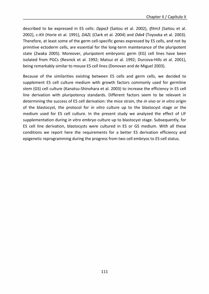
Chapter II / Capítulo II
111
described to be expressed in ES cells: Dppa3 (Saitou et al. 2002), Ifitm3 (Saitou et al.
2002), c-Kit (Horie et al. 1991), DAZL (Clark et al. 2004) and Ddx4 (Toyooka et al. 2003).
Therefore, at least some of the germ cell-specific genes expressed by ES cells, and not by
primitive ectoderm cells, are essential for the long-term maintenance of the pluripotent
state (Zwaka 2005). Moreover, pluripotent embryonic germ (EG) cell lines have been
isolated from PGCs (Resnick et al. 1992; Matsui et al. 1992; Durcova-Hills et al. 2001),
being remarkably similar to mouse ES cell lines (Donovan and de Miguel 2003).
Because of the similarities existing between ES cells and germ cells, we decided to
supplement ES cell culture medium with growth factors commonly used for germline
stem (GS) cell culture (Kanatsu-Shinohara et al. 2003) to increase the efficiency in ES cell
line derivation with pluripotency standards. Different factors seem to be relevant in
determining the success of ES cell derivation: the mice strain, the in vivo or in vitro origin
of the blastocyst, the protocol for in vitro culture up to the blastocyst stage or the
medium used for ES cell culture. In the present study we analyzed the effect of LIF
supplementation during in vitro embryo culture up to blastocyst stage. Subsequently, for
ES cell line derivation, blastocysts were cultured in ES or GS medium. With all these
conditions we report here the requirements for a better ES derivation efficiency and
epigenetic reprogramming during the progress from two cell embryos to ES cell status.

Chapter II / Capítulo II
112
Results
Figure 1. Experimental design.
LIF supplementation increases the ICM/Total cells ratio.
In order to analyze the possible beneficial effect of LIF supplementation in in vitro embryo
culture at different stages, 2-cell embryos were cultured in KSOMaa or KSOMaa
supplemented with LIF up to blastocyst stage (Fig 1). At that point differential cell
counting data were compared with that obtained from in vivo blastocysts. Embryos
cultured in KSOMaa-LIF had a significantly lower total trophectoderm cells number than
embryos cultured in KSOMaa or than blastocysts obtained in vivo (Fig. 2a). Furthermore,
although the blastocysts obtained in the three different conditions did not show
significant differences in the total number of ICM cells, those blastocysts cultured with LIF
supplementation showed a significantly higher ICM/Total ratio (41,4%) than in vivo
produced blastocysts (29,3%) and in vitro cultured blastocysts without LIF
supplementation (27%) (Fig. 2b).
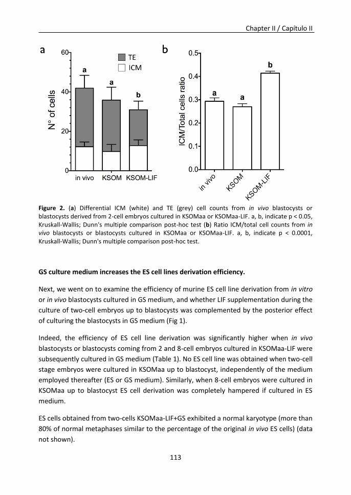
Chapter II / Capítulo II
113
Figure 2. (a) Differential ICM (white) and TE (grey) cell counts from in vivo blastocysts or blastocysts derived from 2-cell embryos cultured in KSOMaa or KSOMaa-LIF. a, b, indicate p < 0.05, Kruskall-Wallis; Dunn's multiple comparison post-hoc test (b) Ratio ICM/total cell counts from in vivo blastocysts or blastocysts cultured in KSOMaa or KSOMaa-LIF. a, b, indicate p < 0.0001, Kruskall-Wallis; Dunn's multiple comparison post-hoc test.
GS culture medium increases the ES cell lines derivation efficiency.
Next, we went on to examine the efficiency of murine ES cell line derivation from in vitro
or in vivo blastocysts cultured in GS medium, and whether LIF supplementation during the
culture of two-cell embryos up to blastocysts was complemented by the posterior effect
of culturing the blastocysts in GS medium (Fig 1).
Indeed, the efficiency of ES cell line derivation was significantly higher when in vivo
blastocysts or blastocysts coming from 2 and 8-cell embryos cultured in KSOMaa-LIF were
subsequently cultured in GS medium (Table 1). No ES cell line was obtained when two-cell
stage embryos were cultured in KSOMaa up to blastocyst, independently of the medium
employed thereafter (ES or GS medium). Similarly, when 8-cell embryos were cultured in
KSOMaa up to blastocyst ES cell derivation was completely hampered if cultured in ES
medium.
ES cells obtained from two-cells KSOMaa-LIF+GS exhibited a normal karyotype (more than
80% of normal metaphases similar to the percentage of the original in vivo ES cells) (data
not shown).

Chapter II / Capítulo II
114
Table 1. ES cells derivation efficiency in different culture medium conditions.
Embryo culture
condition
ES cells derivation
medium
No. embryo No. ES cell lines (%)
In vivo blastocysts ES 35 3 (8.6%)a
GS 91 12 (13.2%)b
2-cell KSOMaa ES 67 0 (0%)c
GS 48 0 (0%)c
2-cell KSOMaa-LIF ES 55 5 (9.1%)a
GS 60 17 (28.3%)d
8-cell KSOMaa ES 54 1 (1.9%)c
GS 58 1 (1.7%)c
8-cell KSOMaa-LIF ES 65 0 (0%)c
GS 59 10 (16.9%)e a,b,c,d,e
Different superscript letters indicate significant differences (P = <0,05), One-way Anova, Holm-Sidak method.
Thus, blastocysts cultured in GS medium showed a significantly higher efficiency to derive
ES cell lines compared with ES medium culture. Moreover, LIF supplementation during 2
or 8-cell culture up to blastocyst is also a crucial aspect on ES cell derivation.
GS culture medium produces a better epigenetic reprogramming pattern.
In vivo blastocysts or blastocysts obtained by two-cell stage embryos cultured in KSOMaa
or KSOMaa-LIF, or their outgrowths, cultured either with ES or GS medium on supportive
MEF monolayers for four days, were collected to perform gene expression analysis during
the early steps of the ES cells derivation process. No significant differences in
pluripotency markers Nanog, Pou5f1 and Slc2a1 were observed in blastocysts of the
different groups performing a second level of normalization of data considering the
ICM/total cells (Fig. 3a). Furthermore, when pluripotency markers were analyzed in cell
clumps, no significant differences were found in Slc2a1 expression among the different
groups. However, a significantly lower expression of Pou5f1 was observed in the in
vivo+ES group (Fig. 3b). In vivo+GS and KSOMaa+GS groups showed a significantly higher
expression of Nanog than in vivo+ES and KSOMaa+ES groups. In contrast, KSOMaa-LIF+ES
and KSOMaa-LIF+GS groups showed an intermediate expression that was not significantly
different with the other groups.
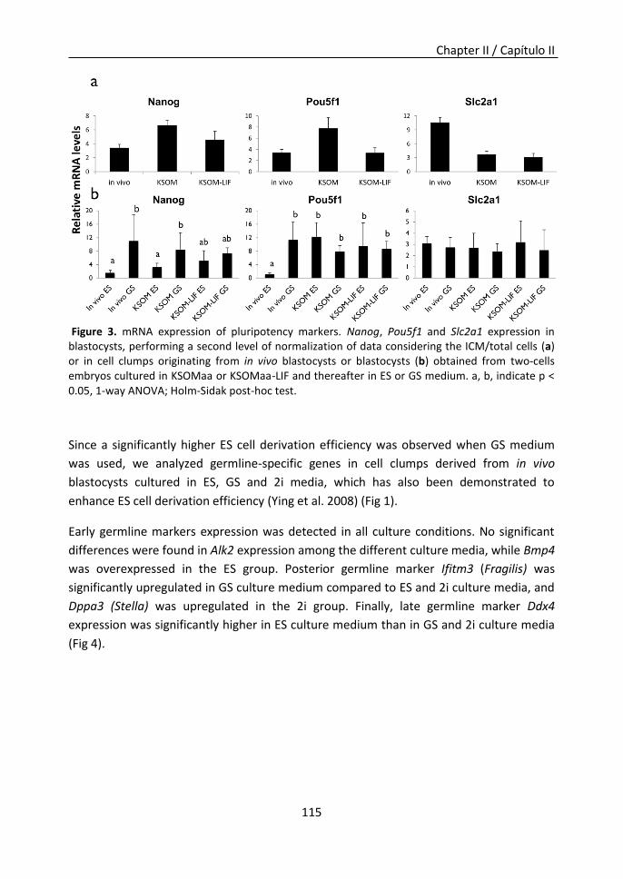
Chapter II / Capítulo II
115
Figure 3. mRNA expression of pluripotency markers. Nanog, Pou5f1 and Slc2a1 expression in blastocysts, performing a second level of normalization of data considering the ICM/total cells (a) or in cell clumps originating from in vivo blastocysts or blastocysts (b) obtained from two-cells embryos cultured in KSOMaa or KSOMaa-LIF and thereafter in ES or GS medium. a, b, indicate p < 0.05, 1-way ANOVA; Holm-Sidak post-hoc test.
Since a significantly higher ES cell derivation efficiency was observed when GS medium
was used, we analyzed germline-specific genes in cell clumps derived from in vivo
blastocysts cultured in ES, GS and 2i media, which has also been demonstrated to
enhance ES cell derivation efficiency (Ying et al. 2008) (Fig 1).
Early germline markers expression was detected in all culture conditions. No significant
differences were found in Alk2 expression among the different culture media, while Bmp4
was overexpressed in the ES group. Posterior germline marker Ifitm3 (Fragilis) was
significantly upregulated in GS culture medium compared to ES and 2i culture media, and
Dppa3 (Stella) was upregulated in the 2i group. Finally, late germline marker Ddx4
expression was significantly higher in ES culture medium than in GS and 2i culture media
(Fig 4).

Chapter II / Capítulo II
116
Figure 4. mRNA expression of genes controlling germline establishment. Bmp4, Alk2, Ifitm3, Dppa3 and Ddx4 markers expression in cell clumps from in vivo blastocysts cultured in ES, GS or 2i media. a, b, indicate p < 0.05, 1-way ANOVA; Holm-Sidak post-hoc test.
It has been demonstrated that a few transcripts encoded by the imprinted Dlk1-Dio3
gene cluster, including Meg3 (also known as Gtl2), are aberrantly silenced in iPS cell lines
with poor contribution to chimaera (Stadtfeld et al. 2010). Consequently, we analyzed
Meg3 expression as a quality indicator in our ES cell lines. Quantitative PCR (qPCR)
analysis of Meg3 confirmed that GS culture conditions increase the expression of Meg3.
Thus, KSOMaa-LIF+GS group showed a significantly higher Meg3 expression than in
vivo+ES, KSOMaa+ES, KSOMaa+GS and KSOMaa-LIF+ES groups (Fig. 5a). Furthermore, a
correlation was found between the efficiency to obtain new ES cell lines and Meg3
expression levels (spearman r 0.7827) (Fig. 5b).

Chapter II / Capítulo II
117
Figure 5. Meg3 expression. (a) Meg3 marker expression from cell clumps originating from in vivo blastocysts or blastocysts obtained from two-cells embryos cultured in KSOMaa or KSOMaa-LIF. *P < 0.05, 1-way ANOVA; Holm-Sidak post-hoc test. (b) Correlation between the efficiency of ES cell derivation in different culture conditions and the expression levels of Meg3 marker. Spearman correlation. r = 0.7827.
We also found significant differences in other imprinted genes expression levels such as
Rhox5, U2af1-rs1, and in the retrotransposable element IAP (Fig. 6). In vivo+ES, in vivo+GS
and KSOMaa-LIF+GS groups showed a significantly lower Rhox5 expression than
KSOMaa+ES, KSOMaa+GS and KSOMaa-LIF+ES groups (Fig. 6). U2af1-rs1 and IAP
expression in in vivo+ES and in vivo+GS groups was significantly lower than in
KSOMaa+ES, KSOMaa+GS and KSOMaa-LIF+ES groups.
These expression analyses further confirmed that GS medium and the addition of LIF
during blastocyst culture produce a better pluripotency pattern and a higher expression
of Meg3 that correlated with ES derivation efficiency.

Chapter II / Capítulo II
118
Figure 6. mRNA expression of imprinted genes. Rhox5, U2af1-rs1 and IAP expression from cell clumps originating from in vivo blastocysts or blastocysts obtained from two-cells embryos cultured in KSOMaa or KSOMaa-LIF and later in ES or GS medium. a, b, indicate p < 0.05, 1-way ANOVA; Holm-Sidak post-hoc test.
ES cells from KSOMaa-LIF+GS medium culture showed an increased chimerism capacity
We next studied the ability of chimera formation in different ES cell lines. We analyzed
three different ES cell lines obtained from the three in vitro conditions which showed a
significantly higher derivation efficiency: 2-cell KSOMaa-LIF+ES (ES/ES(2)2 cell line); 2-cell
KSOMaa-LIF+GS (ES/GS(2)7 cell line) or 8-cell KSOMaa-LIF+GS (ES/GS(8)4 cell line) (shaded
rows in Table 1). The ES cell line ES/GS(2)7 showed a significantly higher percentage of
mice with more than 75% of ES-cell coat color after injection in blastocysts, compared to
ES/ES(2)2 (35.7% vs 3.8%) (Table 2). Furthermore, germline transmission was also higher
in chimeric mice with more than 75% ES-cell coat color obtained from ES cell line
ES/GS(2)7 injection (Fig 7a) than in those produced by ES/ES(2)2 (Fig 7b) (100% vs 14.3%
respectively).
Figure 7. Germline transmission in chimeric mice from ES/GS(2)7 cell line (a) or ES/ES (2)2 cell line (b), with more than 75% ES cell coat color.
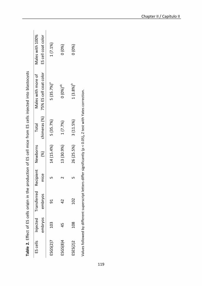
Chapter II / Capítulo II
119
Tab
le 2
. Ef
fect
of
ES c
ells
ori
gin
in
th
e p
rod
uct
ion
of
ES c
ell
mic
e fr
om
ES
cells
in
ject
ed
in
to b
last
ocy
sts
Val
ues
fo
llow
ed b
y d
iffe
ren
t su
per
scri
pt
lett
ers
dif
fer
sign
ific
antl
y (p
< 0
.05
), Z
te
st w
ith
Yat
es c
orr
ecti
on
.
ES c
ells
In
ject
ed
emb
ryo
s
Tran
sfer
red
emb
ryo
s
Rec
ipie
nt
mic
e
New
bo
rns
(%)
Tota
l
chim
eras
(%
)
Mal
es w
ith
mo
re o
f
75
% E
S ce
ll co
at c
olo
r
Mal
es w
ith
10
0%
ES c
ell
coat
co
lor
ESG
S(2
)7
10
3 9
1 5
1
4 (
15.4
%)
5 (
35
.7%
) 5
(3
5.7
%)a
1 (
7.1
%)
ESG
S(8
)4
45
42
2
1
3 (
30
.9%
) 1
(7
.7%
) 0
(0
%)ab
0
(0
%)
ESES
(2)2
1
08
102
5
2
6 (
25
.5%
) 3
(1
1.5
%)
1 (
3.8
%)b
0
(0
%)
Chapter II / Capítulo II
120
Discussion
In this study we investigated the effect of LIF supplementation on the in vitro culture of
murine embryos destined to derive ES cells, as well as the addition of germline-related
factors in the culture medium used for ES cell derivation, in order to improve its efficiency
and quality.
LIF has been demonstrated to play an important role in in vivo embryo development and
implantation in mice (Stewart et al. 1992). However, contradictory information exists
regarding the effect of LIF in embryo development in vitro. Some studies have reported
that LIF increases blastocyst formation rates in mice and human (Dunglison et al. 1996;
Tsai et al. 2000; Rungsiwiwut et al. 2008), while other reports showed no differences in
the development efficiency of human embryos (Jurisicova et al. 1995). Rungsiwiwut et al.
observed an increment in the total cell number of the blastocysts after KSOMaa-LIF
culture in mice (Rungsiwiwut et al. 2008). However, no differential ICM/TE cell number
counting was performed in any of the preceding articles. In our study, although KSOMaa-
LIF cultured embryos showed a lower total cell number compared to in vivo and KSOMaa
cultured blastocysts, the ratio of ICM/total cells was significantly higher than in the other
groups, which indicates the importance of performing a differential cell count. These
blastocysts proved to be more suited for ES cell derivation, as they showed a higher EScell
derivation efficiency.
In addition, the highest ES cell line derivation efficiency was attained when embryos were
first cultured in KSOMaa-LIF, followed by culture in GS medium. There may be a synergy
between LIF supplementation during in vitro embryo culture and the posterior addition of
other mouse growth factors present in the GS medium ie. EGF, GDNF and bFGF (Kanatsu-
Shinohara et al. 2003), that favors the establishment of new ES cell lines. ES-like cells have
been established in vitro from neonatal gonocytes and adult Spermatogonial Stem Cells
(SSCs) in the presence of EGF, GDNF, bFGF and LIF (Kanatsu-Shinohara et al. 2003;
Kanatsu-Shinohara et al. 2004). These factors are also known to affect the proliferation
and maintenance of other stem cell populations: EGF enhances tissue regeneration in
various adult organs such as skin, liver and intestinal epithelium (Reynolds and Weiss
1992); GDNF has been shown to stimulate SSC self-renewal in vivo (Meng et al. 2000), and
bFGF and LIF are necessary for EG cell derivation and culture, being these cell lines
remarkably similar to ES cell lines (Donovan and de Miguel 2003).
Our overall performance was lower than that reported in the literature in recent years,
probably because we have employed the standard ES cell derivation method (Evans and
Kaufman 1981) that supplements culture medium with fetal bovine serum (FBS), which
shows variations between different batches. A defined media supplement, knockout

Chapter II / Capítulo II
121
serum replacement (KSR) with knockout DMEM (KSR-KDMEM) has been shown to
facilitate the generation of ES cell lines, yielding higher efficiency for inbred mouse strains
ES cell derivation in comparison to DMEM with FBS (Cheng et al. 2004a). More recently,
the use of serum-free N2B27 medium supplemented with small-molecule inhibitors of
MEK (2i medium) [MAPK (mitogen activated protein kinase)/ERK (extracellular-signal-
regulated kinase) kinase] and GSK3β (glycogen synthase kinase 3β) proved to be very
efficient for ES cell derivation (Ying et al. 2008). Future experiments would have to assess
whether our implementations provide further efficiency enhancement when combined
with these optimized media.
The efficiency of ES cell derivation is greatly strain dependent. Conventional methods for
deriving ES cell lines are suitable for mouse strain 129, but not for other refractory
strains, so that very few ES cell lines are currently available from inbred strains (mostly
C57BL/6). In addition, ES cells derived from C57BL/6 blastocysts are more difficult to
propagate in vitro, less efficient for chimera generation, and contribute less frequently to
the germline (Brook and Gardner 1997; Auerbach et al. 2000). In the present study, we
derived ES cell lines from CBAxC57BL/6 hybrid mice, which are widely used in genetic
studies.
Although no differences were observed in Nanog expression during blastocyst stage,
higher expression was observed in outgrowths coming from blastocysts cultured in GS
medium. Nanog is considered a core element of the pluripotent transcriptional network,
but it is also specifically required for germ cell formation (Chambers et al. 2007). Thus,
two possibilities could arise for explaining Nanog upregulation: that GS medium enhances
ES cell pluripotency, or that culture supplementation with germline-related factors makes
ES cells adopt an early germ cell pattern. This second possibility led us to investigate if
germline pathways were involved in ES cell establishment when GS medium was used.
PGC specification in the embryo is critically reliant on the signaling pathways elicited by
bone morphogenetic proteins (BMPs), mainly by Bmp4, which activates Alk2 (Hayashi et
al. 2002). These signals direct a small group of proximal epiblast cells to enter the germ
lineage and to express other germline markers as Ifitm3 (Fragilis), Dppa3 (Stella) (Saitou
et al. 2002) and Ddx4 (Young et al. 2010). It has been suggested that during the ES cell
derivation process, early germ cell specification-related genes are expressed under
standard serum culture conditions (Tang et al. 2010). However, this germ cell-like state is
facultative for the stabilization of pluripotency in vitro, since culture in 2i conditions
enables the effective direct recruitment of ES cells skipping this germ cell-like state (Chu
et al. 2011). In our study, germ cell specification-related genes were expressed in all
culture media conditions and we could not observe a marked shift towards the germinal
lineage with GS medium. The upregulation of Stella, which was observed in 2i conditions

Chapter II / Capítulo II
122
could be associated to Tet1 and Tet2-mediated demethylation (Ficz et al. 2011), since the
2i culture conditions have been reported to induce a global demethylation through Tet-
driven hydroxylation (Leitch et al. 2013). Furthermore, the upregulation of Stella could be
related with a different proportion of the subpopulations present in ESCs lines generated
in 2i conditions (ICM-like and epiblast-like subpopulations) (Hayashi et al. 2008). The
higher level of Stella and the lower level of other germ cell-related genes observed in 2i
condition suggest that these efficient conditions for ESCs derivation could produce ESCs
that are richer in the ICM-like subpopulation, meanwhile the ESCs derived in ES and GS
conditions with lower levels of Stella and higher levels of germ cell specification-related
genes could be rich in epiblast/germline-like subpopulations.
According to our observations, some imprinted genes could constitute ES cell quality
indicators. The Dlk1-Dio3 gene cluster consists of multiple imprinted genes, including
Mirg, Rian and Meg3 (Miyoshi et al. 2000). Recently, Stadtfeld et al. (Stadtfeld et al. 2010)
reported that mouse induced pluripotent stem cells (iPSCs) with repressed expression of
Meg3, contributed poorly to chimeras and failed to generate all-iPSC mice. In our study,
we could observe that the highest Meg3 expressions were triggered by the culture with
GS medium, and interestingly, a positive correlation between Meg3 expression and ES cell
derivation efficiency was indeed demonstrated. In vitro culture can induce epigenetic
instability in embryos and in ES cell lines derived from them, leading to biallelic
expression of imprinted genes (Horii et al. 2010; Mann et al. 2004). Highest expression of
imprinted genes Rhox5 and U2af1-rs1 was found in those groups with lowest ES cells
derivation efficiencies, which could be caused by epigenetic instability or biallelic
expression. Epigenetic instability in ES cells can also affect endogenous retrotransposable
elements (REs). Accordingly, we observed upregulation of IAP in those groups with the
lowest ES derivation efficiencies. Reactivation of IAP has been previously described in an
ES cell line that lost the ability of germline transmission and started inducing epigenetic
alterations in chimeric animals (Ramirez et al. 2006).
In summary, our observations suggest that the combined use of LIF during the in vitro
embryo culture up to blastocyst stage and GS culture medium during the transition from
blastocyst to ES cells synergically increase ES cell derivation efficiency. ESCs lines
generated in these conditions accomplish pluripotency criteria and show an increased
chimerism capacity. In addition, a correlation was observed between ES cell derivation
efficiency and some imprinted genes expression.

Chapter II / Capítulo II
123
Material and Methods
Embryo Collection
Mice were bred on a 14-h light/10-h dark cycle. Eight to ten week-old B6D2 F1 hybrid
mice (C57Bl/6 x DBA) were superovulated by intraperitoneal injections of 7.5 IU of
pregnant mare serum gonadotropin (PMSG) (Folligon; Intervet, Boxmeer, Holland)
followed 48 h later by 7.5 IU of hCG (Chorulon; Intervet). On the same day of hCG
injection (day 0), they were paired with male mice of the same strain. The mice were
divided randomly into three groups: one in vivo study group (blastocysts collected at
3.5d) and two in vitro study groups for 2-cell embryo culture (collected at 1.5d) and 8-cell
embryo culture (collected at 2.5d). Oviducts were excised by clamping the cornua,
dissecting the peritoneum and fat between the ovary and tube and cutting the whole
oviduct from the proximal end. After washing and flushing the oviduct from the proximal
end with a 30-gauge needle, two-cell and eight-cell embryos were selected and collected
by 100x stereoscopy.
In Vitro Embryo Culture
Two-cell and eight-cell embryos were placed in a droplet of KSOMaa or KSOMaa
supplemented with 1000 U/ml LIF (KSOMaa-LIF) under oil in an environment of 5% CO2 at
37°C. After 48 h (2-cell embryos) or 24 h (8-cell embryos), in vitro cultured blastocysts and
in vivo blastocysts were plated individually in 96-well plates containing mitomycin-C
(Sigma-Aldrich corporation St. Louis, MO, USA) treated mouse embryonic fibroblast (MEF)
cells on 0.1% gelatin coated tissue plates in different media: (i) ES medium: Dulbecco’s
modified Eagle medium (DMEM plus 4500 mg/l glucose, glutaMAX, and pyruvate;
Invitrogen, Carlsbad, CA, USA) supplemented with 20% FBS (PAA Laboratories Cölbe
Germany), 2 mM glutamine, 1 mM MEM nonessential amino acids solution, 1 mM β-
mercaptoethanol, 1000 U/ml LIF (ORF Genetics Iceland), and antibiotics (100 U/ml
penicillin, 100 μg/ml streptomycin); (ii) GS medium: ES medium supplemented with 20
ng/ml Epidermal growth factor (EGF) (Peprotech, France), 10 ng/ml basic fibroblasts
growth factor (bFGF) (Peprotech, France) and 10 ng/ml recombinant rat glial cell line-
derived neurotrophic factor (GDNF) (Peprotech, France); and (iii) 2i medium: ES medium
supplemented with 3 μM GSK3Beta inhibitor (StemoleculeTM CHIR99021, Stemgent, San
Diego, CA, USA) and 0.5 μM MEK inhibitor (StemoleculeTM PD0325901, Stemgent, San
Diego, CA, USA).
The blastocysts were allowed to attach to supportive MEFs, hatched and expanded. After
4d, outgrowths were recovered for gene expression analyses or trypsinized to derive ES
cell lines by incubation in 0.05% Trypsin / 0.02% EDTA in Ca2+-free and Mg2+-free

Chapter II / Capítulo II
124
Dulbecco’s phosphate-buffered saline (PBS) at 37ºC and seeded directly into a well of a
96-well plate containing MEFs and ES, GS or 2i medium. Approximately 4 d after
trypsinization, ESC colonies could be detected and expanded to a 24- or 96w depending
on their confluency. Half of the cells were frozen when passage to a 35-mm dish was
performed. For cell line expansion, cells were trypsinized every three days.
Differential ICM and TE Cell Counts
Blastocysts from the different groups were stained using a modification of the method
originally described by Biggers et al. (Biggers et al. 2000). Blastocysts (3–5 at a time) were
transferred from culture drops to acid Tyrode solution under constant observation for 5–
15 s, until the zonae pellucidae were completely dissolved. They were next transferred to
three rapid, successive washes in KSOMaa and then for 30 min into 10% rabbit antiserum
to mouse red blood cells (Organon Teknika Corp., Durham, NC) in KSOMaa at 37ºC. After
30 min, embryos were transferred through three successive 5-min washes with KSOMaa
and then into KSOMaa with 10% guinea pig complement (Gibco), 1 mg/ml bisbenzimide
(Hoechst 33258), and 1 mg/ml propidium iodide (PI; Sigma) for 30 min at 37ºC. Each
stained blastocyst was transferred to a clean glass slide, compressed under a glass
coverslip, and visualized at 400x magnification in an inverted Nikon microscope with
epifluorescence. Upon these treatments ICM cells were Hoechst positive, while PI marked
TE cells. The numbers of nonmitotic, mitotic, and dead (i.e., degenerate and fragmented
nuclei) cells were counted; being the total number of surviving cells the sum of
nonmitotic and mitotic cells.
Analysis of marker gene expression by RT-PCR
The techniques for analysis of marker gene expression by RT-PCR have been described in
detail previously (Bermejo-Alvarez et al. 2008) Total RNA was extracted using Dynabeads
mRNA Direct Extraction KIT (Dynal Biotech, Oslo, Norway) following the manufacturer’s
instructions. RT reaction was performed immediately according to the manufacturer’s
instructions (Gibco-BRL, Grand Island, NY, USA). 0.2 µM oligo (dT) and 0.5 µM random
primers were added to RNA and heated 5 min, at 70ºC. RNA was reverse-transcribed at
42 °C for 60 min in a final volume of 40 µl containing 0.375 mM dNTPs (Biotools, Madrid,
Spain), 6.25 U RNasin RNAse inhibitor (Promega, Madison, WI, USA), 10x MMLV-RT buffer
with 8 mM DTT and 5 U MMLV high performance reverse transcriptase (Epicentre,
Madison, WI, USA). Reaction was inactivated at 70ºC for 10 min.
The quantification of mRNA transcripts was performed by real-time quantitative (q)RT-
PCR using a Rotorgene 6000 Real Time CyclerTM (Corbett Re- search, Sydney, Australia)
and SYBR Green (Molecular Probes, Eugene, OR) as a double-stranded DNA-specific

Chapter II / Capítulo II
125
fluorescent dye. PCR was performed by adding 2-µl of each sample to the PCR mix
(Quantimix Easy Sig Kit, Biotools) containing the specific primers to amplify Gapdh as
housekeeping; Nanog, Pou5f1 (Oct3/4) and Slc2a1 as pluripotency markers (Ramirez et al.
2007); Bmp4, Alk2, Ifitm3, Dppa3 and Ddx4 as germ line differentiation markers (Cyril
Ramathal 2011) and Meg3, Rhox5, IAP and U2af1-rs1 as imprinted genes. Primer
sequences and the approximate sizes of the amplified transcripts are given in
Supplementary Table 1. Details of the qRT-PCR procedure have been described else-
where (Ramirez et al. 2006). The PCR protocol included an initial step of 94°C (2 min),
followed by 35 cycles of 94°C (15 s), 56°C (30 s) and 72°C (30 s). Fluorescent data
acquisition temperature for each primer is detailed in Supplementary Table 1. The
melting protocol consisted of 60 s at 40°C for and then heating from 50 to 94°C, holding
at each temperature for 5 s while monitoring fluorescence. For qRT-PCR, three groups of
cDNA per experimental group were used in two repetitions for all genes of interest.
Table 2: Primer sequences, annealing temperatures, and the approximate sizes of the
amplified fragments of all transcripts.
Gene Primer Sequences 5’-3’ Size GeneBank Accesion No.
Acquisition Temperature (ºC)
Gapdh ACCCAGAAGACTGTGGATGG ATGCCTGCTTCACCACCTTC
247 BC102589 86
Nanog AGGGTCTGCTACTGAGATGCTCTG CAACCACTGGTTTTTCTGCCACCG
363 NM_028016 86
Pou5f1 GGAGAGGTGAAACCGTCCCTAGG AGAGGAGGTTCCCTCTGAGTTGC
312 NM_013633 87
Slc2a1 CCAGCTGGGAATCGTCGTT CAAGTCTGCATTGCCCATGAT
688 NM_011400.3 86
Ifitm3 TGCCTTTGCTCCGCACCAT GGGTGAAGCACTTCAGGACC
531 NM_025378.2 84
Alk2 GGAGTAATGATCCTTCCTGTGC TCTTACACGTCATCTTCCCCTG
216 NM_007394.3 84
Stella GCAATCTTGTTCCGAGCTAG CTGGATCGTTGTGCATCCTA
312 AY082485.1 83
Ddx4 GCTCAAACAGGGTCTGGGAAG GGTTGATCAGTTCTCGAG
564 JN951543.1 83
Bmp4 TGTGAGGAGTTTCCATCACG TTATTCTTCTTCCTGGACCG
547 BC013459.1 86
Meg3 TCTGCCTGGCTTTCGCCCCA AAGCACCATGAGCCACTAGG
932 NR_003633.3 86
Rhox5 AGAGATGAGCAAGGTGCACA CGAACCTAGAGCCCTGGAG
237 NM_008818.2 86
U2af1-rs1
CGTGGGAAAGGTGATTCAGT GCTGCCTTAGCTGGGCTCAGG
566 NM_011663.3 84
IAP GGGTATTGTTGAGCGTGCGC TCGGGTGAGTCTTTCTGGTAC
333 XM_001477167.1
82
Chapter II / Capítulo II
126
The comparative CT method was used to quantify expression levels (Miranda et al. 2011).
Quantification was normalized to the endogenous control Gapdh. Fluorescence was
acquired in each cycle to determine the threshold cycle in the cycle during the log-linear
phase of the reaction at which fluorescence increased above background for each sample.
According to the comparative CT method, the ∆CT value was determined by subtracting
the Gapdh CT value for each sample from each gene CT value. Calculation of ∆∆CT
involved using the highest sample ∆CT value (i.e., the sample with the lowest target
expression) as an arbitrary constant to subtract from all other ∆CT sample values. Fold
changes in the relative gene expression of the target were determined using the formula
2-∆∆CT. For those genes specifically expressed in the ICM, a second normalization of the
expression levels was performed taking into account the ratio ICM/Total cells. Data on
mRNA expression were analyzed using Prism 5 (Graph pad software, CA, USA) software
package. Differences in mRNA expression were analyzed using the SigmaStat (Jandel
Scientific, San Rafael, CA) package by one-way repeated-measures ANOVA with arcsine
data transformation, and significance determined using Holm-Sidak post hoc test. A P
value ≤ 0.05 was considered statistically significant.
Karyotype analysis.
To prepare chromosome spreads of the ES cell lines, cells were arrested in metaphase by
supplementing the culture medium with 0.1 μg/ml colcemid for 4 h at 37°C in a 5% CO2
air atmosphere. Cells were then treated with trypsin-EDTA for 2 min at 37°C and after
pipetting, the single cell suspension was washed twice with PBS by centrifugation at 200
G for 5 min. The resulting pellet was exposed to a hypotonic shock by resuspension in
0.075 M KCl for 15 min at 37°C. After a second centrifugation step the hypotonic solution
was removed and the pellet fixed with a methanol/acetic acid solution (3:1; vol/vol) by
gently pipetting. Ten min later, cells were pelleted and fixed again. Before slide mounting,
cells were washed twice with PBS and dropped into slides, which were dried overnight at
55°C, stained with freshly made 10% Giemsa solution for 30 min and rinsed with distilled
water. Finally, chromosome number was counted in at least 30 metaphase cells for each
cell preparation.
Chimera formation
For chimera formation, we used blastocyst ES cell injection procedure (Ramírez et al.
2009). Ten cells were injected into the blastocoel of 3.5 dpc blastocysts of CD1 mice using
Eppendorf micromanipulators (Eppendorf TransferMan NK 2, Hamburg, Germany). The
blastocysts cultured overnight in KSOM, were returned to the oviducts of 0.5 dpc
pseudopregnant CD1 foster mothers on the next day of microinjection. To generate
chimeric mice, the embryos were allowed to develop to term.

Chapter II / Capítulo II
127
Acknowledgements
This work was funded by Grant AGL2012-39652-C02-01 from the Spanish Ministry of
Science and Innovation. P.R.-I. was supported by an FPI grant from the Spanish Ministry of
Science and Innovation.
We are grateful to Dr. María Yáñez-Mó (Hospital Universitario Santa Cristina, Instituto de
Investigaciones Sanitarias Princesa. Madrid 28009, Spain) and Dr. Dimitrios Rizos
(Departamento de Reproducción Animal, INIA, Avenida Puerta de Hierro N. 12, local 10,
28040 Madrid, Spain) for reviewing the manuscript.
Chapter II / Capítulo II
128
References
Auerbach W, JH Dunmore, V Fairchild-Huntress, Q Fang, AB Auerbach, D Huszar, AL Joyner. 2000. Establishment and chimera analysis of 129/SvEv- and C57BL/6-derived mouse embryonic stem cell lines. Biotechniques 5:1024-8, 1030, 1032.
Bermejo-Alvarez P, D Rizos, D Rath, P Lonergan, A Gutierrez-Adan. 2008. Epigenetic differences between male and female bovine blastocysts produced in vitro. Physiol Genomics 2:264-272.
Biggers JD, LK McGinnis, M Raffin. 2000. Amino acids and preimplantation development of the mouse in protein-free potassium simplex optimized medium. Biol Reprod 1:281-293.
Brook FA, RL Gardner. 1997. The origin and efficient derivation of embryonic stem cells in the mouse. Proc Natl Acad Sci U S A 11:5709-5712.
Chambers I, J Silva, D Colby, J Nichols, B Nijmeijer, M Robertson, J Vrana, K Jones, L Grotewold, A Smith. 2007. Nanog safeguards pluripotency and mediates germline development. Nature 7173:1230-1234.
Cheng J, A Dutra, A Takesono, L Garrett-Beal, PL Schwartzberg. 2004a. Improved generation of C57BL/6J mouse embryonic stem cells in a defined serum-free media. genesis 2:100-104.
Cheng T-C, C-C Huang, C-I Chen, C-H Liu, Y-S Hsieh, C-Y Huang, M-S Lee, J-Y Liu. 2004b. Leukemia inhibitory factor antisense oligonucleotide inhibits the development of murine embryos at preimplantation stages. Biol Reprod 5:1270-1276.
Chiquoine AD. 1954. The identification, origin, and migration of the primordial germ cells in the mouse embryo. Anat Rec 2:135-146.
Chu LF, MA Surani, R Jaenisch, TP Zwaka. 2011. Blimp1 expression predicts embryonic stem cell development in vitro. Curr Biol 20:1759-1765.
Clark AT, MS Bodnar, M Fox, RT Rodriquez, MJ Abeyta, MT Firpo, RA Pera. 2004. Spontaneous differentiation of germ cells from human embryonic stem cells in vitro. Hum Mol Genet 7:727-739.
Cyril Ramathal RRPaPT. 2011. Embryonic Stem Cells and the Germ Cell Lineage. In Embryonic Stem Cells - Basic Biology to Bioengineering. editor. Intech, 1-34.
Donovan PJ, MP de Miguel. 2003. Turning germ cells into stem cells. Curr Opin Genet Dev 5:463-471.
Dunglison GF, DH Barlow, IL Sargent. 1996. Leukaemia inhibitory factor significantly enhances the blastocyst formation rates of human embryos cultured in serum-free medium. Hum Reprod 1:191-196.
Durcova-Hills G, J Ainscough, A McLaren. 2001. Pluripotential stem cells derived from migrating primordial germ cells. Differentiation 4-5:220-226.
Evans MJ, MH Kaufman. 1981. Establishment in culture of pluripotential cells from mouse embryos. Nature 5819:154-156.
Ficz G, MR Branco, S Seisenberger, F Santos, F Krueger, TA Hore, CJ Marques, S Andrews, W Reik. 2011. Dynamic regulation of 5-hydroxymethylcytosine in mouse ES cells and during differentiation. Nature 7347:398-402.
Ginsburg M, MH Snow, A McLaren. 1990. Primordial germ cells in the mouse embryo during gastrulation. Development 2:521-528.
Hayashi K, T Kobayashi, T Umino, R Goitsuka, Y Matsui, D Kitamura. 2002. SMAD1 signaling is critical for initial commitment of germ cell lineage from mouse epiblast. Mech Dev 1-2:99-109.
Hayashi K, Lopes SM, Tang F, Surani MA. 2008. Dynamic equilibrium and heterogeneity of mouse pluripotent stem cells with distinct functional and epigenetic states. Cell Stem Cell 3: 391–401.
Horie K, K Takakura, S Taii, K Narimoto, Y Noda, S Nishikawa, H Nakayama, J Fujita, T Mori. 1991. The expression of c-kit protein during oogenesis and early embryonic development. Biol Reprod 4:547-552.

Chapter II / Capítulo II
129
Horii T, E Yanagisawa, M Kimura, S Morita, I Hatada. 2010. Epigenetic differences between embryonic stem cells generated from blastocysts developed in vitro and in vivo. Cell Reprogram 5:551-563.
Jurisicova A, A Ben-Chetrit, SL Varmuza, RF Casper. 1995. Recombinant human leukemia inhibitory factor does not enhance in vitro human blastocyst formation. Fertility and Sterility 5:999-1002.
Kanatsu-Shinohara M, K Inoue, J Lee, M Yoshimoto, N Ogonuki, H Miki, S Baba, T Kato, Y Kazuki, S Toyokuni, M Toyoshima, O Niwa, M Oshimura, T Heike, T Nakahata, F Ishino, A Ogura, T Shinohara. 2004. Generation of pluripotent stem cells from neonatal mouse testis. Cell 7:1001-1012.
Kanatsu-Shinohara M, N Ogonuki, K Inoue, H Miki, A Ogura, S Toyokuni, T Shinohara. 2003. Long-term proliferation in culture and germline transmission of mouse male germline stem cells. Biol Reprod 2:612-616.
Lawson KA, NR Dunn, BA Roelen, LM Zeinstra, AM Davis, CV Wright, JP Korving, BL Hogan. 1999. Bmp4 is required for the generation of primordial germ cells in the mouse embryo. Genes Dev 4:424-436.
Leitch HG, KR McEwen, A Turp, V Encheva, T Carroll, N Grabole, W Mansfield, B Nashun, JG Knezovich, A Smith, MA Surani, P Hajkova. 2013. Naive pluripotency is associated with global DNA hypomethylation. Nat Struct Mol Biol 3:311-316.
Mann MR, SS Lee, AS Doherty, RI Verona, LD Nolen, RM Schultz, MS Bartolomei. 2004. Selective loss of imprinting in the placenta following preimplantation development in culture. Development 15:3727-3735.
Matsui Y, K Zsebo, BL Hogan. 1992. Derivation of pluripotential embryonic stem cells from murine primordial germ cells in culture. Cell 5:841-847.
Meng X, M Lindahl, ME Hyvonen, M Parvinen, DG de Rooij, MW Hess, A Raatikainen-Ahokas, K Sainio, H Rauvala, M Lakso, JG Pichel, H Westphal, M Saarma, H Sariola. 2000. Regulation of cell fate decision of undifferentiated spermatogonia by GDNF. Science 5457:1489-1493.
Miranda A, E Pericuesta, MÁ Ramírez, A Gutierrez-Adan. 2011. Prion Protein Expression Regulates Embryonic Stem Cell Pluripotency and Differentiation. PLoS ONE 4:e18422-12.
Miyoshi N, H Wagatsuma, S Wakana, T Shiroishi, M Nomura, K Aisaka, T Kohda, MA Surani, T Kaneko-Ishino, F Ishino. 2000. Identification of an imprinted gene, Meg3/Gtl2 and its human homologue MEG3, first mapped on mouse distal chromosome 12 and human chromosome 14q. Genes Cells 3:211-220.
Nichols J, J Silva, M Roode, A Smith. 2009. Suppression of Erk signalling promotes ground state pluripotency in the mouse embryo. Development 19:3215-3222.
Pera MF, PP Tam. 2010. Extrinsic regulation of pluripotent stem cells. Nature 7299:713-720. Ramirez MA, E Pericuesta, R Fernandez-Gonzalez, P Moreira, B Pintado, A Gutierrez-Adan. 2006.
Transcriptional and post-transcriptional regulation of retrotransposons IAP and MuERV-L affect pluripotency of mice ES cells. Reprod Biol Endocrinol 55.
Ramirez MA, E Pericuesta, R Fernandez-Gonzalez, B Pintado, A Gutierrez-Adan. 2007. Inadvertent presence of pluripotent cells in monolayers derived from differentiated embryoid bodies. The International Journal of Developmental Biology 5:397-408.
Ramírez MÁ, R Fernandez-Gonzalez, M Pérez-Crespo, E Pericuesta, A Gutierrez-Adan. 2009. Effect of stem cell activation, culture media of manipulated embryos, and site of embryo transfer in the production of F0 embryonic stem cell mice. Biol. Reprod. 6:1216-1222.
Resnick JL, LS Bixler, L Cheng, PJ Donovan. 1992. Long-term proliferation of mouse primordial germ cells in culture. Nature 6395:550-551.
Reynolds BA, S Weiss. 1992. Generation of neurons and astrocytes from isolated cells of the adult mammalian central nervous system. Science 5052:1707-1710.

Chapter II / Capítulo II
130
Rungsiwiwut R, S Rungarunlert, P Numchaisrika, K Pruksananonda, M Techakumphu, P Virutamasen. 2008. Effect of leukemia inhibitory factor (LIF) on the quality of in vitro produced mouse blastocysts and subsequent derivation of embryonic stem (ES) cells. Journal of the Medical Association of Thailand = Chotmaihet thangphaet 5:608-614.
Saitou M, SC Barton, MA Surani. 2002. A molecular programme for the specification of germ cell fate in mice. Nature 6895:293-300.
Stadtfeld M, E Apostolou, H Akutsu, A Fukuda, P Follett, S Natesan, T Kono, T Shioda, K Hochedlinger. 2010. Aberrant silencing of imprinted genes on chromosome 12qF1 in mouse induced pluripotent stem cells. Nature 7295:175-181.
Stewart CL, P Kaspar, LJ Brunet, H Bhatt, I Gadi, F Kontgen, SJ Abbondanzo. 1992. Blastocyst implantation depends on maternal expression of leukaemia inhibitory factor. Nature 6390:76-79.
Tang F, C Barbacioru, S Bao, C Lee, E Nordman, X Wang, K Lao, MA Surani. 2010. Tracing the derivation of embryonic stem cells from the inner cell mass by single-cell RNA-Seq analysis. Cell Stem Cell 5:468-478.
Toyooka Y, N Tsunekawa, R Akasu, T Noce. 2003. Embryonic stem cells can form germ cells in vitro. Proceedings of the National Academy of Sciences 20:11457-11462.
Tsai HD, CC Chang, YY Hsieh, HY Lo, LW Hsu, SC Chang. 1999. Recombinant human leukemia inhibitory factor enhances the development of preimplantation mouse embryo in vitro. Fertil Steril 4:722-725.
Tsai HD, CC Chang, YY Hsieh, LW Hsu, SC Chang, HY Lo. 2000. Effect of different concentrations of recombinant leukemia inhibitory factor on different development stage of mouse embryo in vitro. J Assist Reprod Genet 6:352-355.
Vejlsted M, B Avery, JO Gjorret, P Maddox-Hyttel. 2005. Effect of leukemia inhibitory factor (LIF) on in vitro produced bovine embryos and their outgrowth colonies. Molecular Reproduction and Development 4:445-454.
Ying QL, J Nichols, I Chambers, A Smith. 2003. BMP induction of Id proteins suppresses differentiation and sustains embryonic stem cell self-renewal in collaboration with STAT3. Cell 3:281-292.
Ying QL, J Wray, J Nichols, L Batlle-Morera, B Doble, J Woodgett, P Cohen, A Smith. 2008. The ground state of embryonic stem cell self-renewal. Nature 7194:519-523.
Ying Y, XM Liu, A Marble, KA Lawson, GQ Zhao. 2000. Requirement of Bmp8b for the generation of primordial germ cells in the mouse. Mol Endocrinol 7:1053-1063.
Young JC, VL Dias, KL Loveland. 2010. Defining the window of germline genesis in vitro from murine embryonic stem cells. Biol Reprod 2:390-401.
Zwaka TP. 2005. A germ cell origin of embryonic stem cells? Development 2:227-233.

Chapter III / Capítulo III
131
Dazl-GFP mice model generated by a two-step ESC-based
strategy to identify pluripotent and germ cells
Ramos-Ibeas Priscila, Pericuesta Eva, Fernández-González Raúl, Gutiérrez-Adán Alfonso,
and Ramírez Miguel Ángel
Chapter III / Capítulo III

Chapter III / Capítulo III
132

Chapter III / Capítulo III
133
Summary
Numerous germ cell and premeiotic markers have been found to be expressed in several
pluripotent cell types, which may indicate a common germ cell origin for them.
Deleted in azoospermia like (Dazl) is a gene expressed in germ cells through different
developmental stages, whose ablation results in infertility in both sexes. However, recent
studies suggest that Dazl could have another role related to pluripotency. The aim of this
experiment was to track Dazl expression in different tissues along development, and to
investigate the existence of pluripotent cells from the fertilized embryo up to adulthood.
For this purpose we generated a Dazl-eGFP-transgenic mouse and assayed the ability of
Dazl-driven-eGFP to analyze preimplantational embryo development, foetal, neonatal
and adult tissues, and in vitro differentiation from embryonic stem cells (ESCs) to
embrioid bodies (EBs) and to primordial germ cells (PGCs).
During preimplantational embryo stages, eGFP was detected from zygote to blastocyst.
Later, eGFP was expressed in genital ridges and in neonatal gonads of both sexes. In adult
mice, Dazl-eGFP expression was found during spermatogenesis from spermatogonia to
elongating spermatid and in the cytoplasm of oocytes along follicular development. eGFP
mRNA was detected also in intestine and bone marrow. Fluorescence was maintained
along in vitro Dazl-eGFP ESCs culture and differentiation to EBs and to PGCs, indicating
that Dazl marks both pluripotent and germ cells in vitro.
In conclusion, besides its largely known function in germ cell development, Dazl-eGFP
expression in other tissues harboring pluripotent cells, as well as during embryo
development and in vitro ESCs differentiation, confirms Dazl role in pluripotency.
Therefore, Dazl-eGFP transgene could be used to explore the presence of pluripotent
cells in different tissues.
Chapter III / Capítulo III
134
Resumen
Numerosos marcadores de linaje germinal y premeióticos se expresan también en varios
tipos de células pluripotentes, lo que podría indicar un origen germinal común para todos
ellos.
Deleted in azoospermia like (Dazl) es un gen que se expresa en células germinales a lo
largo de diferentes estadios del desarrollo, y cuya ausencia produce infertilidad en ambos
sexos. El objetivo de este experimento fue hacer un seguimiento de la expresión de Dazl
en diferentes tejidos a lo largo del desarrollo, e investigar la presencia de células
pluripotentes desde el embrión fertilizado hasta la edad adulta. Para esto se generó un
ratón transgénico Dazl-eGFP y se analizó el desarrollo del embrión preimplantacional y su
expresión en tejidos fetales, neonatales y adultos, además de la diferenciación in vitro
desde células troncales embrionarias (ESCs) hacia cuerpos embrioides (EBs) y hacia
células primordiales germinales (PGCs).
Durante los estadios embrionarios preimplantacional, eGFP fue detectado desde el cigoto
hasta el blastocisto. Posteriormente, se observó expresión de eGFP en anillos germinales
y en gónadas neonatales de ambos sexos. En el ratón adulto, Dazl-eGFP se expresó
durante la espermatogénesis desde el estadio de espermatogonia hasta el de
espermátida elongada, y durante el desarrollo folicular en el citoplasma de los ovocitos.
Además se detectó ARNm de eGFP en el intestino y en la médula ósea. La fluorescencia se
mantuvo a lo largo del cultivo y la diferenciación in vitro de las ESCs Dazl-eGFP hacia EBs y
PGCs, indicando Dazl se expresa tanto en células pluripotentes como en células
germinales in vitro.
En conclusión, aparte de su conocida función durante el desarrollo de las células
germinales, la expresión de Dazl-eGFP en otros tejidos en los que existen células
pluripotentes, así como durante el desarrollo embrionario y en la diferenciación in vitro
de ESCs, confirman que Dazl tiene un rol relacionado con la pluripotencia. Por lo tanto, el
transgen Dazl-eGFP podría ser usado para analizar la presencia de células pluripotentes
en diferentes tejidos.
Chapter III / Capítulo III
135
Dazl-GFP mice model generated by a two-step ESC-based strategy to
identify pluripotent and germ cells
Ramos-Ibeas Priscila, Pericuesta Eva, Fernández-González Raúl, Gutiérrez-Adán Alfonso,
and Ramírez Miguel Ángel
Departamento de Reproduccion Animal, INIA, Av. Puerta de Hierro n 12, Local 10 Madrid
28040, Spain
Submitted to Reproductive Biology and Endocrinology
Abstract
Background: Deleted in azoospermia like (Dazl) gene is preferentially expressed in germ
cells; however, recent studies indicate that it may have pluripotency-related functions.
Methods: We have generated Dazl-GFP-transgenic mice and assayed the ability of Dazl-
driven-GFP to mark preimplantation embryo development, foetal, neonatal and adult
tissues, and in vitro differentiation from embryonic stem cells (ESCs) to embryoid bodies
(EBs) and to primordial germ cells (PGCs). Dazl-GFP mice were generated by a two-step
ESC-based strategy, which enabled primary and secondary screening of stably transfected
clones before embryo injection, obtaining Dazl reporter mice with physiologically
appropriate regulation of GFP expression.
Results: During preimplantational embryo stages, GFP was detected from zygote to
blastocyst. At e12.5, GFP was expressed in gonadal ridges and in neonatal gonads of both
sexes. In adult mice, Dazl- driven GFP expression was found during spermatogenesis from
spermatogonia to elongating spermatid and in the cytoplasm of oocytes along follicular
development. However, GFP mRNA was also detected in other tissues harboring
multipotent cells as intestine and bone marrow. Fluorescence was maintained along in
vitro Dazl-GFP ESCs differentiation to EBs in ESC medium without LIF, and in PGCs.
Conclusions: Besides its largely known function in germ cell development, Dazl has an
additional role in pluripotency, supporting these transgenic mice as a valuable tool for the
prospective identification of stem cells from several tissues.

Chapter III / Capítulo III
136
Background
Deleted in azoospermia like (Dazl) gene is expressed in germ cells through different
developmental stages. Several studies based on knock-out mouse models have concluded
that Dazl ablation results in infertility in both male and female mice, which has been
explained mainly by a meiotic failure [1-4]. Thus, in males, Dazl deficiency results in
severe disruption of testicular histology, including an almost complete absence of germ
cells due to a failure of most Aal spermatogonia to differentiate into A1 spermatogonia,
and to an arrest in the pachytene stage of the meiotic prophase [1]. In females, tiny
ovaries with marked deficiency of oocytes can be detected as early as e19 female fetus,
which is also explained by a failure to complete meiotic prophase [2, 3].
However, the fact that Dazl is not only expressed in premeiotic cells suggests that it also
exerts other functions different to meiosis regulation. To assess putative specific roles of
Dazl in processes such as germ cell differentiation, it is crucial to determine its expression
pattern in diverse cell types at different stages of development. In the germline, Dazl
expression begins as early as e11.5 in primordial germ cells (PGCs) located in gonadal
ridges [5, 6], whereas in adult mice, Dazl is expressed in B-spermatogonia, preleptotene,
zygotene and pachytene spermatocytes in males; and in the cytoplasm of oocytes and in
follicular cells in maturing follicles in females [2]. However, recent studies have linked
Dazl expression with pluripotent cells out of the germ cell lineage such as embryonic stem
cells (ESCs), induced pluripotent stem cells (iPSCs), embryonal carcinoma cells (ECCs) [7],
embryoid bodies (EBs) [8, 9], mouse bone marrow mesenchymal stem cells [10, 11] and
human amniotic fluid cells [12], suggesting a possible broader role of Dazl in pluripotency.
Transgenic mice where a reporter gene expression is driven by a known gene promoter
constitute an ideal model to study gene localization in several tissues along different
developmental periods, as not only allows easy identification of cells expressing the
transgene, but also the reporter gene does not interact with biological processes. In this
study we have developed a transgenic mouse expressing GFP under Dazl promoter to
determine the expression pattern of Dazl during mouse development in vivo and in
differentiation studies in vitro, and we have found that Dazl marks not only germ cells,
but it is also expressed in pluripotent cells.

Chapter III / Capítulo III
137
Methods
Culture and transformation of ESCs
Using previously described methods, a XY ESC line was derived in our laboratory from
fertilized embryos in hybrid female mice (C57BL/6 x DBA/2) [13]. The ESCs line was
initially checked for pluripotency based on the expression of molecular markers specific
for ESCs [14] and for its ability to successfully produce chimeras showing germline
transmission. Karyotype analysis was performed as previously described [15].
ESCs were maintained in mitomycin-C treated (Sigma-Aldrich corporation, St. Louis, MO,
USA) mouse embryonic fibroblast (MEF) cells on 0.1% gelatin-coated tissue plates and
cultured in ESC medium composed of Dulbecco’s modified Eagle medium (DMEM plus
4500 mg/l glucose, glutaMAX, and pyruvate; Invitrogen, Carlsbad, CA, USA) supplemented
with 20% FBS (PAA Laboratories, Cölbe, Germany), 2 mM glutamine, 1 mM MEM
nonessential amino acids solution, 1 mM β-mercaptoethanol, 1000 U/ml leukemia
inhibitory factor (LIF), and an antibiotic mixture containing 100 U/ml penicillin and 100
mg/ml streptomycin.
ESCs were transfected with the linearized Dazl-GFP transgene [16] by electroporation as
follows: 10 µg of the linearized Dazl-GFP construction were electroporated into 3x10⁶
cells using a Multiporator (Eppendorf, Hamburg, Germany) and a pulse of 300 V for 500
µs in a 4 mm cuvette. Cells were allowed to recover for 24 h before Geneticin 418 (G418)
was added to a final concentration of 150 µg/ml. Cellular clones were selected by
geneticin resistance over 7 days and screened for GFP expression under fluorescence
microscopy. DNA from the transformed ESCs was processed for PCR analysis to confirm
transgene integration using standard protocols [15].
Production of Dazl-GFP transgenic mice
For Dazl-GFP transgenic mice production, eight-cell stage embryos from CD1 mice were
used for ESCs microinjection using Eppendorf micromanipulation equipment with vario
cell injectors to produce healthy, full germline-competent mice from hybrid ESCs [17].
Mice were kept on a 14-h light / 10-h dark cycle. CD1 female mice (8–10 wk old) were
superovulated by intraperitoneal injection of 7.5 IU of equine chorionic gonadotropin
(eCG; Intervet, Boxmeer, Holland), followed 48 h later by 7.5 IU of human Chorionic
Gonadotropin (hCG; Lepori, Farma-Lepori, Barcelona, Spain). On the day of hCG injection,
female mice were paired with male mice from the same strain to allow mating. Eight-cell
embryos were collected 2.5 days post coitum (dpc) as previously described [18].
A single ESC was injected into eight-cell stage CD1 mouse embryos using Eppendorf
micromanipulators (Eppendorf TransferMan NK 2, Hamburg, Germany). Embryos were
cultured for 24 h in potassium simplex optimization medium (KSOM) supplemented with

Chapter III / Capítulo III
138
aminoacids and 4% ESC medium in droplets overlaid with mineral oil in a humidified
atmosphere of 5% CO2 at 37°C. After culture, microinjected blastocysts and compacted
morulae were transferred into the left uterus of a 2.5 dpc pseudopregnant CD1 female
[17]. DNA from newborn mice was then processed for PCR analysis to identify GFP
integration using standard protocols.
All the animals were kept in an animal house and handled using procedures and protocols
approved by the Animal Care and Ethics Committee (Informe CEEA 2009/009) of the
Instituto Nacional de Investigación y Tecnología Agraria y Alimentaria (INIA, Madrid) and
conducted in accordance with the European Union Directive and Spanish legislation.
RNA isolation, cDNA synthesis and PCR
Total RNA was extracted from testis, ovary, brain, kidney, small intestine, large intestine,
bone marrow, liver, skin, heart and lung of transgenic mice at different ages (e16.5 fetus,
newborn and adult), and from EBs using the UltraspectTM RNA Isolation System following
the manufacturer´s instructions (Biotecx Lab. Inc., Houston, Texas, USA) [19]. Three
samples of each group were processed. RNA was extracted and then dissolved in
nuclease-free water (Promega Corporation, Madison, WI, USA) and treated with 2U of RQ
DNAse I (Promega Corporation, Madison, WI, USA) at 37 °C for 30 min to ensure that the
only source of DNA in the polymerase chain reaction (PCR) was cDNA from cellular RNA.
Immediately after extraction, the retrotranscription (RT) reaction was carried out
following the manufacturer's instructions to produce cDNA. Tubes were heated to 70°C
for 5 min to denature the secondary RNA structure, allowing Random Primer and Oligo dT
annealing, and the RT mix was then completed with the addition of 0.375 mM dNTPs
(Biotools, Madrid, Spain), 6.25 U RNAsin RNAse inhibitor (Promega, Madison, WI, USA),
MMLV HP RT 10X reaction buffer, 5 mM DTT and 50 U MMLV high performance reverse
transcriptase (Epicentre, Madison, WI, USA). Tubes were first incubated at room
temperature for 10 min and then at 42°C for 60 min to allow the reverse transcription of
RNA, followed by 70°C for 10 min to denature the RT enzyme [20].
Qualitative analysis of mRNA transcripts was performed by PCR, by adding a 2-μL aliquot
of each sample to the PCR mix containing the specific primers to amplify glyceraldehyde-
3-phosphate dehydrogenase (Gapdh: 5´-AGGTCGGTGTGAACGGATTTG /
TGTAGACCATGTAGTTGAGGTCA) and GFP (5´-TGAACGGCATCGAGCTGAAGG /
TCCAGCAGGACCATGTGATCGC). PCR conditions were 94 °C for 3 min followed by 40
cycles (94 °C 15 s, 56 °C 20 s, 72 °C 30 s) and 72 ºC for 5 min.
The quantification of mRNA transcripts was carried out by real-time quantitative PCR,
contrasting relative levels of GFP and glyceraldehyde-3-phosphate dehydrogenase

Chapter III / Capítulo III
139
(Gapdh) as housekeeping, in two repetitions per sample. PCR was performed by adding a
2-μL aliquot of each sample to the PCR mix containing the specific primers to amplify
target genes. PCR conditions were tested to achieve efficiencies close to 1, which were 94
°C for 3 min followed by 40 cycles (94 °C 10 s, 56 °C 30 s, 72 °C 10 s, and 10 s of
fluorescence acquisition – SYBR -) (Rotor Gene 2000, Corbett Research), and then the
comparative cycle threshold (CT) method was used to quantify expression levels.
Quantification was normalized to the endogenous control, Gapdh. To avoid primer dimers
artifacts, fluorescence was acquired in each cycle at a temperature higher than the
melting temperature of primer dimers (specific for each product). Then, the threshold
cycle or the cycle during the log-linear phase of the reaction at which fluorescence
increased above background was determined for each sample. According to the
comparative CT method, the ΔCT value was determined by subtracting the endogenous
control (Gapdh) CT value for each sample from GFP gene CT value of the sample.
Calculation of ΔΔCT involved using the highest sample ΔCT value (i.e., the sample with the
lowest target expression) as a constant to subtract from all other ΔCT sample values. Fold
changes in the relative gene expression of the target were determined using the formula
2–ΔΔCT [21].
Immunohistochemistry
Tissues were fixed in Bouin’s fluid for 6 h, this fluid replaced with 70% ethanol and the
tissue then paraffin embedded and sectioned (5 µm). Slides were deparaffinated by
incubating four times for 4 min in xylene, and rehydrated (100, 96, 70% ethanol, water; 4
min each). Antigens were retrieved by heating the slides immersed in trisodic citrate
buffer 10 mM for 3 min in a pressure cooker. Next, endogen peroxidase was blocked in
0.3% hydrogen peroxide (Sigma-Aldrich, Germany) for 30 min at room temperature, and
the tissue sections permeabilized with PBS-0.1% Tween ® 20 (MERCK-Schuchardt,
Germany) for 5 min, blocked in 10% normal goat serum (Vector, Burlingame, CA, USA) for
30 min and treated with the Avidin/Biotin blocking kit (Vector, Burlingame, CA, USA). The
slides were then incubated overnight at 4ºC with rabbit anti-GFP antibody diluted 1:500
(Genetex, San Antonio, TX, USA). This was followed by incubation for: 1 h at room
temperature with a 1:300 dilution of biotinylated goat anti-rabbit IgG secondary antibody
(BA-1000, Vector, Burlingame, CA, USA); 30 min at room temperature with the Vectastain
Elite ABC Kit (Vector, Burlingame, CA, USA); and 10 min at room temperature with the
Vector NovaRED substrate Kit (Vector, Burlingame, CA, USA). Coverslips were mounted
using Vectamount mounting medium (Vector, Burlingame, CA, USA) and observed by
brightfield microscopy (Nikon OPTISHOT-2).
Chapter III / Capítulo III
140
Immunofluorescence
To localize Dazl-GFP expression, blastocysts were washed in phosphate-buffered saline
(PBS) supplemented with 1% bovine serum albumin (BSA) and fixed in 4%
paraformaldehyde (Panreac, Barcelona, Spain) for 10 min at room temperature. Then,
they were permeabilized by incubation in PBS with 5% normal goat serum (Vector,
Burlingame, CA, USA) and 1% Triton X-100 (Sigma, Steinheim, Germany) for 45 min at
room temperature. After permeabilization, blastocysts were incubated overnight at 4 ºC
in PBS containing 1% BSA, 1% normal goat serum and 1:500 rabbit monoclonal anti-GFP
antibody (E385, Abcam, England). Following incubation, blastocysts were washed twice in
PBS - 1% BSA and incubated in PBS supplemented with 1%BSA, 1% normal goat serum,
1:300 goat polyclonal Secondary Antibody (Alexa Fluor® 488, Abcam, England) and 0.01
mg/ml DAPI (Sigma, Steinheim, Germany) for 2 h at room temperature [22]. Finally,
embryos were washed three times in PBS - 1% BSA, mounted in microdrops with
Fluoromount G (EMS, Hatfield, UK), and examined by confocal microscopy. Negative
controls were performed in the same way using wild type blastocysts, and with omission
of the primary antibody before secondary antibody addition.
Isolation and culture of Dazl-GFP primordial germ cells (PGCs)
C57/DBA2 female mice were mated with Dazl-GFP males, and 12.5 dpc embryos were
recovered and dissected. Gonadal ridges expressing the transgene were collected under
fluorescence stereoscopy and were dissociated into single cells by incubation at 37 ºC in
0.05% Trypsin/0,02% EDTA in Ca2+-free and Mg2+-free Dulbecco’s phosphate-buffered
saline for 10 min with gentle pipetting. Cells were seeded into a well containing
inactivated MEF in ES medium supplemented with 10 ng/ml basic fibroblast growth
factor (bFGF) and 50 ng/ml stem cell factor (SCF) [23, 24]. The medium was replaced
every 24-48 hours and cells were dissociated by 0.05% Trypsin / 0.02% EDTA incubation
and replated on a fresh MEF feeder layer when macroscopic colonies appeared.
Dazl-GFP ESCs differentiation
ES cells were differentiated to embryoid bodies (EBs) following standard methods [15].
Briefly, ESCs were trypsinized and back-plated for 15 minutes to deplete fibroblasts and
then placed in non-adherent 10 cm bacterial-grade Petri dishes (5x10⁵ cells per dish) in
ESC medium without LIF supplementation.
EBs differentiation towards primordial germ cells (PGCs) was performed at day 7 of EBs
formation. Medium with floating EBs was centrifuged at 290g for 5 min and the resulting
pellet was incubated first at 37 ºC in collagenase (1mg/ml) during 30 min, and then in
0.05% Trypsin / 0,02% EDTA in Ca2+-free and Mg2+-free Dulbecco’s phosphate-buffered

Chapter III / Capítulo III
141
saline at 37 ºC during 20 min. The reaction was blocked with ESC medium, and single cells
were cultured onto inactivated MEFs for 8 days in the presence of 2 µM Retinoic acid
[25].

Chapter III / Capítulo III
142
Results
Production of Dazl-GFP transgenic mice
The promoter activity of a 1.7 Kb sequence located upstream of the mouse Dazl
translation start site has been described as sufficient to drive robust germ cell-specific
expression in transgenic mice produced by pronuclear microinjection but failed to show
strong reporter gene expression in embryonic or adult tissues [16]. To ensure strong
transgene expression,we used an alternative method to generate transgenic mice with
the goal to screen for optimal level of GFP expression before the production of live
animals. Two Dazl-GFP transfected ESC lines were selected for high GFP expression and
were injected into 60 eight-cell stage embryos of CD1 mice. Fifty-nine of them developed
to blastocysts and were transferred to the uterus of 3.5 dpc pseudopregnant CD1 female.
Pregnancies were allowed to develop to term and 18 newborn mice were obtained (10
and 8 animals for each line). The efficiency of the generation of F0 ESC mice was
quantified by the percentage of ESC coat contribution and germline transmission. Two
chimeric mice with more than 75% ES cell coat color and germline transmission were
produced per line. Similar expression pattern was found between both transgenic lines
(not shown); thus, all the experiments were performed with one of them.
Transgene Expression in vivo
Preimplantation embryos were observed by fluorescence microscopy and a cytoplasmic
localization of the transgene was evident. Fluorescence could be detected from zygote to
blastocyst stage, although it was weaker in the eight-cell stage (Fig 1a). Dazl-GFP
expression in expanded blastocysts was analyzed by immunofluorescence and it was
detected in the cytoplasm of both the inner cell mass and the trophectoderm cells with a
granular pattern in some areas (Fig 1d,e).
Dazl-GFP expression could be detected in male and female fetal gonads by fluorescence
stereoscopy as early as e12.5 (Fig 1b,c), resembling endogenous Dazl expression pattern
[5]. Newborn and adult testis also showed strong transgene expression (Fig 3 d,e); in
contrast, fluorescence could not be detected above background in the ovary.

Chapter III / Capítulo III
143
Figure 1. Dazl-GFP transgene expression in preimplantation embryos and gonadal ridges. (a) Brightfield and fluorescence micrographs of zygote, 2-cells embryo, 4-cells embryo, 8-cells embryo, morula and expanded blastocyst. Bar = 20 µm. (b) Brightfield and fluorescence micrographs of e12.5 female and (c) male gonadal rings. Bars = 500 µm. Immunofluorescence confocal sections of (d) Dazl-GFP and (e) DAPI and Dazl-GFP merged channels of a blastocyst. Bar = 15 µm
Transgene expression was next analyzed by qualitative PCR analysis performed in a panel
of tissues: testis, ovary, brain, kidney, small intestine, liver, skin, heart and lung, isolated
from fetus (e16.5), newborn and adult mice; and large intestine and bone marrow
isolated from newborn and adult Dazl-GFP mice. GFP transcripts were detected in testis
and ovary at all stages, in intestine in fetus, and in bone marrow in newborn and adult.
Subsequently, we quantified GFP mRNA in those tissues expressing the transgene by real-
time qRT-PCR (Fig 2). Highest expression was found in testis, being much higher in adult
than in fetus and newborn. GFP transcript expression in ovaries was higher in fetus and
newborn, but very low in adult. Considerable expression was found in newborn large
intestine, as well as in fetal and newborn small intestine and in newborn and adult bone
marrow.

Chapter III / Capítulo III
144
Figure 2. Relative GFP mRNA expression in tissues from e16.5 fetus, newborn and adult transgenic Dazl-GFP mice. Different letters indicate statistical differences in gene transcription at P<0.05; 1-way ANOVA, Holm-Sidak post-hoc test.
Subsequently, GFP protein location was analyzed by immunostaining in sections from
transgenic mice tissues. All seminiferous tubules in adult testis stained positive for GFP
(Fig 3a,b,c). Dazl-GFP expression was restricted to germ cells, being absent in interstitial
Leydig cells and in Sertoli cells lining the basement membrane of the seminiferous
tubules. Initial expression was found in spermatogonia, followed by strongest expression
in spermatocyte and also high expression in round and elongating spermatid, although
spermatozoa were not stained (Fig 3 f). Strong GFP expression was also observed in some
small round cells located near the basement membrane in certain seminiferous tubules.
Although their distribution pattern was similar to Spermatogonial Stem Cells (SSC),
further analysis would be necessary for a better characterization (Fig 3g). In fetal sections,
GFP staining was restricted to primordial germ cells in the gonadal ridges from male and
female as early as e12.5 (Fig 3h). Expression was also restricted to gonocytes in e15.5
fetal testis (Fig 3i).

Chapter III / Capítulo III
145
Figure 3. Male germ cell-specific Dazl-GFP expression in vivo. (a) Immunohistochemistry (IHC) staining of adult testis sections from transgenic Dazl-GFP mouse with anti-GFP antibody, (b) control with secondary antibody only, and (c) control of adult testis section from WT mouse with anti-GFP antibody. Bar = 100 µ. (d) Brightfield and (e) fluorescence micrographs of a testicle from an adult Dazl-GFP transgenic mouse. Bar = 400 µm. (f) IHC staining of GFP in a seminiferous tubule section from an adult transgenic Dazl-GFP mouse. Bar = 20 µm. (g) Seminiferous tubule section showing strongly stained spermatogonial stem cells. Bar = 50 µm. (h) IHC anti-GFP staining of an e12.5 gonadal ridge section and (i) e15.5 testis section from transgenic Dazl-GFP mouse. Bar = 200 µm.

Chapter III / Capítulo III
146
In adult (Fig 4a,b) and newborn (Fig 4c) ovary sections, Dazl-GFP was detected in germ
cells but not in somatic cells. Most intense signal was observed in the cytoplasm of
oocytes of primordial follicles. Signal persisted in oocytes of primary, secondary and
antral follicles and also appeared in the antrum of antral follicles. In contrast, staining was
not detected in granulosa and thecal cells, or in corpora lutea.
Figure 4. Female germ cell-specific Dazl-GFP expression in vivo. Immunohistochemistry anti-GFP staining of adult ovary sections from transgenic Dazl-GFP mouse showing (a) secondary and antral follicles, and (b) preovulatory and primordial follicles. Bars = 100 µm. (c) IHC anti-GFP staining of a neonatal ovary section from transgenic Dazl-GFP mouse. Bar = 50 µm.
Transgene expression in vitro
Fluorescence microscopy allowed us to isolate e12.5 male and female GFP-positive
gonadal ridges (Fig 1b,c), which were processed separately to establish male and female
GFP-positive Primordial Germ Cells (PGCs) primary cultures. PGCs colonies appeared at
day 6 of culture, showing a flattened morphology, with strong and uniform Alkaline
Phosphatase (AP) staining (Fig 5a). Transgene expression could still be observed by
fluorescence microscopy after 1 month and three cell passages from isolation (Fig 5b).
Dazl-GFP ESCs presented strong fluorescence, which was also maintained in culture along
passages. When ESCs were differentiated towards embryoid bodies (EBs) in ESC medium
without LIF for up to 19 days, fluorescence was retained along differentiation (Fig 5c).
Then, EBs were differentiated towards the germinal lineage by disaggregation and culture
onto mouse embryonic fibroblasts with retinoic acid supplementation, which promotes
rapid ESCs differentiation into PGCs [25]. Two different types of colonies appeared that

Chapter III / Capítulo III
147
showed Dazl-GFP expression by fluorescence microscopy: ESCs, and flattened and densely
packed AP-positive colonies resembling PGCs (Fig 5 d), which maintained Dazl-GFP
expression in culture (Fig 5e,f).
Figure 5. Dazl-GFP expression in vitro. (a) PGCs colony obtained from gonadal ridges showing Alkaline Phosphatase staining. (b) Brightfield and fluorescence micrographs of a PGC colony derived from gonadal ridges. (c) Dazl-GFP expression during ESCs differentiation: brightfield and fluorescence micrographs of EBs at days 1, 3, 7, 11 and 19 of differentiation. (d) Alkaline Phosphatase staining and (e) brightfield and fluorescence micrographs of PGCs derived from EBs.

Chapter III / Capítulo III
148
Discussion
The Dazl-GFP transgenic mouse generated herein by injection of highly expressing
transgenic ESCs into eight-cell embryos constitutes a powerful and reliable tool to
determine the expression of Dazl along development. To ensure transgene expression in
pluripotent cells, we used a two-step ES cell-based visual screening method, and we
produced two transgenic lines. GFP was expressed in vivo in the germline from PGCs
through maturation into gametes, and in non-gonadal adult tissues harboring multipotent
stem cells, as well as during preimplantation embryo development. These transgenic lines
are a good model to identify and enrich for putative stem cell populations and to isolate
germ cell populations derived from ESCs.
GFP protein expression was observed by fluorescence as early as e12.5 in both male and
female gonadal ridges restricted to primordial germ cells, in agreement with previous
studies, where Dazl mRNA expression was consistently detected in gonadal ridges by
e12.5 [5, 6]. However, another study using a transgenic mice generated by pronuclear
microinjection, in which GFP expression is also driven by the Dazl promoter reported that
although GFP mRNA was first detected at e12.5, fluorescence could not be observed at
this time [16]. Dazl expression is known to be maintained through PGCs maturation into
gonocytes, and up to spermatogenic cells in adult testis, as also supported by our
observations [26]. However, extended Dazl expression along the different stages of
spermatogenesis is a matter of controversy. Dazl-driven expression of GFP in a previous
study was only detected from pachytene spermatocyte up to elongating spermatid, while
weak expression was detected in some spermatogonia [16]. In that article, non-uniform
germ cell staining was described, as some tubules did not stain positive for GFP, and
others exhibited partial staining. In contrast, we observed uniform GFP staining in all
epithelial cycle stages of the seminiferous tubules, and continued expression from
spermatogonia through maturation into spermatocyte, round and elongating spermatid.
These discrepancies may be explained by the stronger expression of the reporter gene
(GFP) in our model. In agreement, Dazl expression was initially described in B-
spermatogonia, preleptotene and zygotene spermatocytes, and more intensely in
pachytene spermatocyte [2], but later it was shown that Dazl also persists in spermatids
and even spermatozoa, and that it is expressed in the nucleus of SSCs as well [27, 28].
Recently, it has been reported the existence of an alternatively spliced isoform of murine
Dazl with a deletion of exon8, referred to as Dazl_Δ8 isoform, which is expressed in SSCs
[7]. We could observe that some cells stained strongly for GFP near the basement
membrane of the seminiferous tubules resembling SSCs, but further experiments would
be necessary to confirm their identity.
Chapter III / Capítulo III
149
In female gonads, DAZL has also been observed in other locations besides germ cell
lineage as granulose cells [29], theca interna [30] and corpus luteum [31] in human. In
mice, Dazl expression seems to be limited to the cytoplasm of oocytes and follicular cells
in embryonic and prepubertal ovary, and to a peripheral cytoplasmic localization in
oocytes from adult ovaries [2]. However, in our murine model, transgene expression was
restricted to germ cells as it was only expressed in the cytoplasm of oocytes with no
variations from embryonic to adult ovaries.
Although GFP was expressed from sex determination in primordial germ cells through
maturation into gametes in testis and ovary, expression levels varied considerably
between these tissues and at different stages. This can be explained by the existence of
different proportions of germ cells in these tissues, as the percentage of germ cells
respect to the total is higher in testicles than in ovaries, and shows variations at different
stages. Thus, fetal ovaries contain a higher germ cell: somatic cell ratio than adult ovaries,
whereas the opposite situation occurs in testicles. In this regard, in adult ovaries, the low
proportion of germ cells did not allow the detection of the transgene by fluorescence
stereoscopy. Furthermore, highest Dazl expression in females is detected at 17 dpc, when
pachytene cells are present in the fetal ovary [2], and transcription levels decrease after
birth, as the number of oocytes is depleted [6], which was reflected in our qPCR results.
Dazl expression in other tissues besides gonads is a matter of controversy and raises two
questions: 1) Could non-gonadal tissues be a potential germ cell source, and 2) Could Dazl
have another role distinct to germline development, related to pluripotency. In 2005,
Johnson et al. reported Dazl detection in bone marrow of adult male and female mice,
together with other germline markers as Stella, Ifitm3 and Ddx4. They also reported
oocyte production restoration in sterilized mice by bone marrow and peripheral blood
transplantation [11], supporting the hypothesis that non-gonadal tissues could be used as
a germ cell source, although this finding has been criticized [32, 33]. Later, another study
described that mouse bone marrow mesenchymal stem cells were positive for Dazl, but in
lower levels of expression than the total bone marrow cells tested [10, 34]. In agreement
with these articles, transgene mRNA was detected in total bone marrow cells in our
study. Furthermore, transgene mRNA was detected in small and large intestine.
Multipotent cells identified in small intestine and in colon crypts [35] could explain Dazl-
GFP expression. To contrast these observations, Dazl mRNA was also detected in bone
marrow, as well as in small and large intestine (data not shown). Furthermore, another
study reporting DAZL location in human amniotic fluid cells [12] indicates that this gene
could be expressed in other locations containing adult stem cells. The recently reported
alternatively spliced isoform Dazl_Δ8is expressed together with the full-length isoform of
Dazl in different pluripotent cell types: ESCs, mouse adult germline stem cells (maGSCs),
Chapter III / Capítulo III
150
induced pluripotent stem cells (iPSCs), embryonic germ cells (EGCs) and embryonal
carcinoma cells (ECCs) [7]. Additionally, another recent study associates Dazl with Tet-
dependent DNA demethylation in ESCs [36]. Taking into account all these studies, it
seems that Dazl could have another role distinct to germline development, related to
pluripotency.
Supporting this notion, Dazl is also expressed in preimplantation embryos. In non-
mammal species, zDazl has been localized in early zebrafish embryos [37, 38] and XDazl in
Xenopus embryos until stage 10 [39]. After gastrulation, XDazl expression is abolished,
and is later turned on in PGCs at the gonadal ridge [40]. In mouse, Dazl expression has
been reported from zygote to blastocyst [41], playing a role in transcriptional activation
necessary for oocyte maturation and early embryonic development [42, 43]. In our
transgenic model, Dazl-GFP expression could be detected by fluorescence microscopy in
preimplantation embryos up to expanded blastocysts. Its expression, not only in inner cell
mass cells, but also in trophectoderm cells, might imply other unknown functions of Dazl.
During postimplantation development, Dazl is robustly silenced by methylated CpG-dense
promoters to prevent ectopic activation that may drive malignant tumour growth, and it
is specifically activated by demethylation in the germline [44]. In agreement, transgene
expression was not found by immunohistochemistry in e9.5 (data not shown), but was
later observed in e12.5 PGCs by fluorescence microscopy and immunohistochemistry.
A significant difficulty of in vitro germ cell generation from ESCs lies in the fact that many
PGCs markers are shared with ESCs. Interestingly, several germline-specific genes
expressed in ESCs, including Dazl, have been found to be comparable to that of
pluripotent cells originated from the germ line, i.e. mouse adult germline stem cells
(maGSCs). Furthermore, the expression of germ cell markers has been shown to precede
pluripotency markers appearance during the time course of iPSCs generation [45]. In fact,
we observed robust GFP expression in ESCs and in EBs by fluorescence, in agreement with
several studies describing Dazl expression in mouse and human ESCs and EBs [8, 9, 25,
46]. Fluorescence was maintained along differentiation through EBs and two different cell
populations expressing GFP appeared after EBs differentiation towards germline: PGC-like
cells and ESCs. The fact that some ESCs remain in the EBs along differentiation has been
previously described [15, 47, 48]. Thus, Dazl-GFP expression in EBs is not only restricted
to cells committed to a germinal lineage, as fluorescent pluripotent cells remain in the
EBs along differentiation.

Chapter III / Capítulo III
151
Conclusions
Our Dazl-GFP transgene was expressed in vivo in the germline from PGCs through
maturation into gametes, and in non-gonadal adult tissues harboring multipotent stem
cells, as well as during preimplantation embryo development. In vitro, Dazl-GFP was
expressed in ESCs, in EBs and in PGCs. Thus, our transgene marked both pluripotent and
germ cells in vivo and in vitro, which supports the idea that besides its main function in
the germline, Dazl plays a role in pluripotency.
List of abbreviations
AP: Alkaline Phosphatase
bFGF: Basic Fibroblast Growth Factor
CT: Cycle Threshold
Dazl: Deleted in azoospermia like
EBs: Embryoid Bodies
ECCs: Embryonal Carcinoma Cells
eCG: Equine Chorionic Gonadotropin
ESCs: Embryonic Stem Cells
Gapdh: Glyceraldehyde-3-Phosphate Dehydrogenase
GFP: Green Fluorescent Protein
hCG: Human Chorionic Gonadotropin
iPSCs: Induced Pluripotent Stem Cells
LIF: Leukemia Inhibitory Factor
maGSCs: Mouse Adult Germline Stem Cells
MEF: Mouse Embryonic Fibroblast
mRNA: Messenger RNA
PGCs: Primordial Germ Cells
SCF: Stem Cell Factor
SSCs: Spermatogonial Stem Cells
Competing interests
The authors declare that they have no competing interests
Chapter III / Capítulo III
152
Author´s contributions
PRI carried out the genetic expression, immunohistochemistry and immunofluorescence
analyses and drafted the manuscript. EP participated in in vitro experiments and
reviewed the manuscript. RFG produced the transgenic mice and reviewed the
manuscript. AGA designed the study and helped to draft the manuscript. MAR carried out
the in vitro experiments, participated in the design of the study and reviewed the
manuscript. All authors read and approved the final manuscript.
Acknowledgements
This work was funded by Grant AGL2012-39652-C02-01 from the Spanish Ministry of
Science and Innovation. P.R.-I. was supported by an FPI grant from the Spanish Ministry of
Science and Innovation.
We are grateful to Dr. Ekaitz Agirregoitia Marcos (University of the Basque Country,
Bilbao, Spain) for confocal microscopy images; to A. Reijo Pera (Institute for Stem Cell
Biology and Regenerative Medicine, Stanford University, Palo Alto, CA) for providing a
plasmid with the construction mouse Dazl-eGFP; to Dr. Pablo Bermejo-Álvarez (INIA,
Animal Reproduction Department, Madrid, Spain) for his assistance in writing this
manuscript; and to Dr. María Yáñez-Mó (Hospital Universitario Santa Cristina, Instituto de
Investigaciones Sanitarias Princesa. Madrid, Spain) for critical reading of this manuscript.

Chapter III / Capítulo III
153
References
1. Schrans-Stassen BH, Saunders PT, Cooke HJ, de Rooij DG: Nature of the spermatogenic
arrest in Dazl -/- mice. Biol Reprod 2001, 65:771-776. 2. Ruggiu M, Speed R, Taggart M, McKay SJ, Kilanowski F, Saunders P, Dorin J, Cooke HJ: The
mouse Dazla gene encodes a cytoplasmic protein essential for gametogenesis. Nature 1997, 389:73-77.
3. Saunders PT, Turner JM, Ruggiu M, Taggart M, Burgoyne PS, Elliott D, Cooke HJ: Absence of mDazl produces a final block on germ cell development at meiosis. Reproduction 2003, 126:589-597.
4. Haston KM, Tung JY, Reijo Pera RA: Dazl functions in maintenance of pluripotency and genetic and epigenetic programs of differentiation in mouse primordial germ cells in vivo and in vitro. PLoS One 2009, 4:e5654.
5. Lin Y, Page DC: Dazl deficiency leads to embryonic arrest of germ cell development in XY C57BL/6 mice. Dev Biol 2005, 288:309-316.
6. Seligman J, Page DC: The Dazh gene is expressed in male and female embryonic gonads before germ cell sex differentiation. Biochem Biophys Res Commun 1998, 245:878-882.
7. Xu X, Tan X, Lin Q, Schmidt B, Engel W, Pantakani DV: Mouse Dazl and its novel splice variant functions in translational repression of target mRNAs in embryonic stem cells. Biochim Biophys Acta 2013, 1829:425-435.
8. Clark AT, Bodnar MS, Fox M, Rodriquez RT, Abeyta MJ, Firpo MT, Pera RA: Spontaneous differentiation of germ cells from human embryonic stem cells in vitro. Hum Mol Genet 2004, 13:727-739.
9. Moore FL, Jaruzelska J, Fox MS, Urano J, Firpo MT, Turek PJ, Dorfman DM, Pera RA: Human Pumilio-2 is expressed in embryonic stem cells and germ cells and interacts with DAZ (Deleted in AZoospermia) and DAZ-like proteins. Proc Natl Acad Sci U S A 2003, 100:538-543.
10. Katsara O, Mahaira LG, Iliopoulou EG, Moustaki A, Antsaklis A, Loutradis D, Stefanidis K, Baxevanis CN, Papamichail M, Perez SA: Effects of donor age, gender, and in vitro cellular aging on the phenotypic, functional, and molecular characteristics of mouse bone marrow-derived mesenchymal stem cells. Stem Cells Dev, 20:1549-1561.
11. Johnson J, Bagley J, Skaznik-Wikiel M, Lee HJ, Adams GB, Niikura Y, Tschudy KS, Tilly JC, Cortes ML, Forkert R, et al: Oocyte generation in adult mammalian ovaries by putative germ cells in bone marrow and peripheral blood. Cell 2005, 122:303-315.
12. Stefanidis K, Loutradis D, Koumbi L, Anastasiadou V, Dinopoulou V, Kiapekou E, Lavdas AA, Mesogitis S, Antsaklis A: Deleted in Azoospermia-Like (DAZL) gene-expressing cells in human amniotic fluid: a new source for germ cells research? Fertil Steril 2008, 90:798-804.
13. Pericuesta E, Ramirez MA, Villa-Diaz A, Relano-Gines A, Torres JM, Nieto M, Pintado B, Gutierrez-Adan A: The proximal promoter region of mTert is sufficient to regulate telomerase activity in ES cells and transgenic animals. Reprod Biol Endocrinol 2006, 4:5.
14. Ramirez MA, Pericuesta E, Fernandez-Gonzalez R, Moreira P, Pintado B, Gutierrez-Adan A: Transcriptional and post-transcriptional regulation of retrotransposons IAP and MuERV-L affect pluripotency of mice ES cells. Reprod Biol Endocrinol 2006, 4:55.
15. Ramirez MA, Pericuesta E, Fernandez-Gonzalez R, Pintado B, Gutierrez-Adan A: Inadvertent presence of pluripotent cells in monolayers derived from differentiated embryoid bodies. Int J Dev Biol 2007, 51:397-407.

Chapter III / Capítulo III
154
16. Nicholas CR, Xu EY, Banani SF, Hammer RE, Hamra FK, Reijo Pera RA: Characterization of a Dazl-GFP germ cell-specific reporter. Genesis 2009, 47:74-84.
17. Ramirez MA, Fernandez-Gonzalez R, Perez-Crespo M, Pericuesta E, Gutierrez-Adan A: Effect of stem cell activation, culture media of manipulated embryos, and site of embryo transfer in the production of F0 embryonic stem cell mice. Biol Reprod 2009, 80:1216-1222.
18. Miranda A, Pericuesta E, Ramirez MA, Gutierrez-Adan A: Prion protein expression regulates embryonic stem cell pluripotency and differentiation. PLoS One 2011, 6:e18422.
19. Gutierrez-Adan A, Maga EA, Meade H, Shoemaker CF, Medrano JF, Anderson GB, Murray JD: Alterations of the physical characteristics of milk from transgenic mice producing bovine kappa-casein. J Dairy Sci 1996, 79:791-799.
20. Bermejo-Alvarez P, Lonergan P, Rath D, Gutierrez-Adan A, Rizos D: Developmental kinetics and gene expression in male and female bovine embryos produced in vitro with sex-sorted spermatozoa. Reprod Fertil Dev 2010, 22:426-436.
21. Bermejo-Alvarez P, Rizos D, Rath D, Lonergan P, Gutierrez-Adan A: Sex determines the expression level of one third of the actively expressed genes in bovine blastocysts. Proc Natl Acad Sci U S A 2010, 107:3394-3399.
22. Gutierrez-Adan A, Pintado B: Effect of flanking matrix attachment regions on the expression of microinjected transgenes during preimplantation development of mouse embryos. Transgenic Res 2000, 9:81-89.
23. Matsui Y, Zsebo K, Hogan BL: Derivation of pluripotential embryonic stem cells from murine primordial germ cells in culture. Cell 1992, 70:841-847.
24. Resnick JL, Bixler LS, Cheng L, Donovan PJ: Long-term proliferation of mouse primordial germ cells in culture. Nature 1992, 359:550-551.
25. Geijsen N, Horoschak M, Kim K, Gribnau J, Eggan K, Daley GQ: Derivation of embryonic germ cells and male gametes from embryonic stem cells. Nature 2004, 427:148-154.
26. Reijo R, Seligman J, Dinulos MB, Jaffe T, Brown LG, Disteche CM, Page DC: Mouse autosomal homolog of DAZ, a candidate male sterility gene in humans, is expressed in male germ cells before and after puberty. Genomics 1996, 35:346-352.
27. Rocchietti-March M, Weinbauer GF, Page DC, Nieschlag E, Gromoll J: Dazl protein expression in adult rat testis is up-regulated at meiosis and not hormonally regulated. Int J Androl 2000, 23:51-56.
28. Reijo RA, Dorfman DM, Slee R, Renshaw AA, Loughlin KR, Cooke H, Page DC: DAZ family proteins exist throughout male germ cell development and transit from nucleus to cytoplasm at meiosis in humans and mice. Biol Reprod 2000, 63:1490-1496.
29. Dorfman DM, Genest DR, Reijo Pera RA: Human DAZL1 encodes a candidate fertility factor in women that localizes to the prenatal and postnatal germ cells. Hum Reprod 1999, 14:2531-2536.
30. Nishi S, Hoshi N, Kasahara M, Ishibashi T, Fujimoto S: Existence of human DAZLA protein in the cytoplasm of human oocytes. Mol Hum Reprod 1999, 5:495-497.
31. Pan HA, Tsai SJ, Chen CW, Lee YC, Lin YM, Kuo PL: Expression of DAZL protein in the human corpus luteum. Mol Hum Reprod 2002, 8:540-545.
32. Bukovsky A: Can ovarian infertility be treated with bone marrow- or ovary-derived germ cells? Reprod Biol Endocrinol 2005, 3:36.
33. Telfer EE, Gosden RG, Byskov AG, Spears N, Albertini D, Andersen CY, Anderson R, Braw-Tal R, Clarke H, Gougeon A, et al: On regenerating the ovary and generating controversy. In Cell. Volume 122. United States; 2005: 821-822
Chapter III / Capítulo III
155
34. Katsara O, Mahaira LG, Iliopoulou EG, Moustaki A, Antsaklis A, Loutradis D, Stefanidis K, Baxevanis CN, Papamichail M, Perez SA: Effects of donor age, gender, and in vitro cellular aging on the phenotypic, functional, and molecular characteristics of mouse bone marrow-derived mesenchymal stem cells. Stem Cells Dev 2011, 20:1549-1561.
35. Barker N, van Es JH, Kuipers J, Kujala P, van den Born M, Cozijnsen M, Haegebarth A, Korving J, Begthel H, Peters PJ, Clevers H: Identification of stem cells in small intestine and colon by marker gene Lgr5. Nature 2007, 449:1003-1007.
36. Blaschke K, Ebata KT, Karimi MM, Zepeda-Martínez JA, Goyal P, Mahapatra S, Tam A, Laird DJ, Hirst M, Rao A, et al: Vitamin[thinsp]C induces Tet-dependent DNA demethylation and a blastocyst-like state in ES cells. Nature 2013, 500:222-226.
37. Maegawa S, Yasuda K, Inoue K: Maternal mRNA localization of zebrafish DAZ-like gene. Mech Dev 1999, 81:223-226.
38. Hashimoto Y, Maegawa S, Nagai T, Yamaha E, Suzuki H, Yasuda K, Inoue K: Localized maternal factors are required for zebrafish germ cell formation. Dev Biol 2004, 268:152-161.
39. Houston DW, King ML: A critical role for Xdazl, a germ plasm-localized RNA, in the differentiation of primordial germ cells in Xenopus. Development 2000, 127:447-456.
40. Mita K, Yamashita M: Expression of Xenopus Daz-like protein during gametogenesis and embryogenesis. Mech Dev 2000, 94:251-255.
41. Pan HA, Liao RW, Chung CL, Teng YN, Lin YM, Kuo PL: DAZL protein expression in mouse preimplantation embryo. Fertil Steril 2008, 89:1324-1327.
42. Chen J, Melton C, Suh N, Oh JS, Horner K, Xie F, Sette C, Blelloch R, Conti M: Genome-wide analysis of translation reveals a critical role for deleted in azoospermia-like (Dazl) at the oocyte-to-zygote transition. Genes Dev, 25:755-766.
43. Chen J, Melton C, Suh N, Oh JS, Horner K, Xie F, Sette C, Blelloch R, Conti M: Genome-wide analysis of translation reveals a critical role for deleted in azoospermia-like (Dazl) at the oocyte-to-zygote transition. Genes Dev 2011, 25:755-766.
44. Hackett JA, Zylicz JJ, Surani MA: Parallel mechanisms of epigenetic reprogramming in the germline. Trends Genet 2012, 28:164-174.
45. Xu X, Pantakani DV, Luhrig S, Tan X, Khromov T, Nolte J, Dressel R, Zechner U, Engel W: Stage-specific germ-cell marker genes are expressed in all mouse pluripotent cell types and emerge early during induced pluripotency. PLoS One 2011, 6:e22413.
46. Cauffman G, Van de Velde H, Liebaers I, Van Steirteghem A: DAZL expression in human oocytes, preimplantation embryos and embryonic stem cells. Mol Hum Reprod 2005, 11:405-411.
47. Attia WA, Abd El Aziz OM, Spitkovsky D, Gaspar JA, Droge P, Suhr F, Sabour D, Winkler J, Meganathan K, Jagtap S, et al: Evidence for self-maintaining pluripotent murine stem cells in embryoid bodies. Stem Cell Rev 2013.
48. Ensenat-Waser R, Santana A, Vicente-Salar N, Cigudosa JC, Roche E, Soria B, Reig JA: Isolation and characterization of residual undifferentiated mouse embryonic stem cells from embryoid body cultures by fluorescence tracking. In Vitro Cell Dev Biol Anim 2006, 42:115-123.

156
Chapter IV / Capítulo IV
157
An efficient system to establish biopsy-derived trophoblastic
cell lines from bovine embryos
Ramos-Ibeas Priscila, Calle Alexandra, Pericuesta Eva, Laguna-Barraza Ricardo, Moros-
Mora Rommel, Lopera-Vásquez Ricaurte, Maillo Verónica, Yáñez-Mó María1, Gutiérrez-
Adán Alfonso, Rizos Dimitrios, Ramírez Miguel Ángel
Biology of Reproduction, paper in press
Chapter IV / Capítulo IV

Chapter IV / Capítulo IV
158
Chapter IV / Capítulo IV
159
Summary
Trophoblastic cells lines provide a useful model to study implantation and
placentogenesis. In the same way, these cell lines could reflect the characteristics or
alterations carried by the embryos, and could be used as a model for their study.
However, only a reduced number of cell lines are currently used for research due to the
difficulty to establish new cell lines.
In this chapter we established a system to easily derive new trophoblastic cell lines from
bovine embryo biopsies cultured in microdrops in different conditioned media, being the
medium conditioned by mouse embryonic fibroblasts the most succesfull. Morphological
features and in vitro behaviour of the generated trophoblastic cell lines were consistent
with those described by other authors. Trophoblast-specific genes were analyzed, and
different expression patterns were observed among the cell lines that varied with
passages. Moreover, trophoblastic cell lines proliferated for more than two years and
expressed different pluripotency-related genes.
Variability in gene expression patterns among trophoblastic cell lines that came from the
same derivation conditions could be due to the embryonic source, and variations
observed along passages could be caused by long-term culture. Furthermore, bovine
trophoblastic cell lines showed certain self-renewal capacity that could reflect the
presence of a pluripotent cells population.

Chapter IV / Capítulo IV
160
Resumen
Las líneas de células trofoblásticas pueden ser usadas como modelos de implantación y
placentogénesis. Del mismo modo, estas líneas celulares podrían reflejar las
características o alteraciones presentes en los embriones de los que proceden, pudiendo
así ser usadas para su análisis. Sin embargo, tan sólo un reducido número de líneas de
células trofoblásticas bovinas se utiliza actualmente en investigación debido a la dificultad
de producir nuevas líneas.
En este capítulo hemos establecido un sistema eficiente para obtener nuevas líneas
celulares trofoblásticas a partir de biopsias embrionarias bovinas cultivadas en
microgotas de diferentes medios condicionados, siendo el medio condicionado por
fibroblastos embrionarios murinos el más eficiente. Las características morfológicas y el
comportamiento in vitro de las líneas de células trofoblásticas generadas fueron
comparables a lo descrito previamente por otros autores. Además se observaron
diferentes patrones de expresión de genes específicos del trofoblasto en las líneas
celulares, que también variaron a lo largo de los pases. Las líneas de células trofoblásticas
se mantuvieron en cultivo durante más de dos años, y se pudo observar expresión de
diferentes genes relacionados con la pluripotencia.
La variabilidad en los patrones de expresión génica observados en líneas de células
trofoblasticas obtenidas en idénticas condiciones podría deberse a la fuente embrionaria,
y las variaciones observadas a lo largo de los pases podrían estar causadas por el cultivo a
largo plazo. Además, las líneas de células trofoblásticas mostraron cierta capacidad de
auto-renovación que podría ser reflejo de la presencia de una población de células
pluripotentes.

Chapter IV / Capítulo IV
161
An efficient system to establish biopsy-derived trophoblastic cell
lines from bovine embryos
Ramos-Ibeas Priscila, Calle Alexandra, Pericuesta Eva, Laguna-Barraza Ricardo, Moros-
Mora Rommel, Lopera-Vásquez Ricaurte, Maillo Verónica, Yáñez-Mó María1, Gutiérrez-
Adán Alfonso, Rizos Dimitrios, Ramírez Miguel Ángel
Departamento de Reproduccion Animal, INIA, Av. Puerta de Hierro n 12, Local 10 Madrid
28040, Spain
1 Hospital Universitario Santa Cristina, Instituto de Investigaciones Sanitarias Princesa.
Madrid 28009, Spain.
Accepted in Biology of Reproduction.
Abstract
Trophoblastic cells play a crucial role in implantation and placentogenesis and can be
used as a model to provide substantial information on the peri-implantation period.
Unfortunately, there are few cell lines for this purpose in cattle because of the difficulty
of raising successive cell stocks in the long term. Our results show that the combination of
a monolayer culture system in microdrops on a surface treated with gelatin, and the
employment of conditioned media from mouse embryonic fibroblasts, support the
growth of bovine trophoblastic cells lines from an embryo biopsy. Expression profiles of
mononucleate (MNC) and binucleate (BNC)-specific genes in established trophoblastic
cells lines represented various stages of gestation. Moreover, the ability to expand
trophoblastic cell lines for more than two years together with pluripotency-related gene
expression patterns revealed certain self-renewal capacity. In summary, we have
developed a system to expand in vitro trophoblastic cells from an embryo biopsy which
solves the limitations of using amplified DNA from a small number of cells for bovine
embryo genotyping and epigenotyping, and on the other hand facilitates the
establishment of trophoblastic cell lines that can be useful as peri-implantation in vitro
models.

Chapter IV / Capítulo IV
162
Introduction
The application of new technologies for genotyping bovine embryos using last generation
high-density marker chips or complete genome sequencing, represents an alternative to
reduce the generational interval, and to limit the high cost of producing large number of
offspring and progeny testing in multi-character genetic selection programs (for a
comprehensive review, see [1]). Although these genetic analyses have been successfully
performed on embryo biopsies, their main limitation is the reduced number of cells
obtained, and therefore the limited amount of genomic DNA as starting material [2].
Preimplantation embryo development is a crucial period in which epigenetic marks are
established and persist throughout posterior life. It has been described that epigenetic
alterations occasionally occur in embryos produced by assisted reproductive technologies
(ARTs) [3] and the identification of early markers of these alterations would avoid
posterior epigenetic syndromes or diseases in the animals and would improve livestock
efficiency. However, embryo genotyping and epigenotyping present the same critical
limitations: reduced sample amount and embryo destruction. To overcome these
problems we aimed at developing an efficient system of in vitro trophoblastic cell culture
from an embryo biopsy which would enable the production of a good quality genomic
DNA without pre-amplification.
In cattle, a mononuclear trophoblastic cell (MNC) may differentiate into a binuclear cell
(BNC), which then fuses with an endometrial epithelial cell to form a trinucleated cell [4].
In ruminants, the trophectoderm-derived factor, interferon-tau (IFNT), produced by MNC,
is responsible for sustaining the pregnant state by restricting the pulsatile release of
prostaglandin F2α from the endometrial epithelium and thereby stimulating the
maintenance of the corpus luteum during pregnancy [5][6]. IFNT also controls the
expression of several uterine-derived factors that prepare the uterus for placental
attachment, modifies the uterine immune system, and regulates early conceptus
development [7]. Therefore, the insufficient production of IFNT or the absence of
maternal recognition of this signal leads to pregnancy failures in cattle [8]. Moreover,
BNCs produce placental lactogen (CSH2), prolactin-related protein (PRPs), and pregnancy-
associated glycoproteins (PAGs), among other genes required for placental development
[9]. Therefore, trophoblastic cells play a crucial role in implantation and placentogenesis
and further analyses would be necessary to understand molecular mechanisms by which
trophoblastic-specific genes are regulated.
However, the physiology of trophoblastic cells has remained unclear because of lack of
definitive information on cell lineages. Talbot et al. [10] derived for the first time two
trophoblastic cell lines from the culture of 7-8-days in vitro-produced blastocysts (CT-1
Chapter IV / Capítulo IV
163
and CT-5), using culture dishes containing STO feeder cells. However, the co-culture with
mouse feeder cells has the detrimental risk of contaminating the bovine trophoblastic
cells, while separation of both cell lines would involve an additional effort. Later, Shimada
et al. [11] reported the establishment of a bovine trophoblastic cell line derived from in
vitro matured and fertilized blastocyst (BT-1) in the absence of feeder cells, using bovine
endometrial fibroblast-conditioned medium, but attaining very low derivation efficiency.
Since then, a large number of studies regarding trophectoderm (TE) signaling and
differentiation have been performed with only two trophoblastic cell lines: BT-1
[4][12][13] and CT-1 [14][15][16][17][18]{Michael, 2006, Granulocyte-macrophage
colony-stimulating-factor increases interferon-tau protein secretion in bovine
trophectoderm cells;Das, 2008, Combinatorial roles of protein kinase A`, Ets2`, and 3'`,5'-
cyclic-adenosine monophosphate response element-binding protein-binding
protein/p300 in the transcriptional control of interferon-tau expression in a trophoblast
cell line;Nakaya, 2009, The characterization of DNA methylation-mediated regulation of
bovine placental lactogen and bovine prolactin-related protein-1 genes;Bai, 2011,
Regulation of trophoblast-specific factors by GATA2 and GATA3 in bovine trophoblast CT-
1 cells;Schiffmacher, 2012, Optimization of a lipitoid-based plasmid DNA transfection
protocol for bovine trophectoderm CT-1 cells;Schiffmacher, 2013, CDX2 regulates
multiple trophoblast genes in bovine trophectoderm CT-1 cells}. In addition, cotyledonary
trophoblastic cell lines have been derived from placental stages [19][20][21]. A larger
number of trophoblastic cell lines suitable for in vitro studies would be helpful for future
studies.
The aim of the present study was to establish an adequate in vitro culture system to
support the growth of bovine trophoblastic cells from an embryo biopsy using different
cell sources of conditioned media, eliminating the risk of contamination with feeder cells.
This in vitro culture system facilitates the establishment of trophoblastic cell lines, which
can be expanded for more than two years and can be useful to study implantation and
placentogenesis processes. Moreover, this approach could also be employed to produce a
relatively large amount of good quality genomic DNA for bovine embryo genotyping and
epigenotyping.

Chapter IV / Capítulo IV
164
Materials and Methods
In vitro embryo production
Unless otherwise stated, all chemicals were purchased from Sigma Chemical Company
(Madrid, Spain). The techniques for in vitro embryo production have been described in
detail previously [22]. Briefly, immature cumulus oocyte complexes (COCs) were obtained
by aspirating follicles from the ovaries of heifers and cows collected at slaughterhouse.
COCs were matured for 24 h in TCM-199 supplemented with 10% (v/v) foetal calf serum
(FCS) (Sigma, Aldrich, Spain), and 10 ng/ml epidermal growth factor, at 38.5°C under an
atmosphere of 5% CO2 in air, with maximum humidity. For in vitro fertilization (IVF),
matured COCs were inseminated with frozen-thawed Bovi-Pure (Nidacon, Mölndal,
Sweden) separated bull sperm at a final concentration of 1 x 10⁶ spermatozoa/ml.
Gametes were co-incubated at 38.5°C in an atmosphere of 5% CO2 and maximum
humidity. Semen from the same bull was used for all experiments. At approximately 20 h
post-insemination (p.i.), presumptive zygotes were denuded and cultured in groups of 25
in 25μl droplets under mineral oil at 38.5°C under an atmosphere of 5% CO2, 5% O2 and
90% N2. The basal medium for all embryo culture was synthetic oviduct fluid (SOF)
supplemented with 5% FCS. Cleavage rate was recorded at 48 h p.i. and blastocyst
development from day 6 to 10 p.i.
A total number of 9174 in vitro matured and fertilized presumptive zygotes were
produced in 23 replicates. Zygotes showed a cleavage rate of 85.4±2.1% and a blastocyst
yield of 6.5±1.5%, 26.3±2.2%, 28.9±1.7%, and 30.1±1.7% on days 6, 7, 8 and 9 p.i.,
respectively (mean±SEM). The cumulative hatching rate was 1.1±0.4%, 22.0±3.4%,
46.0±3.3% and 54.0±3.5% on day 7, 8, 9 and 10 p.i., respectively.
Biopsy and embryo survival analyses
Good quality day 8-10 hatched blastocysts were selected under a stereomicroscope
based on their morphology according to the criteria of International Embryo Transfer
Society (IETS) Manual [23] and placed in groups of five in M2 medium (M7167, Sigma
Aldrich Company, Ayrshire, UK) at 38.5°C. Embryo biopsies were performed individually in
microdrops of M2. First an incision was made in the plastic surface with a scalpel blade
(no. 18) to enable immobilization of the embryo without any bonding substance. Then
embryo was cut at the opposed pole to the inner cell mass (ICM) to avoid its damage
(Figure 1). Each TE biopsy was washed in corresponding culture medium and transferred
for cultivation in microdrop.
For evaluating embryo survival, biopsied embryos (ICM with the rest of TE cells) were
washed in SOF medium supplemented with 5% FCS and 5.56 mM glucose, and cultured in

Chapter IV / Capítulo IV
165
group of 10-15 in droplets of 25 l of SOF under mineral oil for 24 h at 38.5 °C, 5% CO2, and
5% O2.
Figure 1. Experimental design. Arrow points to ICM.
Microdrop cultures
Microdrops in corning 35-mm tissue culture dishes (24 x 10-µL droplets) (Figure 1) were
coated for 30 min with 5 % gelatin (Sigma Chemical Company, Madrid, Spain). The
different media assessed were placed in the droplets under mineral oil and equilibrated
for 3 h in the incubator at 38.5°C, 5% CO2 and saturated humidity. Intact embryos or
biopsies were seeded on the microdrops containing the different media.
Preparation of mouse embryonic fibroblast (MEFs)
MEFs were derived from 12.5 day-old foetuses of Swiss mice [24]. The visceral organs and
head of the foetuses were removed and the remaining tissue was cut into small pieces.
The tissue pieces were incubated in 0.04% (v/v) trypsin-EDTA for 15 min with agitation,
and subsequently centrifuged at 110g for 2 min. The supernatants were diluted in control

Chapter IV / Capítulo IV
166
media consisting of Dulbecco’s modified Eagle medium (DMEM) plus 4500 mg/l glucose,
glutaMAX, and pyruvate (Invitrogen, Carlsbad, CA, USA), 2 mM glutamine, 1 mM MEM
nonessential amino acids solution, and an antibiotic mixture containing 100 U/ml
penicillin and 100 mg/ml streptomycin, and supplemented with 10% FBS (PAA
Laboratories Cölbe Germany) and centrifuged at 300g for 5 min. The pellets were re-
suspended in control media for monolayer formation.
Preparation of bovine embryonic fibroblasts (BEFs)
BEFs were derived from foetal skin collected at approximately 2 to 3 months of gestation
following the same process described above for MEF isolation.
Preparation of bovine oviductal cells (BOECs)
Ipsilateral oviducts at the mid-luteal phase of the estrous cycle were collected from
slaughtered heifers. Each oviduct was flushed with 10 ml of SOF, 5% FCS. BOECs were
centrifuged at 750 g for 9 min and the resulting pellet was washed by centrifugation at
100 g for 5 min in 5 ml of the same medium. The final pellet was re-suspended in control
medium by pipetting. Oviductal cells were counted in a hemocytometer and diluted in the
appropriate volume of culture medium to give a final concentration of 1x10⁶ cells/ml [25].
Preparation of conditioned media
The media from MEFs, BEFs and BOECs cultures were collected after 72 h of confluent
culture, centrifuged at 10000g for 10 min at 4°C, filtered through a 0.22 μm nitrocellulose
membrane and stored at -20°C until use. These conditioned media were mixed 1:1 with
control media. All media were supplemented with 20 ng/ml of epidermal growth factor
(Peprotech, France).
Trophoblastic cell lines long-term culture
Based on our results indicating that Cm is the most appropriate medium for trophoblastic
cell lines derivation, long term culture of trophoblastic cell lines was performed in mouse
embryonic fibroblasts conditioned medium (Cm) mixed 1:1 with control media, and
supplemented with 20 ng/ml epidermal growth factor (Peprotech, France). Passage was
done by physical dissociation, removing the monolayer of cells from the tissue culture
plate by shooting jets of medium onto the monolayer and shearing by repeated pipette
aspirations. Trypsin-EDTA treatment was found to be deleterious to the cells.
Trophoblastic cells lines were routinely plated at a 1:2 dilution from confluent cell culture
every 7-10 d.
Chapter IV / Capítulo IV
167
Cell count of biopsies and biopsied embryos
Biopsies and biopsied blastocysts were allowed to recover for 3 h after the biopsy in SOF
+ 5% FCS and 5.56 mM glucose, and stained with Hoechst 33342 (10 mg/mL in 2.3% (w/v)
sodium citrate) to be visualized in an epifluorescence microscope.
Mononucleated and binucleated cell counting
For cell count in a monolayer, glass coverslips were coated with 5% gelatin (Sigma,
Madrid, Spain) and placed into a tissue culture plate to allow trophoblastic cells
attachment and growth. Once the coverslips were covered by a confluent monolayer,
cells were washed with PBS and fixed with 4% paraformaldehyde (Panreac, Barcelona,
Spain) for 1 h. Samples were incubated with 5 µg/ml Hoechst 33342 (Sigma, Madrid,
Spain) in PBS for 15 min in the dark and washed with PBS. Then, glass coverslips were
removed from the tissue culture plates and mounted with a 1:1 PBS: Glycerol (Sigma,
Madrid, Spain) solution containing 1 µg/ml Hoechst 33342 onto slides to be visualized in
an epifluorescence microscope.
For cell counts in spheroids, they were collected from the supernatant of TE culture
dishes, washed with PBS and fixed with 4% paraformaldehyde (Panreac, Barcelona, Spain)
for 1 h, washed with Tris-buffered Saline and permeabilized by incubation with 0.5%
Triton X-100 for 5 min. Samples were stained overnight with AlexaFluor488-Phalloidin
(Invitrogen) and mounted with Prolong (Invitrogen) supplemented with DAPI (1μg/ml).
Images were acquired in a Leica TCS-SP5 confocal laser-scanning unit equipped with Ar
and He/Ne laser beams and attached to a Leica DMIRBE inverted epifluorescence
microscope (Leica Microsystems, Heidelberg, Germany) [26].
TUNEL apoptosis assay
The TUNEL assay for apoptotic cell detection was performed using the In Situ Cell Death
Detection Kit (Roche, Mannheim, Germany) according to the manufacturer’s instructions
with minor modification [24]. Briefly, three confluent cell plates of each trophoblastic cell
line were fixed with 4% Paraformaldehyde in PBS for 1 h at room temperature. Then, cells
were incubated in permeabilization solution containing 0.1% Triton X-100 in PBS for 15
min at 37 ºC, followed by incubation in TUNEL reaction mixture for 1 h at 37ºC. Finally, a
solution containing 1 µg/ml Hoechst 33342 was added and cells were analyzed by
fluorescence microscopy. The number of TUNEL positive cells in four different
microscopic fields was counted, and apoptotic index determined by calculating the ratio
of the TUNEL positive cells/total cells.
Chapter IV / Capítulo IV
168
Reverse transcription-polymerase chain reaction (RT-PCR) analysis
Poly (A) RNA was extracted from ten different trophoblastic cell lines using the
Dynabeads mRNA Direct Extraction KIT (Dynal Biotech) following the manufacturer’s
instructions with minor modifications [27]. Immediately after extraction, the reverse
transcription (RT) reaction was carried out following the manufacturer’s instructions
(Epicentre, Madison, WI, USA). Briefly, OligodT primer and random primers (Biotools,
Madrid, Spain) were added to the samples and heated to 70°C for 5 min to denature the
secondary RNA structure. Then, the RT mix was completed by the addition of 0.375 mM
dNTPs (Biotools, Madrid, Spain), 6.25 U RNAsin RNAse inhibitor (Promega, Madison, WI,
USA), MMLV HP RT 10X Reaction Buffer, 5 mM DTT and 50 U MMLV High Performance
Reverse Transcriptase (Epicentre, Madison, WI, USA). Tubes were incubated at 42°C for
60 min to allow the reverse transcription of RNA, followed by incubation at 70°C for 10
min to denature the RT enzyme [28].
After reverse transcription, PCR amplification was performed by adding a 2-μL aliquot of
each sample in a total volume of 25 μL of PCR mix containing 10 μM specific primers, 0.1
mM dNTPs (Biotools, Madrid, Spain), 2 mM MgCl2, 10X Buffer and 1U Biotools DNA
Polymerase (Biotools, Madrid, Spain). For PAG1, CSH2 and PRP1 amplification,
heminested and nested PCRs were performed by adding a 2-μL aliquot of PCR product in
a total volume of 25 μL of PCR mix containing 10 μM specific primers, 0.1 mM dNTPs
(Biotools, Madrid, Spain), 2 mM MgCl2, 5X GoTaq Flexi Buffer and 1U GoTaq DNA
Polymerase (Promega, Madison, WI, USA). Primer sequences and amplification
temperatures are listed on Supplementary Table 1. The PCR products were subjected to
electrophoresis in a 2% agarose gel and recognized by ethidium bromide staining.
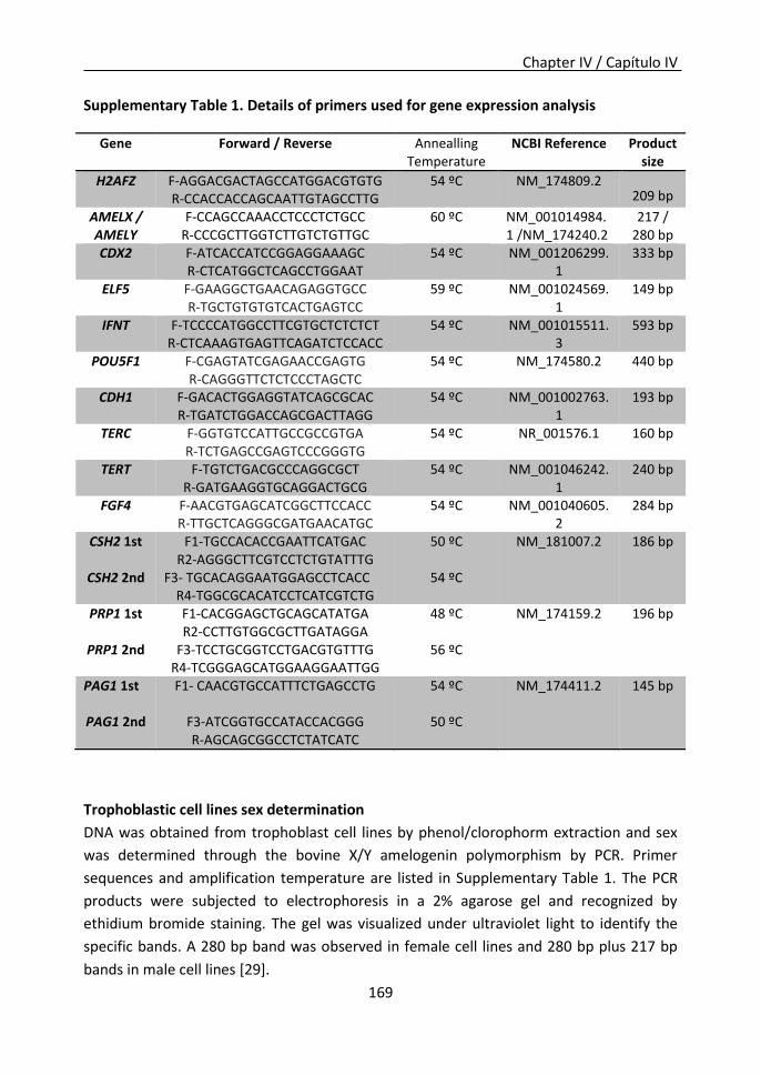
Chapter IV / Capítulo IV
169
Supplementary Table 1. Details of primers used for gene expression analysis
Gene Forward / Reverse Annealling Temperature
NCBI Reference Product size
H2AFZ F-AGGACGACTAGCCATGGACGTGTG R-CCACCACCAGCAATTGTAGCCTTG
54 ºC NM_174809.2 209 bp
AMELX / AMELY
F-CCAGCCAAACCTCCCTCTGCC R-CCCGCTTGGTCTTGTCTGTTGC
60 ºC NM_001014984.1 /NM_174240.2
217 / 280 bp
CDX2 F-ATCACCATCCGGAGGAAAGC R-CTCATGGCTCAGCCTGGAAT
54 ºC NM_001206299.1
333 bp
ELF5 F-GAAGGCTGAACAGAGGTGCC R-TGCTGTGTGTCACTGAGTCC
59 ºC NM_001024569.1
149 bp
IFNT F-TCCCCATGGCCTTCGTGCTCTCTCT R-CTCAAAGTGAGTTCAGATCTCCACC
54 ºC NM_001015511.3
593 bp
POU5F1 F-CGAGTATCGAGAACCGAGTG R-CAGGGTTCTCTCCCTAGCTC
54 ºC NM_174580.2 440 bp
CDH1 F-GACACTGGAGGTATCAGCGCAC R-TGATCTGGACCAGCGACTTAGG
54 ºC NM_001002763.1
193 bp
TERC F-GGTGTCCATTGCCGCCGTGA R-TCTGAGCCGAGTCCCGGGTG
54 ºC NR_001576.1 160 bp
TERT F-TGTCTGACGCCCAGGCGCT R-GATGAAGGTGCAGGACTGCG
54 ºC NM_001046242.1
240 bp
FGF4 F-AACGTGAGCATCGGCTTCCACC R-TTGCTCAGGGCGATGAACATGC
54 ºC NM_001040605.2
284 bp
CSH2 1st
CSH2 2nd
F1-TGCCACACCGAATTCATGAC R2-AGGGCTTCGTCCTCTGTATTTG
F3- TGCACAGGAATGGAGCCTCACC R4-TGGCGCACATCCTCATCGTCTG
50 ºC
54 ºC
NM_181007.2 186 bp
PRP1 1st
PRP1 2nd
F1-CACGGAGCTGCAGCATATGA R2-CCTTGTGGCGCTTGATAGGA
F3-TCCTGCGGTCCTGACGTGTTTG R4-TCGGGAGCATGGAAGGAATTGG
48 ºC
56 ºC
NM_174159.2 196 bp
PAG1 1st
PAG1 2nd
F1- CAACGTGCCATTTCTGAGCCTG
F3-ATCGGTGCCATACCACGGG R-AGCAGCGGCCTCTATCATC
54 ºC
50 ºC
NM_174411.2 145 bp
Trophoblastic cell lines sex determination
DNA was obtained from trophoblast cell lines by phenol/clorophorm extraction and sex
was determined through the bovine X/Y amelogenin polymorphism by PCR. Primer
sequences and amplification temperature are listed in Supplementary Table 1. The PCR
products were subjected to electrophoresis in a 2% agarose gel and recognized by
ethidium bromide staining. The gel was visualized under ultraviolet light to identify the
specific bands. A 280 bp band was observed in female cell lines and 280 bp plus 217 bp
bands in male cell lines [29].
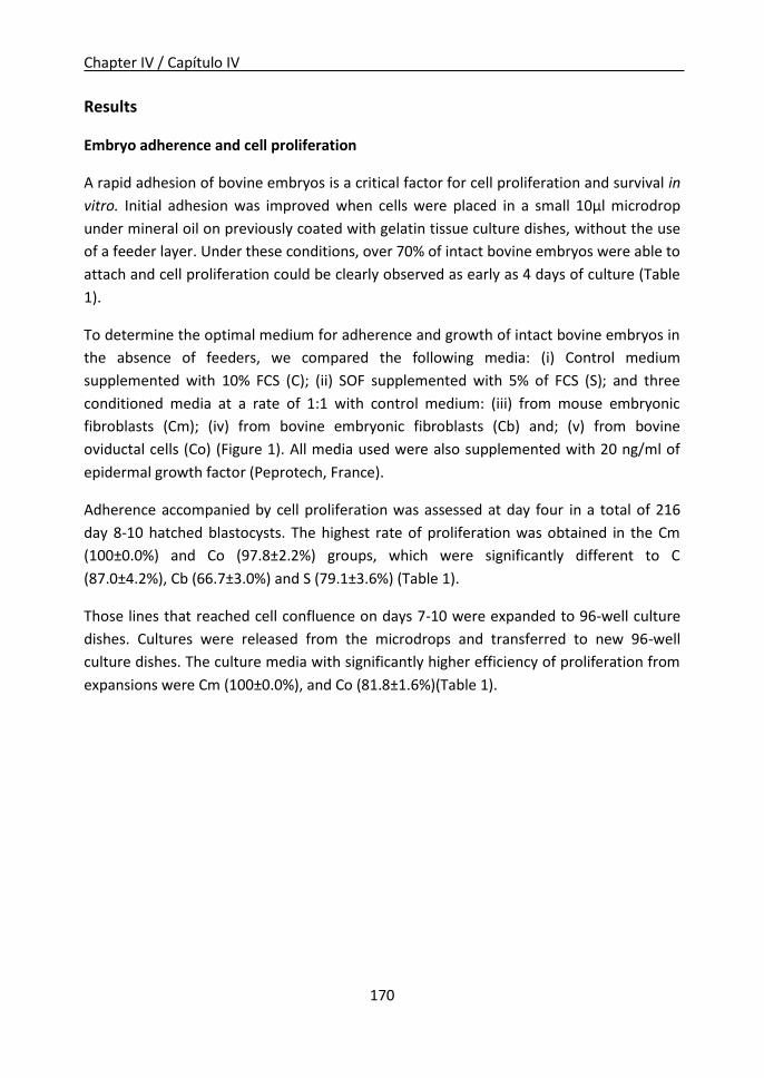
Chapter IV / Capítulo IV
170
Results
Embryo adherence and cell proliferation
A rapid adhesion of bovine embryos is a critical factor for cell proliferation and survival in
vitro. Initial adhesion was improved when cells were placed in a small 10μl microdrop
under mineral oil on previously coated with gelatin tissue culture dishes, without the use
of a feeder layer. Under these conditions, over 70% of intact bovine embryos were able to
attach and cell proliferation could be clearly observed as early as 4 days of culture (Table
1).
To determine the optimal medium for adherence and growth of intact bovine embryos in
the absence of feeders, we compared the following media: (i) Control medium
supplemented with 10% FCS (C); (ii) SOF supplemented with 5% of FCS (S); and three
conditioned media at a rate of 1:1 with control medium: (iii) from mouse embryonic
fibroblasts (Cm); (iv) from bovine embryonic fibroblasts (Cb) and; (v) from bovine
oviductal cells (Co) (Figure 1). All media used were also supplemented with 20 ng/ml of
epidermal growth factor (Peprotech, France).
Adherence accompanied by cell proliferation was assessed at day four in a total of 216
day 8-10 hatched blastocysts. The highest rate of proliferation was obtained in the Cm
(100±0.0%) and Co (97.8±2.2%) groups, which were significantly different to C
(87.0±4.2%), Cb (66.7±3.0%) and S (79.1±3.6%) (Table 1).
Those lines that reached cell confluence on days 7-10 were expanded to 96-well culture
dishes. Cultures were released from the microdrops and transferred to new 96-well
culture dishes. The culture media with significantly higher efficiency of proliferation from
expansions were Cm (100±0.0%), and Co (81.8±1.6%)(Table 1).

Chapter IV / Capítulo IV
171
Table 1. Embryo adherence and cell proliferation in different media.
abc P<0.05. Values with different superscripts letter in the same row are significant different. One-
way ANOVA. Data were analyzed using the SigmaStat (Jandel Scientific, San Rafael, CA) software package.
Embryo biopsy and survival
Our next goal was to isolate trophoblastic cells by biopsy from bovine embryos without
affecting the final survival of the remaining embryo. Each TE biopsy was transferred for
cultivation in microdrop on gelatin-coated surfaces, as described above for full embryos
(Figure 1). To determine the rate of in vitro embryo survival post biopsy, TE biopsies were
performed in six replicates from a total of 190 grade 1 and 2 Day-8 hatched blastocysts,
based on the guidelines of the International Embryo Transfer Society [23], showing a
survival rate at 24 h post biopsy of 82.2±2.6% (mean±SEM). 16 h after biopsy, the
biopsied blastocysts (which included ICM) re-expanded and recovered their typical
morphology (Figure 2a). TE biopsies (devoided of ICM) also re-expanded and adopted a
round morphology 16 h after the biopsy procedure (Figure 2b).
From a total of 25 grade 1 and 2 Day-8 hatched blastocysts, produced in six replicates, cell
count on biopsies and biopsied embryos showed an average of 72.44±3.82 cells (42.37%
of total cells), and 98.52±3,27 cells (57.63% of total cells) respectively, while the average
number of total cells in an intact embryo was 170.96±4,59 (mean±SEM).
Culture media
C S Cm Co Cb
N 48 71 25 39 33
Proliferation µdroplet (4d)
n %(Mean+SEM)
42 87.0+4.2c
56 79.1+3.6bc
25 100.0+0.0a
38 97.8+2.2a
22 66.7+3.0b
Proliferation P96 (reexpansion, 10d)
n %(Mean+SEM)
21 48.9+5.1b
0 0.0+0.0b
25 100.0+0.0a
31 81.8+1.6a
7 31.6+3.0b

Chapter IV / Capítulo IV
172
Figure 2. Phase-contrast micrograph examples of (a) biopsied embryos after 16 h of culture, and (b) TE biopsies after 16 h of culture. Magnification ≈ 30x. (c) Phase-contrast micrograph examples of IVF bovine blastocyst’s primary trophoblastic outgrowth only 48 h after initiating the culture without feeder layer. Magnification 200x. Arrows indicate the borders of the primary colony. (d) Trophoblastic cell cultures after 7-10 days. Note dome formation (arrow) in the monolayer. Magnification 200x. (e) Trophoblastic cell culture after 21 months and 76 passages. Magnification 100x. Note cavern formation (arrow).
Trophoblastic cells adherence and proliferation
We next evaluated the efficiency attained in our different media conditions of the
adhesion and proliferation of trophoblastic cells derived from biopsies cultured on
gelatin-coated microdrops. A total of 221 TE biopsies from hatched day 8-10 blastocysts
were used. The greatest proliferation was again registered when Cm was used (73.9%)
(Table 2). We could detect adhesion and proliferation of some biopsies as early as 48 h of
culture (Figure 2c). Regarding the expansion of biopsy cultures in P96, we observed no
differences between C (55.6±5.6%), Cm (48.2±4.3%) and Co (43.8±3.4%) groups; while Cb
group presented a significantly lower percentage of expansion (30.0%) and S did not
support any expansion of the biopsy (0.0%) (Table 2).

Chapter IV / Capítulo IV
173
Table 2. TE biopsy adherence and cell proliferation in different media.
Culture media
C S Cm Co Cb N 35 42 42 69 33
Proliferation µdroplet (4d)
n %(Mean+SEM)
18 51.4+1.52b
15 35.7+2.7c
31 73.9+1.6a
41 60.3+1.5b
13 39.4+3.0c
Proliferation P96 (reexpansion 10d)
n %(Mean+SEM)
10 55.6+5.6a
0 0.0+0.0b
15 48.2+4.3ac
18 43.8+3.4ac
4 30.0+21.5c
abc P<0.05. Values with different superscripts letter in the same row are significant different. One-
way ANOVA. Data were analyzed using the SigmaStat (Jandel Scientific, San Rafael, CA) software package.
Because we may loose the activity of relevant soluble factors by freezing/thawing the
conditioned medium, we analyzed proliferation in the presence of fresh Cm conditioned
media, finding no differences between fresh or frozen Cm media at day 4, when
microdrop confluence was reached (71.0±4.9% vs 77.9±4.1% respectively), or at day 10 of
culture in P96 (44.8±8.8% vs 57.6±8.3).
Ten trophoblastic cells lines were routinely plated at a 1:2 dilution from confluent cell
culture every 7-10 d and three of these lines have been cultured for more than 80
passages over two years maintaining trophoblastic morphology and steady proliferation
rate (Figure 2e). Thus, trophoblastic cells are apparently capable of an unlimited number
of population doublings.
We observed that trophoblastic cell lines in continuous culture showed an accumulation
of fluid under the monolayer that elicited the formation of dome-like structure within the
colony (Figure 2d) [11]. Such domes continued to accumulate fluid until discrete vesicles
were formed (Figure 2e). Subsequently, vesicles spontaneously dissociated from the
colony and formed spheroids, which were released to the medium. These spheroids of
trophoblastic were able to attach within 24 h of being transferred to a new dish, giving
rise to new trophoblastic cells outgrowths.
To analyze spheroids, cell-cell boundaries were visualized by phalloidin staining, which
labels the cortical actin cytoskeleton (Figure 3a). In the three-dimensional optical
sectioning of the spheroids, we could clearly observe that cells formed discrete layers of

Chapter IV / Capítulo IV
174
polarized cells. Some spheroids were quite complex in their topology, and frequently
presented several cell layers (Figure 3a).
We next assessed whether TE lines recapitulated the ability of producing binucleated
cells, by counting mononucleated and binucleated cells in both monolayer cultures and
three-dimensional spheroids. In both cultures, the majority of cells of the trophoblastic
cell lines were found to be mononucleated, but 2-9% of binucleated cells were found in
spheroids and 8-12% on monolayer cell cultures (Figure 3b).
Figure 3. (a) Representative confocal image of the phalloidin/DAPI staining used for BNC cell counting in spheroids. Arrow points to a binucleated cell. (b) Proportion of binucleated cells corresponding to spheroids from different trophoblastic cell lines (Mean±SD of over 6000 cells counted in three replicates).
Apoptotic cells were rarely detected in these culture conditions, ranging from 0.45±0.37
to 3.04±1.86 % in the different TE lines (Figure 4). Only when cell cultures were too
confluent abundant apoptotic cells were detected in the wrinkled edges of the
monolayer.
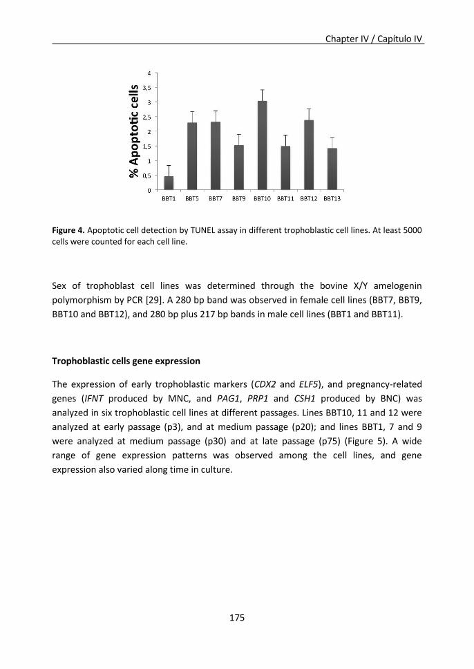
Chapter IV / Capítulo IV
175
Figure 4. Apoptotic cell detection by TUNEL assay in different trophoblastic cell lines. At least 5000 cells were counted for each cell line.
Sex of trophoblast cell lines was determined through the bovine X/Y amelogenin
polymorphism by PCR [29]. A 280 bp band was observed in female cell lines (BBT7, BBT9,
BBT10 and BBT12), and 280 bp plus 217 bp bands in male cell lines (BBT1 and BBT11).
Trophoblastic cells gene expression
The expression of early trophoblastic markers (CDX2 and ELF5), and pregnancy-related
genes (IFNT produced by MNC, and PAG1, PRP1 and CSH1 produced by BNC) was
analyzed in six trophoblastic cell lines at different passages. Lines BBT10, 11 and 12 were
analyzed at early passage (p3), and at medium passage (p20); and lines BBT1, 7 and 9
were analyzed at medium passage (p30) and at late passage (p75) (Figure 5). A wide
range of gene expression patterns was observed among the cell lines, and gene
expression also varied along time in culture.

Chapter IV / Capítulo IV
176
Figure 5. Trophoblastic-specific genes expression in diverse trophoblastic cell lines at different
passages.
Pluripotency-related genes: POU class 5 Homeobox 1 (POU5F1), Cadherin 1 (CDH1),
Telomerase RNA Component (TERC), Telomerase Reverse Transcriptase (TERT) and
Fibroblast Growth Factor 4 (FGF4) expression was also analyzed at the same passages in
six trophoblastic cell lines (Figure 6). In the same way, different expression patterns were
found that varied with time in culture.
Figure 6. Pluripotency-related genes expression in diverse trophoblastic cell lines at different passages.

Chapter IV / Capítulo IV
177
Discussion
In this manuscript we describe an efficient method to obtain a trophoblastic cell line from
a single bovine embryo or from an embryo biopsy.
Using amplified DNA from embryo biopsies for genotyping has many technical limitations
as reduced genome coverage, allele drop-out at heterozygous loci, missing genotypes,
amplification of artifacts or allele drop-in [2]. Likewise, little is known about
preimplantation embryo epigenetics due to the technical limitations derived from
analyzing limited sample amounts. Our procedure for in vitro trophoblastic cell expansion
represents a suitable new method to produce sufficient amount of genomic DNA for the
analysis of multiple production traits with economic interest, overcoming the drawbacks
of DNA pre-amplification. In the same way, our system would allow the analysis of the
epigenetic profile of preimplantation embryos in order to correlate genome-wide
epigenetic characteristics of TE biopsies with epigenetic alterations that could have a
short-term effect in implantation and fetal development, or a long-term effect during
adulthood. Although additional time is required for the culture step in our system, it is an
easy and reliable system and we estimate that in 10 days we can increase the cell number
up to 30000 and it is a reliable approach to obtain abundant genomic DNA.
In addition, few trophoblastic cell lines have been derived in cattle due to the difficulties
found during the long-term culture, so the establishment of different trophoblastic cells
lines provides a valuable tool to explore peri-implantation and placentation processes in
ruminants.
Regarding our methodology, our IVF results showed an average blastocyst yield of around
29.0% between days 7 and 10, being within the average range of in vitro bovine embryo
production [25]. Our hatching rate of 54.3% on day 10 with the largest proportion of
hatched blastocysts occurring on days 8 and 9 is also consistent with previously reported
data for good quality embryos [30] [31]. Embryo biopsy is a reliable technique as previous
reports attained survival rates of 90% 24h after embryo biopsy using micro-section of an
embryo on day 7 [32] [33]. Survival rate and embryonic development after the biopsy
were identical between embryos produced in vitro or in vivo [33]. We achieved a survival
rate of 82.2% of biopsied embryos, confirming that the chosen state (hatched blastocysts
on day 8-10) and quality of the embryos was adequate. Even though the site of the biopsy
has been shown to directly affect the quality of the embryos in terms of survival [32], our
method did not damage the embryo even when 40% of the total cells were biopsied,
since the biopsy was always performed at the opposing pole of the ICM.
Chapter IV / Capítulo IV
178
Regarding bovine trophoblastic cell lines, a very limited number have been reported so
far. Shimada et al [11] started from more than 50 blastocysts, and within a week only a
small proportion attached and began to spread out, while the majority neither attached
nor grew. They finally obtained only one trophoblastic cell line in absence of feeder cells,
using bovine endometrial fibroblast-conditioned medium. The relevance of our work lies
in the possibility of establishing a trophoblastic cell line from each embryo, with 100%
efficiency in Cm medium. Previous attempts to culture biopsied cells from bovine
blastocysts were reported by Le Bourhis et al [34] and by Gamarra et al [35]. However,
none of the biopsied cells attached to the coated dishes, probably because they had a
very low number of cells (from 5 to 10), large culture surfaces were used (4-well culture
dishes) or the media used was not adequate. We found that a rapid adhesion of biopsied
trophoblastic cells is a limiting factor. This step was greatly enhanced when using 10μl
droplets on previously gelatin-coated surfaces under mineral oil. In the case of biopsies,
adhesion began only 48 h after initiating the culture (Figure 2c), and conditioned media
from mouse embryonic fibroblasts supported the growth of 78% of TE biopsies. Once cell
confluence was reached in microdrops (≈3000 cells) at 7-10 days we were able to re-
expand these cultures to P96 plates. From all the initiated trophoblastic cell cultures, 57%
reached cell confluence (≈30000 cells) in P96 re-expansions. The reduction in the
efficiency in trophoblastic cell lines establishment starting from biopsies (57%,) compared
with the 100% proliferation in the case of complete embryo culture, could be explained
by the reduction in the number of starting cells [36].
We supplemented all media with EGF, since earlier studies demonstrated its positive
effect on proliferation of trophoblastic cells [19]. In a recent study, Suzuki et al. used
BMP4 to raise trophoblastic cell lines because exogenous BMP4 stimulated embryonic
stem cells (ES) to become trophoblastic cells [37] but no significant differences were
found between untreated and BMP4-treated groups regarding derivation efficiency.
Most of the studies that aim to investigate the in vitro process of placentation have been
performed with cell lines established by co-cultivation with mouse feeder layers
[10][38][39]. In our study, cell lines were established from mouse fibroblast conditioned
media, in the absence of feeder layer, eliminating the risk of contamination of the
extracted genomic DNA. Endoderm outgrowths have also been shown to frequently
contaminate the primary trophoblastic cell cultures [10, 38], as well as some condensed
and dark cell clumps resembling the ICM [40]. Our methodology avoids contamination by
other cell types because only trophoblastic cells are included in the TE biopsy.
The media conditioned from bovine oviductal cells (Co), bovine embryonic fibroblasts (Cb)
as well as control media (C) allowed the establishment of cell lines, although the initial
growth from the biopsy was significantly lower than in the case of media conditioned by
Chapter IV / Capítulo IV
179
mouse embryonic fibroblasts (Cm). Therefore Cm is an optimal medium for rapid
adhesion and bovine trophoblastic cell growth.
The morphological features and in vitro behavior of the different bovine trophoblastic cell
lines that we have established are consistent with those described by Shimada et al [11].
We observed that trophoblastic cell lines in continuous culture formed dome-like
structures that maturated into vesicles, which spontaneously dissociated from the colony
forming spheroids that floated freely in the medium. In vivo, trophoblastic binucleated
cells are present at the beginning of implantation and throughout pregnancy,
representing about 20% of trophoblastic cells [41]. However, it has been described that
the availability of BNCs in vitro is limited (2-3%) [4] and that their secretory activity cease
within several days in culture [42]. In contrast, in our culture conditions a higher
proportion of BNC appeared. The expression of PRP1, CSH2 and PAG1 confirms the
existence of binucleated cells in our trophoblastic cell lines, and the expression of these
BNC-specific genes was maintained after more than 80 passages.
Thus, our culture system of individual biopsy-derived trophoblast cell lines can serve as a
model for the investigation of placental development and the differentiation of the
bovine embryo in vitro.
The different trophoblastic cell lines established showed diverse gene expression profiles
of early trophoblastic markers (CDX2, ELF5), MNCs (IFNT) and BNCs (PAG1, PRP1 and
CSH2) specific genes that varied along time in culture. Differences in gene expression
patterns could be due to the variability among the starting embryos, as gene expression
in early passages varied among trophoblastic cell lines coming from the same culture
conditions; but also to time term in culture, as variations were observed along passages,
indicating that these cells lines are dynamic populations.
Among the genes studied, CDX2 first specifies the trophoblast versus the ICM cell fate
[43] and is a core regulator of multiple trophoblast genes in bovine trophoblastic cell line
CT-1 [18]. In fact, we could observe CDX2 expression in all trophoblastic cell lines at
different time points. ELF5 transcription factor is detected in bovine blastocysts at the
ovoid stage, peaking at day 12 [43] and decreasing during the elongation process [44]. In
our study, ELF5 was expressed in every trophoblastic cell line, mostly from passage 20
onwards. Its expression was also described in CT-1 cell line, which was compared to the
ovoid trophoblast stage [18].
Expression of interferon-t (IFNT), placental lactogen (CSH2), prolactin-related protein 1
(PRP1) and pregnancy-associated glycoprotein 1 (PAG1) is necessary for pregnancy
establishment in cattle. The transcription of these genes is regulated in a temporal and
Chapter IV / Capítulo IV
180
spatial manner by not completely elucidated molecular mechanisms. IFNT is expressed
from the formation of the TE in the early blastocyst until its attachment to the maternal
uterus after day 19 of pregnancy in cattle [45]. Different factors have been shown to
influence IFNT expression. Long time in in vitro culture of blastocyst or trophoblastic cell
lines has been negatively correlated with IFNT secretion [46], although in our
trophoblastic cell lines IFNT expression was detected up to passage 75. Sexual
dimorphism affects IFNT expression as well, since female blastocysts have been shown to
produce twice as much IFNT as male blastocysts [47], but we could not find any
correlation among sex and IFNT expression in our trophoblastic cell lines.
The co-expression of early and late trophoblastic markers would make difficult to
associate our trophoblastic cell lines to a specific gestational stage. However, the fact that
trophoblastic cell lines express simultaneously early and late trophoblast specific-markers
has been previously described [40].
It has been suggested that bovine trophoblastic cells are not terminal cells and present
some stem cell features [40]. Therefore, we assessed pluripotency-related gene
expression. Telomerase is the enzymatic complex responsible of adding telomeric repeats
to the ends of chromosomes. Its expression is silenced in adult somatic tissues with the
exception of adult stem cell compartments [48]. The core telomerase components are the
telomerase reverse transcriptase (TERT), and the telomerase RNA (TERC) [49]. Acquisition
of indefinite self-renewal capacity in reprogrammed cells is accompanied by induction of
TERT and TERC genes, which is considered a hallmark of the pluripotent state [50] [51]. In
our study, TERC and TERT expression was observed in almost all trophoblastic cell lines
generated, which could be an indicator of self-renewal capacity.
POU5F1 expression is rapidly down-regulated in the TE during blastocyst stage in mouse
[52]. In contrast, in bovine, POU5F1 expression is maintained through the ovoid-stage TE
[53], and may play a role in maintaining TE cells in a “differentiation-delayed” state [43].
POU5F1 expression has been reported in CT-1 trophoblastic cell line [18] and in other
trophoblastic cell lines generated afterwards, although cells expressing POU5F1 were rare
(only about 4% [40]). In our study, POU5F1 expression was detected in some
trophoblastic cell lines, mainly at medium and late passages, which could be an indicator
of cell line immortalization.
In recent years, E-cadherin (CDH1) has been shown to regulate pluripotent and self-
renewal signaling pathways in stem cells. Loss of E-cadherin is associated with ES
differentiation and its expression enhances iPS cell derivation [54]. CDH1 was expressed
in BBT1, BBT5 and BBT9 cell lines. In addition, FGF4, produced by the ICM and to a lesser
extent by TE in ruminant [55], an inductive signal for trophectoderm elongation in porcine

Chapter IV / Capítulo IV
181
embryos [56], and an essential factor for mouse trophectoderm stem cells proliferation in
an undifferentiated state [57] was expressed in BBT11 and BBT12 cell lines, although
expression was lost with time in culture. Thus, the expression of different pluripotency-
related genes, together with long-term steady cell proliferation, indicates certain self-
renewal capacity of the trophoblastic cell lines generated in this study.

Chapter IV / Capítulo IV
182
References
1. Ponsart, C, Le Bourhis, D, Knijn, H, Fritz, S, Guyader-Joly, C, Otter, T, Lacaze, S, Charreaux, F, Schibler, L, Dupassieux, D, Mullaart, E. Reproductive technologies and genomic selection in dairy cattle. Reprod Fertil Dev 2013; 26:12-21.
2. Lauri, A, Lazzari, G, Galli, C, Lagutina, I, Genzini, E, Braga, F, Mariani, P, Williams, JL. Assessment of MDA efficiency for genotyping using cloned embryo biopsies. Genomics 2013; 101:24-29.
3. Fernandez-Gonzalez, R, Moreira, PN, Perez-Crespo, M, Sanchez-Martin, M, Ramirez, MA, Pericuesta, E, Bilbao, A, Bermejo-Alvarez, P, de Dios Hourcade, J, de Fonseca, FR, Gutierrez-Adan, A. Long-term effects of mouse intracytoplasmic sperm injection with DNA-fragmented sperm on health and behavior of adult offspring. Biol Reprod 2008; 78:761-772.
4. Nakano, H, Shimada, A, Imai, K, Takezawa, T, Takahashi, T, Hashizume, K. Bovine trophoblastic cell differentiation on collagen substrata: formation of binucleate cells expressing placental lactogen. Cell Tissue Res 2002; 307:225-235.
5. Demmers, KJ, Derecka, K, Flint, A. Trophoblast interferon and pregnancy. Reproduction 2001; 121:41-49.
6. Spencer, TE, Burghardt, RC, Johnson, GA, Bazer, FW. Conceptus signals for establishment and maintenance of pregnancy. Anim Reprod Sci 2004; 82-83:537-550.
7. Austin, KJ, Ward, SK, Teixeira, MG, Dean, VC, Moore, DW, Hansen, TR. Ubiquitin cross-reactive protein is released by the bovine uterus in response to interferon during early pregnancy. Biol Reprod 1996; 54:600-606.
8. Thatcher, WW, Guzeloglu, A, Mattos, R, Binelli, M, Hansen, TR, Pru, JK. Uterine-conceptus interactions and reproductive failure in cattle. Theriogenology 2001; 56:1435-1450.
9. Hashizume, K, Ushizawa, K, Patel, OV, Kizaki, K, Imai, K, Yamada, O, Nakano, H, Takahashi, T. Gene expression and maintenance of pregnancy in bovine: roles of trophoblastic binucleate cell-specific molecules. Reproduction, Fertility and Development 2007; 19:79-90.
10. Talbot, NC, Caperna, TJ, Edwards, JL, Garrett, W, Wells, KD, Ealy, AD. Bovine blastocyst-derived trophectoderm and endoderm cell cultures: interferon tau and transferrin expression as respective in vitro markers. Biol Reprod 2000; 62:235-247.
11. Shimada, A, Nakano, H, Takahashi, T, Imai, K, Hashizume, K. Isolation and Characterization of a Bovine Blastocyst-Derived Trophoblastic Cell line, BT-1: Development of a Culture System in the Absence of Feeder Cell. Placenta 2001; 22:652-662.
12. Hirata, M, Sato, T, Tsumagari, M, Shimada, A, Nakano, H, Hashizume, K, Ito, A. Differential regulation of the expression of matrix metalloproteinases and tissue inhibitors of metalloproteinases by cytokines and growth factors in bovine endometrial stromal cells and trophoblast cell line BT-1 in vitro. Biol Reprod 2003; 68:1276-1281.
13. Ushizawa, K, Takahashi, T, Kaneyama, K, Tokunaga, T, Tsunoda, Y, Hashizume, K. Gene expression profiles of bovine trophoblastic cell line (BT-1) analyzed by a custom cDNA microarray. The Journal of reproduction and development 2005; 51:211-220.
14. Michael, DD, Alvarez, IM, Ocón, OM, Powell, AM, Talbot, NC, Johnson, SE, Ealy, AD. Fibroblast growth factor-2 is expressed by the bovine uterus and stimulates interferon-tau production in bovine trophectoderm. Endocrinology 2006; 147:3571-3579.
15. Das, P, Ezashi, T, Gupta, R, Roberts, RM. Combinatorial roles of protein kinase A, Ets2, and 3’,5’-cyclic-adenosine monophosphate response element-binding protein-binding protein/p300 in the transcriptional control of interferon-tau expression in a trophoblast cell line. Mol Endocrinol 2008; 22:331-343.
16. Bai, H, Sakurai, T, Someya, Y, Konno, T, Ideta, A, Aoyagi, Y, Imakawa, K. Regulation of trophoblast-specific factors by GATA2 and GATA3 in bovine trophoblast CT-1 cells. J Reprod
Chapter IV / Capítulo IV
183
Dev 2011; 57:518-525. 17. Schiffmacher, AT, Keefer, CL. Optimization of a lipitoid-based plasmid DNA transfection
protocol for bovine trophectoderm CT-1 cells. In vitro cellular & developmental biology. Animal 2012; 48:403-406.
18. Schiffmacher, AT, Keefer, CL. CDX2 regulates multiple trophoblast genes in bovine trophectoderm CT-1 cells. Mol Reprod Dev 2013;
19. Hambruch, N, Haeger, JD, Dilly, M, Pfarrer, C. EGF stimulates proliferation in the bovine placental trophoblast cell line F3 via Ras and MAPK. Placenta 2010; 31:67-74.
20. Zeiler, M, Leiser, R, Johnson, GA, Tinneberg, HR, Pfarrer, C. Development of an in vitro model for bovine placentation: a comparison of the in vivo and in vitro expression of integrins and components of extracellular matrix in bovine placental cells. Cells Tissues Organs 2007; 186:229-242.
21. Landim, LPJ, Miglino, MA, Pfarrer, C, Ambrosio, CE, Garcia, JM. Culture of mature trophoblastic giant cells from bovine placentomes. Anim Reprod Sci 2007; 98:357-364.
22. Clemente, M, Lopez-Vidriero, I, O’Gaora, P, Mehta, JP, Forde, N, Gutierrez-Adan, A, Lonergan, P, Rizos, D. Transcriptome changes at the initiation of elongation in the bovine conceptus. Biol Reprod 2011; 85:285-295.
23. R.E., RIaN. Certification of the embryo. In: SDAaS S.M (ed.), Manual of the International Embryo Transfer Society, Savoy; 1998:103-134.
24. Ramirez, MA, Pericuesta, E, Yáñez-Mó, M, Palasz, A, Gutierrez-Adan, A. Effect of long-term culture of mouse embryonic stem cells under low oxygen concentration as well as on glycosaminoglycan hyaluronan on cell proliferation and differentiation. Cell Proliferation 2010; 44:75-85.
25. Clemente, M, de La Fuente, J, Fair, T, Al Naib, A, Gutierrez-Adan, A, Roche, JF, Rizos, D, Lonergan, P. Progesterone and conceptus elongation in cattle: a direct effect on the embryo or an indirect effect via the endometrium? Reproduction 2009; 138:507-517.
26. Dominguez, F, Simon, C, Quinonero, A, Ramirez, MA, Gonzalez-Munoz, E, Burghardt, H, Cervero, A, Martinez, S, Pellicer, A, Palacin, M, Sanchez-Madrid, F, Yanez-Mo, M. Human endometrial CD98 is essential for blastocyst adhesion. PLoS One 2010; 5:e13380.
27. Bermejo-Alvarez, P, Rizos, D, Lonergan, P, Gutierrez-Adan, A. Transcriptional sexual dimorphism in elongating bovine embryos: implications for XCI and sex determination genes. Reproduction 2011; 141:801-808.
28. Ramirez, MA, Pericuesta, E, Fernandez-Gonzalez, R, Moreira, P, Pintado, B, Gutierrez-Adan, A. Transcriptional and post-transcriptional regulation of retrotransposons IAP and MuERV-L affect pluripotency of mice ES cells. Reproductive biology and endocrinology : RB&E 2006; 4:55.
29. Madrid-Bury, N, Fernandez, R, Jimenez, A, Perez-Garnelo, S, Moreira, PN, Pintado, B, de la Fuente, J, Gutierrez-Adan, A. Effect of ejaculate, bull, and a double swim-up sperm processing method on sperm sex ratio. Zygote 2003; 11:229-235.
30. Nedambale, TL, Dinnyes, A, Groen, W, Dobrinsky, JR, Tian, XC, Yang, X. Comparison on in vitro fertilized bovine embryos cultured in KSOM or SOF and cryopreserved by slow freezing or vitrification. Theriogenology 2004; 62:437-449.
31. Laboratory production of cattle embryos. Biotechnology in agriculture series 2003; 27 32. Peippo, J, Viitala, S, Virta, J, Räty, M, Tammiranta, N, Lamminen, T, Aro, J, Myllymäki, H,
Vilkki, J. Birth of correctly genotyped calves after multiplex marker detection from bovine embryo microblade biopsies. Molecular Reproduction and Development 2007; 74:1373-1378.
33. Humblot, P, Le Bourhis, D, Fritz, S, Colleau, JJ, Gonzalez, C, Guyader Joly, C, Malafosse, A, Heyman, Y, Amigues, Y, Tissier, M, Ponsart, C. Reproductive technologies and genomic
Chapter IV / Capítulo IV
184
selection in cattle. Veterinary medicine international 2010; 2010:192787. 34. Le Bourhis, D, Gamarra, G, Gall, L, Laffont, L, Ruffini, S, Humblot, P. Attempts to culture
biopsied cells from in vitro bovine blastocysts for genotyping. 25th Annual Meeting A.E.T.E. 2009; 210.
35. Gamarra, G, Le Bourhis, D, Gall, L, Laffont, L, Ruffini, S, Humblot, P. Attempts to culture biopsied cells from in vitro bovine blastocysts for genotyping. Reproduction, Fertility and Development 2010; 22:238.
36. Bredbacka, P. Factors affecting cell viability during bisection of bovine embryos. Theriogenology 1995; 44:159-166.
37. Murohashi, M, Nakamura, T, Tanaka, S, Ichise, T, Yoshida, N, Yamamoto, T, Shibuya, M, Schlessinger, J, Gotoh, N. An FGF4-FRS2alpha-Cdx2 axis in trophoblast stem cells induces Bmp4 to regulate proper growth of early mouse embryos. Stem Cells 2010; 28:113-121.
38. Talbot, NC, Caperna, TJ, Powell, AM, Garrett, WM, Ealy, AD. Isolation and characterization of a bovine trophectoderm cell line derived from a parthenogenetic blastocyst. Mol Reprod Dev 2004; 69:164-173.
39. Talbot, N, Powell, A, Camp, M, Ealy, A. Establishment of a bovine blastocyst-derived cell line collection for the comparative analysis of embryos created in vivo and by in vitro fertilization, somatic cell nuclear transfer, or parthenogenetic activation. In Vitro Cellular & Developmental Biology - Animal 2007; 43:59-71.
40. Suzuki, Y, Koshi, K, Imai, K, Takahashi, T, Kizaki, K, Hashizume, K. Bone morphogenetic protein 4 accelerates the establishment of bovine trophoblastic cell lines. Reproduction 2011; 142:733-743.
41. WIMSATT, WA. Observations on the morphogenesis, cytochemistry, and significance of the binocleate giant cells of the placenta of ruminants. Am J Anat 1951; 89:233-281.
42. Nakano, H, Takahashi, T, Imai, K, Hashizume, K. Expression of placental lactogen and cytokeratin in bovine placental binucleate cells in culture. Cell Tissue Res 2001; 303:263-270.
43. Degrelle, SA, Campion, E, Cabau, C, Piumi, F, Reinaud, P, Richard, C, Renard, J-P, Hue, I. Molecular evidence for a critical period in mural trophoblast development in bovine blastocysts. Developmental Biology 2005; 288:448-460.
44. Smith, CS, Berg, DK, Berg, M, Pfeffer, PL. Nuclear transfer-specific defects are not apparent during the second week of embryogenesis in cattle. Cell Reprogram 2010; 12:699-707.
45. Helmer, SD, Hansen, PJ, Anthony, RV, Thatcher, WW, Bazer, FW, Roberts, RM. Identification of bovine trophoblast protein-1, a secretory protein immunologically related to ovine trophoblast protein-1. J Reprod Fertil 1987; 79:83-91.
46. Talbot, NC, Blomberg, LA. The Pursuit of ES Cell Lines of Domesticated Ungulates. Stem Cell Rev 2008; 4:235-254.
47. Larson, MA, Kimura, K, Kubisch, HM, Roberts, RM. Sexual dimorphism among bovine embryos in their ability to make the transition to expanded blastocyst and in the expression of the signaling molecule IFN-tau. Proc Natl Acad Sci U S A 2001; 98:9677-9682.
48. Marion, RM, Blasco, MA. Telomeres and telomerase in adult stem cells and pluripotent embryonic stem cells. Adv Exp Med Biol 2010; 695:118-131.
49. Cao, Y, Bryan, TM, Reddel, RR. Increased copy number of the TERT and TERC telomerase subunit genes in cancer cells. Cancer Sci 2008; 99:1092-1099.
50. Agarwal, S, Loh, YH, McLoughlin, EM, Huang, J, Park, IH, Miller, JD, Huo, H, Okuka, M, Dos Reis, RM, Loewer, S, Ng, HH, Keefe, DL et al. Telomere elongation in induced pluripotent stem cells from dyskeratosis congenita patients. Nature 2010; 464:292-296.
51. Unternaehrer, JJ, Daley, GQ. Induced pluripotent stem cells for modelling human diseases. Philos Trans R Soc Lond B Biol Sci 2011; 366:2274-2285.

Chapter IV / Capítulo IV
185
52. Dietrich, JE, Hiiragi, T. Stochastic patterning in the mouse pre-implantation embryo. Development 2007; 134:4219-4231.
53. Berg, DK, Smith, CS, Pearton, DJ, Wells, DN, Pfeffer, PL, Broadhurst, R, Donnison, M. Trophectoderm lineage determination in cattle. Dev Cell 2011; 20:244-255.
54. Soncin, F, Mohamet, L, Ritson, S, Hawkins, K, Bobola, N, Zeef, L, Merry, CL, Ward, CM. E-cadherin acts as a regulator of transcripts associated with a wide range of cellular processes in mouse embryonic stem cells. PLoS One 2011; 6:e21463.
55. Fujii, T, Moriyasu, S, Hirayama, H, Hashizume, T, Sawai, K. Aberrant expression patterns of genes involved in segregation of inner cell mass and trophectoderm lineages in bovine embryos derived from somatic cell nuclear transfer. Cell Reprogram 2010; 12:617-625.
56. Valdez Magana, G, Rodriguez, A, Zhang, H, Webb, R, Alberio, R. Paracrine effects of embryo-derived FGF4 and BMP4 during pig trophoblast elongation. Dev Biol 2014; 387:15-27.
57. Erlebacher, A, Price, KA, Glimcher, LH. Maintenance of mouse trophoblast stem cell proliferation by TGF-beta/activin. Dev Biol 2004; 275:158-169.

186

General discussion
187
General discussion

General discussion
188
General discussion
189
Over the last few decades, much research has been done on pluripotent cells; however,
optimal conditions for identification, isolation and in vitro culture of pluripotent cells
remain unknown. Thus, several determinant aspects for cell lines derivation, as the effect
of embryonic source or culture conditions, have been analyzed in this thesis.
Furthermore, an efficient system for trophoblastic cell lines derivation from embryo
biopsies has been established that allows the development of placentation studies and
preimplantational genetic and epigenetic diagnosis.
Embryonic source affects pluripotent cell lines derivation
ESCs have been the most commonly used pluripotent cell type since they were first
isolated (Evans and Kaufman 1981). ESCs isolation techniques and culture systems have
evolved in the last years to improve derivation efficiency, but it remains controversial
whether embryonic characteristics have an influence over the process.
ARTs as in vitro culture (IVC), in vitro fertilization (IVF), intracytoplasmic sperm injection
(ICSI) or somatic cell nuclear transfer (SCNT) have contributed to the development of
infertility treatments in humans and of new animal reproduction methods. However,
embryo manipulation and in vitro culture is associated with a number of perturbations of
the embryonic ultrastructure, genetic and epigenetic alterations and syndromes and
diseases during adulthood (Rizos, Fair et al. 2002, Corcoran, Fair et al. 2006, Fernandez-
Gonzalez, Moreira et al. 2008, Calle, Miranda et al. 2012). Following ARTs, the
classification of good quality embryos is based largely on subjective criteria. Furthermore,
it is often overlooked that human ESCs are generated from in vitro cultured, often surplus
or discarded embryos considered unsuitable for transfer in infertility clinics. These
circumstances lead to the question of whether ESCs retain some memory of the embryos
from which they were derived.
In chapter I, we have performed ICSI with DNA fragmented sperm (DFS), a previously
delevoped model in our laboratory (Fernandez-Gonzalez, Moreira et al. 2008), which can
be used as a model to produce bad quality embryos, and we have demonstrated that
these embryos show a reduced potential to generate ESCs lines compared to in vivo-
produced embryos. Furthermore, during early passages, these DFS-ICSI ESCs differ from in
vivo-ESCs in their expression of certain genes related to pluripotency and epigenetic
repression, DNA damage and repair, and de novo DNA methyltransferases and histone
deacetylases. However, at late passages no differences in gene expression were
observed. This is consistent with a previous study that described the retention of
epigenetic differences in ESCs dependent on the in vivo or in vitro fertilization (IVF) origin
of the embryo from which they were derived, and the disappearance of these differences
after several passages (Horii, Yanagisawa et al. 2010). Another study describing
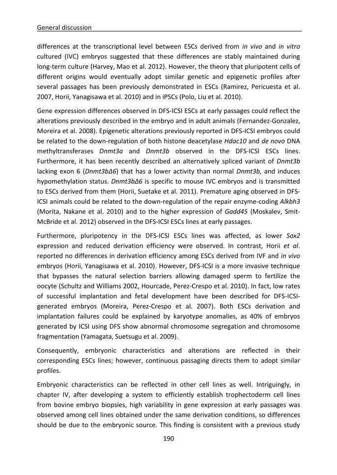
General discussion
190
differences at the transcriptional level between ESCs derived from in vivo and in vitro
cultured (IVC) embryos suggested that these differences are stably maintained during
long-term culture (Harvey, Mao et al. 2012). However, the theory that pluripotent cells of
different origins would eventually adopt similar genetic and epigenetic profiles after
several passages has been previously demonstrated in ESCs (Ramirez, Pericuesta et al.
2007, Horii, Yanagisawa et al. 2010) and in iPSCs (Polo, Liu et al. 2010).
Gene expression differences observed in DFS-ICSI ESCs at early passages could reflect the
alterations previously described in the embryo and in adult animals (Fernandez-Gonzalez,
Moreira et al. 2008). Epigenetic alterations previously reported in DFS-ICSI embryos could
be related to the down-regulation of both histone deacetylase Hdac10 and de novo DNA
methyltransferases Dnmt3a and Dnmt3b observed in the DFS-ICSI ESCs lines.
Furthermore, it has been recently described an alternatively spliced variant of Dnmt3b
lacking exon 6 (Dnmt3bΔ6) that has a lower activity than normal Dnmt3b, and induces
hypomethylation status. Dnmt3bΔ6 is specific to mouse IVC embryos and is transmitted
to ESCs derived from them (Horii, Suetake et al. 2011). Premature aging observed in DFS-
ICSI animals could be related to the down-regulation of the repair enzyme-coding Alkbh3
(Morita, Nakane et al. 2010) and to the higher expression of Gadd45 (Moskalev, Smit-
McBride et al. 2012) observed in the DFS-ICSI ESCs lines at early passages.
Furthermore, pluripotency in the DFS-ICSI ESCs lines was affected, as lower Sox2
expression and reduced derivation efficiency were observed. In contrast, Horii et al.
reported no differences in derivation efficiency among ESCs derived from IVF and in vivo
embryos (Horii, Yanagisawa et al. 2010). However, DFS-ICSI is a more invasive technique
that bypasses the natural selection barriers allowing damaged sperm to fertilize the
oocyte (Schultz and Williams 2002, Hourcade, Perez-Crespo et al. 2010). In fact, low rates
of successful implantation and fetal development have been described for DFS-ICSI-
generated embryos (Moreira, Perez-Crespo et al. 2007). Both ESCs derivation and
implantation failures could be explained by karyotype anomalies, as 40% of embryos
generated by ICSI using DFS show abnormal chromosome segregation and chromosome
fragmentation (Yamagata, Suetsugu et al. 2009).
Consequently, embryonic characteristics and alterations are reflected in their
corresponding ESCs lines; however, continuous passaging directs them to adopt similar
profiles.
Embryonic characteristics can be reflected in other cell lines as well. Intriguingly, in
chapter IV, after developing a system to efficiently establish trophectoderm cell lines
from bovine embryo biopsies, high variability in gene expression at early passages was
observed among cell lines obtained under the same derivation conditions, so differences
should be due to the embryonic source. This finding is consistent with a previous study

General discussion
191
that derived 12 bovine trophoblastic cell lines and quantified the expression of
trophoblastic genes: placental lactogen (CSH1), interferon-t (IFNt), pregnancy-associated
glycoprotein 1 (PAG1), and prolactin-related protein 1 (PRP1); and found different
expression patterns among the cell lines with no correlations with culture conditions
(Suzuki, Koshi et al. 2011). Thus, in this chapter IV it was demonstrated that embryonic
characteristics influence trophoblastic cell lines derivation as well.
Culture conditions affect cell lines derivation
Culture conditions are a main factor affecting cell lines derivation and characteristics. In
this thesis, the effect of culture conditions has been studied in ESCs lines in chapter II, and
in trophoblastic cell lines in chapter IV.
In chapter II, the effect of leukemia inhibitory factor (LIF) supplementation during
preimplantational embryo culture up to blastocyst for ESCs derivation was analyzed due
to the contradictory evidences existing among previous studies (Jurisicova, Ben-Chetrit et
al. 1995, Dunglison, Barlow et al. 1996, Tsai, Chang et al. 2000, Rungsiwiwut,
Rungarunlert et al. 2008). Although the embryos supplemented with LIF showed a lower
total cell number, the ratio of inner cell mass (ICM)/total cells was significantly higher.
Furthermore, these embryos proved to be more suitable for ESCs isolation, as their
derivation efficiency was higher. Additionally, regular medium for ESCs lines derivation
was supplemented with growth factors regularly used for germline stem cells (GSCs)
culture (EGF, GDNF and bFGF), and ESCs derivation efficiency increased in the same way.
In agreement to this observation, the beneficial effect of these factors on other
pluripotent cells populations has been previously reported (Reynolds and Weiss 1992,
Donovan and de Miguel 2003, Kanatsu-Shinohara, Ogonuki et al. 2003).
The vast effect of culture medium supplementation with different factors has been
exhaustively studied in ESCs. In chapter II, ESCs were derived over a feeder layer of mouse
embryonic fibroblasts (EFs) in medium supplemented with: fetal calf serum (FCS) and LIF
(ES medium); FCS, LIF and GSCs-related growth factors (GS medium); or FCS, LIF and
inhibitory factors (2i medium). We could observe an improvement in ESCs derivation
efficiency in GS medium compared to ES medium, and ESCs lines derived in GS medium
fulfilled pluripotency criteria and showed a higher chimeric mice generation ability. Thus,
a possible synergy among LIF supplementation during embryo culture and the posterior
addition of growth factors present in GS medium that favours ESCs derivation. Different
culture systems have been established to avoid the variability induced by FCS, which
shows variations between batches, as knockout serum replacement (KSR) (Cheng, Dutra
et al. 2004) and small-molecule inhibitors (MEK and GSK3β; 2i) (Ying, Wray et al. 2008).
The 2i culture system allows efficient derivation and expansion of germline-competent

General discussion
192
ESCs from different strains of mice and species that were previously recalcitrant for ESCs
derivation (Buehr, Meek et al. 2008, Nichols, Jones et al. 2009). Thus, ESCs derivation
efficiency is highly dependent on culture medium. In the same way, transcriptome is
defined by culture conditions. In chapter II, gene expression in ESCs lines varied according
to the different culture media employed for their derivation (ES, GS and 2i). Indeed, it has
been reported that culture conditions are the major aspect determining gene expression,
over embryonic origin and derivation procedure (Marks, Kalkan et al. 2012, Leitch,
McEwen et al. 2013).
In chapter IV, different media were used with the aim of improving derivation efficiency
of trophoblastic cell lines. Although we found that a derivation procedure allowing a rapid
adhesion of the embryo or biopsy to the culture plate was a critical step and significantly
increased derivation efficiency, culture medium was decisive as well. Conditioned culture
medium contains secreted proteins that include numerous enzymes, growth factors,
cytokines and hormones (Dowling and Clynes 2011). Most of the studies regarding
trophoblast cell lines derivation employed conditioned medium by co-cultivation with
mouse embryonic fibroblasts (Talbot, Caperna et al. 2000, Talbot, Caperna et al. 2004,
Talbot, Powell et al. 2007). However, co-cultivation has the disadvantage of
contaminating the cell lines of interest, so in our study trophoblastic cells were cultured
over a gelatinized culture plate, and different conditioned media from mouse embryonic
fibroblasts (Cm), bovine embryonic fibroblasts (Cb) and bovine oviductal cells (Co) were
assayed. Except for Cb, conditioned media improved derivation efficiency, being Cm the
most efficient medium for trophoblastic cell lines derivation. Other articles previously
used bovine endometrial fibroblast-conditioned medium to derive trophoblastic cell lines
(Shimada, Nakano et al. 2001, Hashizume, Shimada et al. 2006); however, heterologous
conditioned medium by mouse embryonic fibroblasts was the most efficient for rapid
adhesion and cell growth in our study. Furthermore, conditioned media were
supplemented with EGF, since earlier studies demonstrated its positive effect on
trophoblastic cells proliferation (Hambruch, Haeger et al. 2010). In conclusion, our
observations and those from other authors indicate that trophoblastic cell lines derivation
efficiency significantly depends on culture conditions.
A germ cell origin for pluripotent cells
During the transition from blastocyst cells to pluripotent ESCs, the developmental
program in ICM cells is modified in order to acquire the ability to divide indefinitely while
maintaining the potential to differentiate towards any tissue. Several evidences such as
similarities existing among ESCs and EGCs (Matsui, Zsebo et al. 1992, Resnick, Bixler et al.
1992), expression of key pluripotency genes Pou5f1, Sox2 and Nanog in PGCs (Kehler,
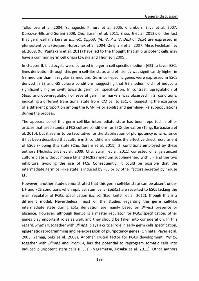
General discussion
193
Tolkunova et al. 2004, Yamaguchi, Kimura et al. 2005, Chambers, Silva et al. 2007,
Durcova-Hills and Surani 2008, Chu, Surani et al. 2011, Zhao, Ji et al. 2012), or the fact
that germ-cell markers as Blimp1, Dppa3, Ifitm3, Piwil2, Dazl or Ddx4 are expressed in
pluripotent cells (Geijsen, Horoschak et al. 2004, Qing, Shi et al. 2007, Mise, Fuchikami et
al. 2008, Xu, Pantakani et al. 2011) have led to the thought that all pluripotent cells may
have a common germ cell origin (Zwaka and Thomson 2005).
In chapter II, blastocysts were cultured in a germ cell-specific medium (GS) to favor ESCs
lines derivation through this germ cell-like state, and efficiency was significantly higher in
GS medium than in regular ES medium. Germ cell-specific genes were expressed in ESCs
derived in ES and GS culture conditions, suggesting that GS medium did not induce a
significantly higher swift towards germ cell specification. In contrast, upregulation of
Stella and downregulation of several germline markers was observed in 2i conditions,
indicating a different transitional state from ICM cell to ESC, or suggesting the existence
of a different proportion among the ICM-like or epiblst and germline-like subpopulations
during the process.
The appearance of this germ cell-like intermediate state has been reported in other
articles that used standard FCS culture conditions for ESCs derivation (Tang, Barbacioru et
al. 2010), but it seems to be facultative for the stabilization of pluripotency in vitro, since
it has been described that culture in 2i conditions enables the effective direct recruitment
of ESCs skipping this state (Chu, Surani et al. 2011). 2i conditions employed by these
authors (Nichols, Silva et al. 2009, Chu, Surani et al. 2011) consisted of a gelatinized
culture plate without mouse EF and N2B27 medium supplemented with LIF and the two
inhibitors, avoiding the use of FCS. Consequently, it could be possible that the
intermediate germ cell-like state is induced by FCS or by other factors secreted by mouse
EF.
However, another study demonstrated that this germ cell-like state can be absent under
LIF and FCS conditions when epiblast stem cells (EpiSCs) are reverted to ESCs lacking the
main regulator of PGCs specification Blimp1 (Bao, Leitch et al. 2012), though this is a
different model. Nevertheless, most of the studies regarding the germ cell-like
intermediate state during ESCs derivation are mainly based on Blimp1 presence or
absence. However, although Blimp1 is a master regulator for PGCs specification, other
genes play important roles as well, and they should be taken into consideration. In this
regard, Prdm14, together with Blimp1, plays a critical role in early germ cells specification,
epigenetic reprogramming and re-expression of pluripotency genes (Ohinata, Payer et al.
2005, Yamaji, Seki et al. 2008). Another crucial factor for PGCs development, Prmt5,
together with Blimp1 and Prdm14, has the potential to reprogram somatic cells into
induced pluripotent stem cells (iPSCs) (Nagamatsu, Kosaka et al. 2011). Other authors

General discussion
194
demonstrated that Prdm14 accelerates the EpiSCs reprogramming process to ESCs when
expressed in combination with Klf2 in classical culture conditions with FCS, and induces
the expression of several germline-associated genes as Blimp1, Dppa3, Ifitm3 and
Nanos3, indicating that the reprogramming process may require progression through the
germ cell-like intermediate. However, reprogramming was not affected in the absence of
Blimp1 (Gillich, Bao et al. 2012). Thus, it is possible that Prdm14 and Blimp1 have
redundant activities during the establishment of the pluripotent state (Geijsen 2012).
Prdm14 also ensures naïve pluripotency in ESCs in 2i conditions by antagonizing FGF
receptor signaling and by repressing expression of de novo DNA methyltransferases that
modify the epigenome to a primed state (Yamaji, Ueda et al. 2013).
Studies reporting that germline, premeiotic or meiotic markers such as Blimp1, Dppa3,
Ifitm3, Piwil2, Dazl, Ddx4, Stra8, Rnf17, Rnh2, Sycp3, Pgk2 or Creb3/4 are expressed in
ESCs or in other pluripotent populations as iPSCs, GSCs and EGCs, were performed under
classical culture conditions over a mouse EF feeder layer and medium supplemented with
LIF and FCS (Geijsen, Horoschak et al. 2004, Qing, Shi et al. 2007, Mise, Fuchikami et al.
2008, Tang, Barbacioru et al. 2010, Xu, Pantakani et al. 2011). Consequently, according to
our results in chapter II and to those reported by other authors, it seems that during ESCs
derivation, cells undergo a germ cell-like intermediate state under commonly used
culture conditions, although pluripotency can be directly captured from the epiblast by
MEK and GSK3β inhibitors supplementation without FCS and EF feeder layer.
Thus, ESCs in serum conditions consist of a mixed population of cells with naïve ICM-like
properties and cells with primed epiblast or germline-like properties, and these two
populations would interchange their identities during culture maintaining a dynamic
equilibrium, where Prdm14 is associated with the naïve ICM-like properties (Yamaji, Ueda
et al. 2013). By the contrary, ESCs in 2i conditions appear to be homogeneous and have a
gene expression profile and epigenome different to ESCs cultured in serum conditions
and more similar to those of ICM cells (Ying, Wray et al. 2008, Marks, Kalkan et al. 2012).
Our results also show that ES and GS media produce a higher expression of some
germinal markers during the transition from blastocyst embryonary cells to ESCs, while 2i
medium produces an upregulation of Stella, suggesting that it could exist a different
proportion of the subpopulations present in ESCs, prevailing the epiblast or germline-like
subpopulation in ES or GS culture conditions, while the ICM-like subpopulation would
prevail in 2i conditions. It has been described that culture conditions that preserve ESCs
pluripotency would maintain a balanced proportion among both subpopulations (Hayashi,
Lopes et al. 2008).
In vitro cultured pluripotent cells are not the only ones that seem to retain a germinal
origin, as germline-specific genes have been localized in vivo in different tissues holding
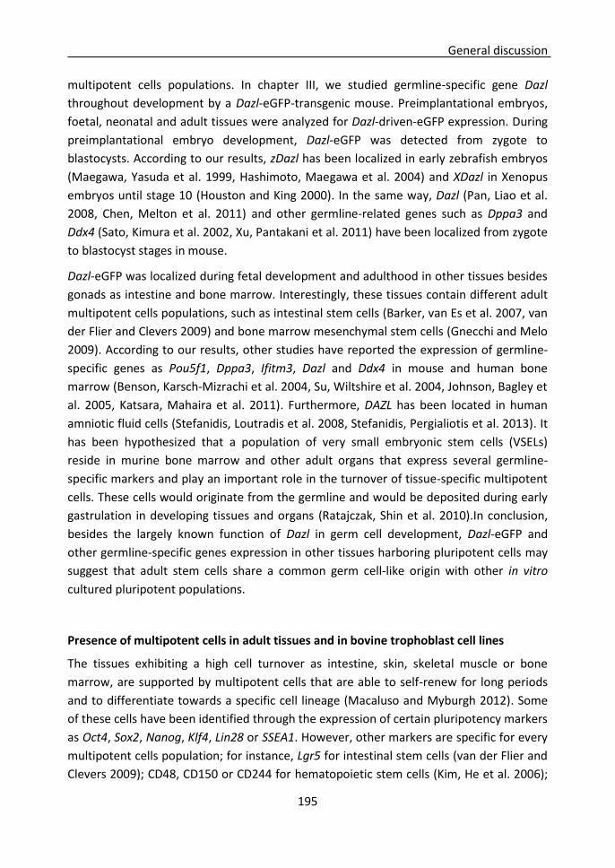
General discussion
195
multipotent cells populations. In chapter III, we studied germline-specific gene Dazl
throughout development by a Dazl-eGFP-transgenic mouse. Preimplantational embryos,
foetal, neonatal and adult tissues were analyzed for Dazl-driven-eGFP expression. During
preimplantational embryo development, Dazl-eGFP was detected from zygote to
blastocysts. According to our results, zDazl has been localized in early zebrafish embryos
(Maegawa, Yasuda et al. 1999, Hashimoto, Maegawa et al. 2004) and XDazl in Xenopus
embryos until stage 10 (Houston and King 2000). In the same way, Dazl (Pan, Liao et al.
2008, Chen, Melton et al. 2011) and other germline-related genes such as Dppa3 and
Ddx4 (Sato, Kimura et al. 2002, Xu, Pantakani et al. 2011) have been localized from zygote
to blastocyst stages in mouse.
Dazl-eGFP was localized during fetal development and adulthood in other tissues besides
gonads as intestine and bone marrow. Interestingly, these tissues contain different adult
multipotent cells populations, such as intestinal stem cells (Barker, van Es et al. 2007, van
der Flier and Clevers 2009) and bone marrow mesenchymal stem cells (Gnecchi and Melo
2009). According to our results, other studies have reported the expression of germline-
specific genes as Pou5f1, Dppa3, Ifitm3, Dazl and Ddx4 in mouse and human bone
marrow (Benson, Karsch-Mizrachi et al. 2004, Su, Wiltshire et al. 2004, Johnson, Bagley et
al. 2005, Katsara, Mahaira et al. 2011). Furthermore, DAZL has been located in human
amniotic fluid cells (Stefanidis, Loutradis et al. 2008, Stefanidis, Pergialiotis et al. 2013). It
has been hypothesized that a population of very small embryonic stem cells (VSELs)
reside in murine bone marrow and other adult organs that express several germline-
specific markers and play an important role in the turnover of tissue-specific multipotent
cells. These cells would originate from the germline and would be deposited during early
gastrulation in developing tissues and organs (Ratajczak, Shin et al. 2010).In conclusion,
besides the largely known function of Dazl in germ cell development, Dazl-eGFP and
other germline-specific genes expression in other tissues harboring pluripotent cells may
suggest that adult stem cells share a common germ cell-like origin with other in vitro
cultured pluripotent populations.
Presence of multipotent cells in adult tissues and in bovine trophoblast cell lines
The tissues exhibiting a high cell turnover as intestine, skin, skeletal muscle or bone
marrow, are supported by multipotent cells that are able to self-renew for long periods
and to differentiate towards a specific cell lineage (Macaluso and Myburgh 2012). Some
of these cells have been identified through the expression of certain pluripotency markers
as Oct4, Sox2, Nanog, Klf4, Lin28 or SSEA1. However, other markers are specific for every
multipotent cells population; for instance, Lgr5 for intestinal stem cells (van der Flier and
Clevers 2009); CD48, CD150 or CD244 for hematopoietic stem cells (Kim, He et al. 2006);
General discussion
196
CD133, CD15 or CD24 for neural stem cells (Rietze, Valcanis et al. 2001, Capela and
Temple 2002, Corti, Nizzardo et al. 2007, Peh, Lang et al. 2009); and CD10, CD13, CD73,
CD105 or CD271 for mesenchymal stem cells (Mafi, Hindocha et al. 2011).
Furthermore, some germline-specific markers such as Dppa3, Ifitm3, Dazl and Ddx4 have
been localized in tissues harboring multipotent cells (Benson, Karsch-Mizrachi et al. 2004,
Su, Wiltshire et al. 2004, Johnson, Bagley et al. 2005, Stefanidis, Loutradis et al. 2008,
Katsara, Mahaira et al. 2011, Stefanidis, Pergialiotis et al. 2013). Similarly, in chapter III
Dazl-eGFP expression was found in bone marrow and intestine, probably due to the
presence of hematopoietic and mesenchymal stem cells residing in bone marrow, and to
intestinal stem cells located in the crypts of Lieberhahn (Schabort, Myburgh et al. 2009).
Therefore, Dazl-GFP transgene could be used to explore the presence of multipotent cells
in different tissues.
Trophoblast stem cells (TSCs) represent another multipotent cells population residing in
the trophectoderm (TE) (Tanaka, Kunath et al. 1998). However, TSCs have been isolated
and well characterized only in mouse and rhesus macaque (Vandevoort, Thirkill et al.
2007, Roberts and Fisher 2011). In contrast, the existence and location of a TSCs niche in
domestic species remains unknown, in part due to a different TE development (Bindon
1971, Geisert, Brookbank et al. 1982, Thatcher, Meyer et al. 1995). Consequently, TSCs
have not been isolated in these species (Roberts and Fisher 2011); although primary
trophectoderm cell lines have been derived from sheep and goat (Miyazaki, Imai et al.
2002), pig (Ramsoondar, Christopherson et al. 1993, Flechon, Laurie et al. 1995, La
Bonnardiere, Flechon et al. 2002) and cattle (Talbot, Caperna et al. 2000, Shimada,
Nakano et al. 2001).
Interestingly, bovine trophoblast cell lines obtained in chapter IV showed a long-term
steady cell proliferation in vitro, and expressed several pluripotency-related genes as
POU5F1, CDH1, FGF4, TERT, and TERC. According to our results, other studies have
suggested that trophoblastic cells are not terminal cells and present some pluripotency
features (Suzuki, Koshi et al. 2011, Schiffmacher and Keefer 2013). Thus, a population of
multipotent cells could be present also in bovine trophoblastic cell lines.

Discusión general
197
Discusión general

Discusión general
198
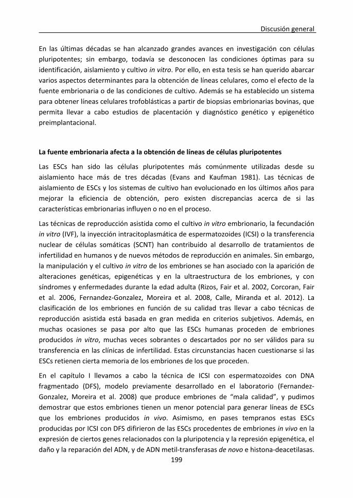
Discusión general
199
En las últimas décadas se han alcanzado grandes avances en investigación con células
pluripotentes; sin embargo, todavía se desconocen las condiciones óptimas para su
identificación, aislamiento y cultivo in vitro. Por ello, en esta tesis se han querido abarcar
varios aspectos determinantes para la obtención de líneas celulares, como el efecto de la
fuente embrionaria o de las condiciones de cultivo. Además se ha establecido un sistema
para obtener líneas celulares trofoblásticas a partir de biopsias embrionarias bovinas, que
permita llevar a cabo estudios de placentación y diagnóstico genético y epigenético
preimplantacional.
La fuente embrionaria afecta a la obtención de líneas de células pluripotentes
Las ESCs han sido las células pluripotentes más comúnmente utilizadas desde su
aislamiento hace más de tres décadas (Evans and Kaufman 1981). Las técnicas de
aislamiento de ESCs y los sistemas de cultivo han evolucionado en los últimos años para
mejorar la eficiencia de obtención, pero existen discrepancias acerca de si las
características embrionarias influyen o no en el proceso.
Las técnicas de reproducción asistida como el cultivo in vitro embrionario, la fecundación
in vitro (IVF), la inyección intracitoplasmática de espermatozoides (ICSI) o la transferencia
nuclear de células somáticas (SCNT) han contribuido al desarrollo de tratamientos de
infertilidad en humanos y de nuevos métodos de reproducción en animales. Sin embargo,
la manipulación y el cultivo in vitro de los embriones se han asociado con la aparición de
alteraciones genéticas, epigenéticas y en la ultraestructura de los embriones, y con
síndromes y enfermedades durante la edad adulta (Rizos, Fair et al. 2002, Corcoran, Fair
et al. 2006, Fernandez-Gonzalez, Moreira et al. 2008, Calle, Miranda et al. 2012). La
clasificación de los embriones en función de su calidad tras llevar a cabo técnicas de
reproducción asistida está basada en gran medida en criterios subjetivos. Además, en
muchas ocasiones se pasa por alto que las ESCs humanas proceden de embriones
producidos in vitro, muchas veces sobrantes o descartados por no ser válidos para su
transferencia en las clínicas de infertilidad. Estas circunstancias hacen cuestionarse si las
ESCs retienen cierta memoria de los embriones de los que proceden.
En el capítulo I llevamos a cabo la técnica de ICSI con espermatozoides con DNA
fragmentado (DFS), modelo previamente desarrollado en el laboratorio (Fernandez-
Gonzalez, Moreira et al. 2008) que produce embriones de “mala calidad”, y pudimos
demostrar que estos embriones tienen un menor potencial para generar líneas de ESCs
que los embriones producidos in vivo. Asimismo, en pases tempranos estas ESCs
producidas por ICSI con DFS difirieron de las ESCs procedentes de embriones in vivo en la
expresión de ciertos genes relacionados con la pluripotencia y la represión epigenética, el
daño y la reparación del ADN, y de ADN metil-transferasas de novo e histona-deacetilasas.
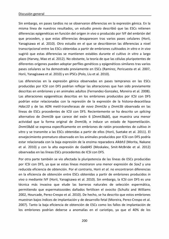
Discusión general
200
Sin embargo, en pases tardíos no se observaron diferencias en la expresión génica. En la
misma línea de nuestros resultados, un estudio previo describió que las ESCs retienen
diferencias epigenéticas en función del origen in vivo o producido por IVF del embrión del
que proceden, y que estas diferencias desaparecen tras varios pases celulares (Horii,
Yanagisawa et al. 2010). Otro estudio en el que se describieron las diferencias a nivel
transcripcional entre las ESCs obtenidas a partir de embriones cultivados in vitro e in vivo
sugirió que estas diferencias se mantienen estables durante el cultivo in vitro a largo
plazo (Harvey, Mao et al. 2012). No obstante, la teoría de que las células pluripotentes de
diferentes orígenes pueden adoptar perfiles genéticos y epigenéticos similares tras varios
pases celulares se ha demostrado previamente en ESCs (Ramirez, Pericuesta et al. 2007,
Horii, Yanagisawa et al. 2010) y en iPSCs (Polo, Liu et al. 2010).
Las diferencias en la expresión génica observadas en pases tempranos en las ESCs
producidas por ICSI con DFS podrían reflejar las alteraciones que han sido previamente
descritas en embriones y en animales adultos (Fernandez-Gonzalez, Moreira et al. 2008).
Las alteraciones epigenéticas descritas en los embriones producidos por ICSI con DFS
podrían estar relacionadas con la represión de la expresión de la histona-deacetilasa
Hdac10 y de las ADN metil-transferasas de novo Dnmt3a y Dnmt3b observada en las
líneas de ESCs procedentes de ICSI con DFS. Recientemente se ha descrito un splicing
alternativo de Dnmt3b que carece del exón 6 (Dnmt3bΔ6), que muestra una menor
actividad que la forma original de Dnmt3b, e induce un estado de hipometilación.
Dnmt3bΔ6 se expresa específicamente en embriones de ratón procedentes de cultivo in
vitro y se transmite a las ESCs obtenidas a partir de ellos (Horii, Suetake et al. 2011). El
envejecimiento prematuro observado en los animales producidos por ICSI con DFS podría
estar relacionada con la baja expresión de la enzima reparadora Alkbh3 (Morita, Nakane
et al. 2010) y con la alta expresión de Gadd45 (Moskalev, Smit-McBride et al. 2012)
observadas en las líneas ESCs procedentes de ICSI con DFS.
Por otra parte también se vio afectada la pluripotencia de las líneas de ESCs producidas
por ICSI con DFS, ya que se estas líneas mostraron una menor expresión de Sox2 y una
reducida eficiencia de obtención. Por el contrario, Horii et al. no encontraron diferencias
en la eficiencia de obtención entre ESCs obtenidas a partir de embriones producidos in
vivo o mediante IVF (Horii, Yanagisawa et al. 2010). Sin embargo, la ICSI con DFS es una
técnica más invasiva que elude las barreras naturales de selección espermática,
permitiendo que espermatozoides dañados fertilicen el ovocito (Schultz and Williams
2002, Hourcade, Perez-Crespo et al. 2010). De hecho, se ha descrito que estos embriones
muestran bajos índices de implantación y de desarrollo fetal (Moreira, Perez-Crespo et al.
2007). Tanto la baja eficiencia de obtención de ESCs como los fallos de implantación de
los embriones podrían deberse a anomalías en el cariotipo, ya que el 40% de los

Discusión general
201
embriones producidos por ICSI con DFS muestran fragmentación cromosómica y una
segregación cromosómica anormal (Yamagata, Suetsugu et al. 2009).
Por lo tanto, las características o alteraciones embrionarias se ven reflejadas en sus
correspondientes líneas de ESCs; sin embargo, el cultivo a largo plazo hace que adopten
perfiles similares.
Las características embrionarias podrían verse reflejadas también en otras líneas
celulares. Curiosamente, en el capítulo IV, tras desarrollar un sistema para establecer de
manera eficiente líneas de células trofoblásticas bovinas a partir de biopsias
embrionarias, pudimos observar una gran variabilidad en la expresión génica en pases
tempranos de líneas celulares procedentes de las mismas condiciones de obtención, por
lo que las diferencias podrían tener su origen en la fuente embrionaria. Este hallazgo se
corresponde con lo observado en un estudio previo en el que se obtuvieron 12 líneas de
células de trofoblásticas bovinas y se cuantificó la expresión de los genes específicos de
células trofoblásticas: interferón tau (IFNt), lactógeno placentario (CSH1), proteína
relacionada con la prolactina 1 (PRP1) y glicoproteína asociada a la gestación (PAG1);
encontrando diferentes patrones de expresión entre las líneas celulares sin relación
alguna con las condiciones de cultivo (Suzuki, Koshi et al. 2011). Por lo tanto, en el
capítulo IV se demostró que las características embrionarias afectan también a la
obtención de líneas de células trofoblásticas bovinas.
Las condiciones de cultivo afectan a la obtención de líneas celulares
Las condiciones de cultivo son un factor fundamental que afecta a la obtención de líneas
celulares y a sus características. En esta tesis, el efecto de las condiciones de cultivo se ha
analizado en líneas de ESCs en el capítulo II, y en líneas de células trofoblásticas en el
capítulo IV.
En el capítulo II, se analizó el efecto de la adición de “leukemia inhibitory factor” (LIF) al
medio durante el cultivo del embrión preimplantacional hasta el estadio de blastocisto
para su posterior uso en la obtención de ESCs, debido a la información contradictoria
procedente de estudios previos (Jurisicova, Ben-Chetrit et al. 1995, Dunglison, Barlow et
al. 1996, Tsai, Chang et al. 2000, Rungsiwiwut, Rungarunlert et al. 2008). A pesar de que
los embriones cultivados con LIF mostraron un menor número total de células, el índice
de células de la masa celular interna (ICM)/células totales fue significativamente mayor
en estos embriones. Además, resultaron ser más aptos para el aislamiento de ESCs, ya
que mostraron una mayor eficiencia de obtención. Asimismo se suplementó el medio
tradicional para el cultivo de ESCs con factores de cultivo normalmente usados para el
cultivo de células germinales embrionarias (GSCs) (EGF, GDNF y bFGF), y la eficiencia de

Discusión general
202
obtención de ESCs también se vio incrementada. En concordancia con nuestro estudio, el
efecto beneficioso de estos factores en otras poblaciones de células pluripotentes ha sido
previamente demostrado (Reynolds and Weiss 1992, Donovan and de Miguel 2003,
Kanatsu-Shinohara, Ogonuki et al. 2003).
El enorme efecto de suplementar el medio de cultivo con diferentes factores ha sido
ampliamente estudiado en las ESCs. En el capítulo II, las ESCs fueron obtenidas sobre una
monocapa de fibroblastos embrionarios de ratón en diferentes medios suplementados
con: suero fetal bovino (FCS) y LIF (medio ES); FCS, LIF y factores de crecimiento de GSCs
(medio GS); o FCS, LIF y factores inhibidores (medio 2i). Se observó un incremento en la
eficiencia de obtención de ESCs en el medio GS en comparación con el medio ES, y las
líneas de ESCs obtenidas en medio GS cumplieron los criterios de pluripotencia y
mostraron una mayor capacidad de formación de ratones quiméricos. Se manifiesta por
tanto una posible sinergia entre la suplementación de LIF durante el cultivo embrionario
in vitro y la adición posterior de otros factores de crecimiento presentes en el medio GS,
que favorece la obtención de nuevas líneas de ESCs. Diferentes sistemas de cultivo como
el “knockout serum replacement” (KSR) (Cheng, Dutra et al. 2004) y los inhibidores (MEK
y GSK3β; 2i) han sido desarrollados para evitar la variabilidad inducida por el uso de FCS,
ya que existen grandes variaciones entre lotes (Ying, Wray et al. 2008). El sistema 2i
permite la eficiente obtención y expansión de ESCs capaces de contribuir al linaje
germinal en animales quiméricos en diferentes cepas de ratones y en especies en las que
previamente no había sido posible (Buehr, Meek et al. 2008, Nichols, Jones et al. 2009).
Por lo tanto, la obtención de ESCs depende enormemente del medio de cultivo. Del
mismo modo, el transcriptoma está definido fundamentalmente por las condiciones de
cultivo. En el capítulo II, la expresión génica de las líneas de ESCs fue diferente en función
del medio de cultivo utilizado para su obtención (ES, GS y 2i). De hecho, se ha descrito
que las condiciones de cultivo son el aspecto que más condiciona la expresión génica, por
encima del origen embrionario y del procedimiento de obtención de las ESCs (Marks,
Kalkan et al. 2012, Leitch, McEwen et al. 2013).
En el capítulo IV se utilizaron diferentes medios de cultivo con el objetivo de mejorar la
eficiencia de obtención de líneas de células trofoblásticas bovinas. A pesar de que
observamos que una rápida adhesión del embrión o biopsia a la placa de cultivo es
primordial, e incrementa significativamente la eficiencia de obtención de líneas celulares,
el medio de cultivo es decisivo también. Los medios de cultivo condicionados contienen
diferentes proteínas secretadas que incluyen enzimas, factores de crecimiento, citoquinas
y hormonas (Dowling and Clynes 2011). En muchos de los estudios en los que se
obtuvieron líneas de células trofoblásticas bovinas se empleó medio condicionado al
llevar a cabo un co-cultivo con fibroblastos embrionarios murinos (Talbot, Caperna et al.
2000, Talbot, Caperna et al. 2004, Talbot, Powell et al. 2007). Sin embargo, el co-cultivo

Discusión general
203
tiene como inconveniente la contaminación de las líneas celulares de interés con otro
tipo celular, por lo que en nuestro trabajo las células trofoblásticas fueron cultivadas en
un sistema en microgota gelatinizada, utilizando diferentes medios de cultivo
condicionados por fibroblastos embrionarios murinos (Cm), fibroblastos embrionarios
bovinos (Cb) y células oviductales ovinas (Co). A excepción del medio Cb, los medios
condicionados mejoraron la eficiencia, siendo el medio Cm el más apropiado para la
obtención de líneas de células trofoblásticas. En otros artículos anteriores se había usado
medio condicionado por fibroblastos endometriales bovinos para obtener líneas de
células trofoblásticas (Shimada, Nakano et al. 2001, Hashizume, Shimada et al. 2006); sin
embargo nuestro medio de cultivo heterólogo condicionado por fibroblastos
embrionarios murinos fue el más eficaz, permitiendo una rápida adhesión del embrión o
biopsia, y el posterior crecimiento celular. Además, los medios de cultivo fueron
suplementados con EGF, ya que estudios anteriores habían demostrado su efecto positivo
en la proliferación de células trofoblásticas (Hambruch, Haeger et al. 2010). En
conclusión, nuestras observaciones junto con las de otros autores indican que la
eficiencia de obtención de líneas de células trofoblásticas bovinas depende en gran
medida de las condiciones de cultivo.
Un origen germinal para las células pluripotentes
Diversas evidencias como la similitud existente entre las ESCs y las EGCs (Matsui, Zsebo et
al. 1992, Resnick, Bixler et al. 1992), la expresión de diversos genes de pluripotencia como
Pou5f1, Sox2 y Nanog en células primordiales germinales (PGCs) (Kehler, Tolkunova et al.
2004, Yamaguchi, Kimura et al. 2005, Chambers, Silva et al. 2007, Durcova-Hills and
Surani 2008, Chu, Surani et al. 2011, Zhao, Ji et al. 2012), o el hecho de que marcadores
específicos de linaje germinal como Blimp1, Dppa3, Ifitm3, Piwil2, Dazl o Ddx4 se
expresen en células pluripotentes (Geijsen, Horoschak et al. 2004, Qing, Shi et al. 2007,
Mise, Fuchikami et al. 2008, Xu, Pantakani et al. 2011) han llevado a pensar que todas las
células pluripotentes podrían tener un origen germinal común (Zwaka and Thomson
2005).
En el capítulo II se cultivaron blastocistos en un medio específico para células germinales
(GS) para favorecer la obtención de líneas de ESCs a través de este posible estado
transitorio similar a una célula germinal, y la eficiencia de obtención fue
significativamente mayor en el medio GS que en el medio ES. Sin embargo, no pudimos
encontrar indicios de una diferenciación más pronunciada hacia el linaje germinal
inducida por el medio GS, ya que encontramos expresión de genes específicos de linaje
germinal en todas las condiciones de cultivo (GS, ES y 2i), lo que apunta a la aparición de
este estado similar a una célula germinal durante el proceso de obtención de ESCs.

Discusión general
204
La aparición de este intermediario común se ha descrito en otros artículos que utilizaron
condiciones de cultivo estándar con FCS para la obtención de ESCs (Tang, Barbacioru et al.
2010), pero parece no ser estrictamente necesario para la estabilización de la
pluripotencia in vitro, ya que el cultivo en condiciones 2i permite el reclutamiento directo
de ESCs de manera efectiva sorteando este estado similar a una célula germinal (Chu,
Surani et al. 2011). En nuestro estudio, los inhibidores de MEK y GSK3β fueron usados
junto con FCS y LIF sobre una monocapa de EF de ratón. Por el contrario, las condiciones
de cultivo 2i utilizadas por otros autores (Nichols, Silva et al. 2009, Chu, Surani et al. 2011)
consistieron en una placa de cultivo gelatinizada sin EF murinos y medio N2B27
suplementado con LIF y los inhibidores de MEK y GSK3β, evitando el uso de FCS.
Consecuentemente, es posible que el estado intermedio similar a una célula germinal
esté inducido por el FCS o por otros factores secretados por los EF murinos.
Sin embargo, un estudio ha demostrado que este estado similar a una célula germinal
puede estar ausente también en condiciones de cultivo con FCS y LIF cuando células
troncales del epiblasto (EpiSCs) carentes del principal regulador de la especificación de las
PGCs, Blimp1, son revertidas a ESCs (Bao, Leitch et al. 2012). No obstante, la mayoría de
los autores que han investigado la existencia de este estado similar a una célula germinal
durante la obtención de ESCs se han basado principalmente en la presencia o ausencia de
Blimp1. Es cierto que Blimp1 es fundamental durante la especificación de las PGCs, pero
existen otros genes que también juegan papeles elementales y deberían ser analizados
igualmente. De hecho, Prdm14, junto con Blimp1, juega un papel esencial en la
especificación temprana de las células germinales (Ohinata, Payer et al. 2005, Yamaji, Seki
et al. 2008). Otro factor fundamental para el desarrollo de las PGCs, Prmt5, junto con
Blimp1 y Prdm14, tiene el potencial de reprogramar células somáticas en iPSCs.
(Nagamatsu, Kosaka et al. 2011). Otros autores han demostrado que Prdm14 acelera la
reprogramación de EpiSCs hacia ESCs cuando se expresa en combinación con Klf2 en
condiciones de cultivo con FCS, e induce la expresión de diversos genes asociados con el
linaje germinal como Blimp1, Dppa3, Ifitm3 y Nanos3, indicando que este proceso de
reprogramación también podría requerir un paso intermedio similar a una célula
germinal. Sin embargo, la ausencia de Blimp1 no afecta a dicho proceso de
reprogramación (Gillich, Bao et al. 2012). Por ello, es posible que Prdm14 y Blimp1 tengan
una actividad redundante durante el establecimiento de la pluripotencia (Geijsen 2012).
Prdm14 también garantiza el estado de pluripotencia naïve en ESCs al antagonizar la
señalización del receptor de FGF y reprimir la expresión de ADN metil-transferasas de
novo que modifican el epigenoma hacia un estado primed (Yamaji, Ueda et al. 2013).
Los estudios que muestran que los marcadores de linaje germinal, premeióticos o
meióticos como Blimp1, Dppa3, Ifitm3, Piwil2, Dazl, Ddx4, Stra8, Rnf17, Rnh2, Sycp3, Pgk2
o Creb3/4 se expresan en ESCs o en otras poblaciones de células pluripotentes como las

Discusión general
205
iPSCs, las GSCs o las EGCs, se han llevado a cabo en condiciones clásicas de cultivo con
FCS (Geijsen, Horoschak et al. 2004, Qing, Shi et al. 2007, Mise, Fuchikami et al. 2008,
Tang, Barbacioru et al. 2010, Xu, Pantakani et al. 2011). Consecuentemente, de acuerdo
con los resultados descritos en el capítulo II de esta tesis y con aquellos descritos por
otros autores, parece que durante la obtención de ESCs bajo ciertas condiciones de
cultivo, las células pasan por un estado intermedio similar a una célula germinal, aunque
la pluripotencia puede ser capturada directamente del epiblasto gracias a los inhibidores
de MEK and GSK3β pero sin utilizar FCS ni una monocapa de fibroblastos murinos.
Por lo tanto, las ESCs cultivadas con FCS están constituidas por una población mixta de
células con propiedades naïve similares a las células de la ICM, y células con propiedades
primed similares a las del epiblasto; y ambas poblaciones intercambiarían sus identidades
durante el cultivo manteniendo un equilibrio dinámico, estando Prdm14 asociado con las
propiedades naïve de la ICM (Yamaji, Ueda et al. 2013). Por el contrario, las ESCs en
condiciones 2i parecen ser una población homogénea que tiene un perfil de expresión
génica y un epigenoma diferente a los de las ESCs cultivadas con FCS y similar a los de las
células de la ICM (Ying, Wray et al. 2008, Marks, Kalkan et al. 2012). Nuestros resultados
muestran además cómo los medios ES y GS producen una mayor expresión de algunos
marcadores germinales durante la transición de las células embrionarias del blastocisto a
ESCs, mientras que el medio 2i produce un aumento de Stella, sugiriendo que existe una
distinta proporción de las subpoblaciones presentes en las ESCs, siendo predominante la
subpoblación similar al epiblasto o a la línea germinal en las condiciones de cultivo ES o
GS, mientras que en condiciones 2i sería predominante la subpoblación similar a la ICM.
Se ha descrito que durante el cultivo en condiciones que mantienen la pluripotencia de
las ESCs, las proporciones ambas subpoblaciones permanecen en equilibrio (Hayashi,
Lopes et al. 2008).
No sólo las células pluripotentes cultivadas in vitro parecen tener un origen germinal, ya
que también se han localizado genes específicos de linaje germinal in vivo en diferentes
tejidos que albergan poblaciones de células pluripotentes. En el capítulo III se ha
estudiado el gen específico de linaje germinal Dazl a lo largo del desarrollo mediante un
ratón transgénico Dazl-eGFP, en el que se analizó la expresión de eGFP inducida por Dazl
en el desarrollo embrionario y en los tejidos fetales, neonatales y adultos. Durante el
desarrollo preimplantacional embrionario, la expresión de Dazl-eGFP fue detectada desde
el estadio de cigoto hasta el de blastocisto. En consonancia con nuestros resultados, se ha
descrito la expresión de zDazl en embriones tempranos de pez cebra (Maegawa, Yasuda
et al. 1999, Hashimoto, Maegawa et al. 2004) y de XDazl en embriones de Xenopus hasta
el estadio 10 (Houston and King 2000). Del mismo modo, en el ratón se ha localizado Dazl
Discusión general
206
entre los estadios de cigoto y blastocisto (Pan, Liao et al. 2008, Chen, Melton et al. 2011),
así como otros genes relacionados con el linaje germinal, como Dppa3 y Ddx4 (Sato,
Kimura et al. 2002, Xu, Pantakani et al. 2011).
La expresión de Dazl-eGFP fue encontrada durante el desarrollo fetal y la edad adulta en
otros tejidos además de las gónadas, como son el intestino y la médula ósea.
Curiosamente, en estos tejidos residen diferentes poblaciones de células pluripotentes
adultas como las células troncales intestinales (Barker, van Es et al. 2007, van der Flier
and Clevers 2009) y las células troncales mesenquimales de la médula ósea (Gnecchi and
Melo 2009). De acuerdo con nuestros resultados, otros estudios han descrito la expresión
de genes específicos de linaje germinal como Pou5f1, Dppa3, Ifitm3, Dazl y Ddx4 en la
médula ósea en humanos y en ratones (Benson, Karsch-Mizrachi et al. 2004, Su, Wiltshire
et al. 2004, Johnson, Bagley et al. 2005, Katsara, Mahaira et al. 2011). Además, DAZL ha
sido localizado en células del fluido amniótico en humanos (Stefanidis, Loutradis et al.
2008, Stefanidis, Pergialiotis et al. 2013). Se ha propuesto que una población de células
troncales embrionarias muy pequeñas (“very small embryonic stem cells”, VSELs) reside
en la médula ósea y en otros órganos adultos en el ratón y expresa varios marcadores
específicos del linaje germinal. Estas células jugarían un papel funcamental en el
reemplazo de las células multipotentes específicas de cada tejido y se originarían a partir
del linaje germinal, siendo depositadas durante la gastrulación en los tejidos y órganos en
desarrollo (Ratajczak, Shin et al. 2010).
En conclusión, además de la ampliamente conocida función de Dazl en el desarrollo de las
células germinales, la expresión de Dazl-eGFP y de otros genes específicos del linaje
germinal en otros tejidos que albergan células pluripotentes podría sugerir que las células
troncales adultas comparten un origen común similar a una célula germinal con otras
poblaciones de células pluripotentes cultivadas in vitro como las ESCs.
Presencia de células multipotentes en tejidos adultos y en líneas de células
trofoblásticas bovinas
Algunos tejidos con una alta tasa de renovación celular como el intestino, la piel, el
músculo esquelético o la médula ósea, son mantenidos por células multipotentes capaces
de auto-renovarse durante largos periodos de tiempo, y de diferenciarse hacia un linaje
celular específico (Macaluso and Myburgh 2012). Algunas de estas células han sido
identificadas gracias a la expresión de marcadores de pluripotencia ampliamente
conocidos como Oct4, Sox2, Nanog, Klf4, Lin28 o SSEA1. Sin embargo, existen otros
marcadores que son específicos de cada población de células multipotentes; por ejemplo,
Lgr5 de células troncales intestinales (van der Flier and Clevers 2009); CD48, CD150 o
CD244 de células troncales hematopoyéticas (Kim, He et al. 2006); CD133, CD15 o CD24

Discusión general
207
de células troncales neuronales (Rietze, Valcanis et al. 2001, Capela and Temple 2002,
Corti, Nizzardo et al. 2007, Peh, Lang et al. 2009); y CD10, CD13, CD73, CD105 o CD271 de
células troncales mesenquimales (Mafi, Hindocha et al. 2011).
Además, algunos marcadores específicos del linaje germinal como Dppa3, Ifitm3, Dazl y
Ddx4 se han localizado en tejidos que albergan células multipotentes (Benson, Karsch-
Mizrachi et al. 2004, Su, Wiltshire et al. 2004, Johnson, Bagley et al. 2005, Stefanidis,
Loutradis et al. 2008, Katsara, Mahaira et al. 2011, Stefanidis, Pergialiotis et al. 2013). De
la misma manera, en el capítulo III se encontró expresión de Dazl-GFP en médula ósea e
intestino; y esto probablemente sea debido a las células troncales hematopoyéticas y
mesenquimales que residen en la médula ósea, y a las células troncales intestinales
localizadas en las criptas de Lieberhahn (Schabort, Myburgh et al. 2009). Por lo tanto, el
transgen Dazl-eGFP podría usarse para analizar la presencia de células multipotentes en
diferentes tejidos.
Las células troncales trofoblásticas (TSCs) conforman otra población de células
multipotentes localizada en el trofoectodermo (TE) (Tanaka, Kunath et al. 1998). Sin
embargo, las TSCs sólo se han aislado y caracterizado en profundidad en el ratón y en el
macaco Rhesus (Vandevoort, Thirkill et al. 2007, Roberts and Fisher 2011). Por el
contrario, en especies domésticas, debido a las diferencias en el desarrollo del TE (Bindon
1971, Geisert, Brookbank et al. 1982, Thatcher, Meyer et al. 1995), se desconoce la
existencia y la localización de un nicho de TSCs. En consecuencia, las TSCs no han podido
ser aisladas (Roberts and Fisher 2011), aunque se han obtenido líneas de células
trofoblásticas en oveja y cabra (Miyazaki, Imai et al. 2002), cerdo (Ramsoondar,
Christopherson et al. 1993, Flechon, Laurie et al. 1995, La Bonnardiere, Flechon et al.
2002) y bovino (Talbot, Caperna et al. 2000, Shimada, Nakano et al. 2001).
Curiosamente, las líneas de células trofoblásticas bovinas obtenidas en el capítulo IV
muestran una capacidad de proliferación in vitro estable a largo plazo y expresan varios
genes relacionados con la pluripotencia como POU5F1, CDH1, FGF4, TERT, y TERC. De
acuerdo con nuestros resultados, otros estudios han sugerido que las células
trofoblásticas no son células terminales y que presentan algunas propiedades de
pluripotencia (Suzuki, Koshi et al. 2011, Schiffmacher and Keefer 2013). Por lo tanto, en
las líneas de células trofoblásticas bovinas podría existir una población de células
multipotentes responsable de estas propiedades.

208

Bibliography / Bibliografía
209
Bibliography / Bibliografía

Bibliography / Bibliografía
210

Bibliography / Bibliografía
211
Bao, S., H. G. Leitch, A. Gillich, J. Nichols, F. Tang, S. Kim, C. Lee, T. Zwaka, X. Li and M. A. Surani (2012). "The germ cell determinant Blimp1 is not required for derivation of pluripotent stem cells." Cell Stem Cell 11(1): 110-117.
Barker, N., J. H. van Es, J. Kuipers, P. Kujala, M. van den Born, M. Cozijnsen, A. Haegebarth, J. Korving, H. Begthel, P. J. Peters and H. Clevers (2007). "Identification of stem cells in small intestine and colon by marker gene Lgr5." Nature 449(7165): 1003-1007.
Benson, D. A., I. Karsch-Mizrachi, D. J. Lipman, J. Ostell and D. L. Wheeler (2004). "GenBank: update." Nucleic Acids Res 32(Database issue): D23-26.
Bindon, B. M. (1971). "Systematic study of preimplantation stages of pregnancy in the sheep." Aust J Biol Sci 24(1): 131-147.
Buehr, M., S. Meek, K. Blair, J. Yang, J. Ure, J. Silva, R. McLay, J. Hall, Q. L. Ying and A. Smith (2008). "Capture of authentic embryonic stem cells from rat blastocysts." Cell 135(7): 1287-1298.
Calle, A., A. Miranda, R. Fernandez-Gonzalez, E. Pericuesta, R. Laguna and A. Gutierrez-Adan (2012). "Male mice produced by in vitro culture have reduced fertility and transmit organomegaly and glucose intolerance to their male offspring." Biol Reprod 87(2): 34.
Capela, A. and S. Temple (2002). "LeX/ssea-1 is expressed by adult mouse CNS stem cells, identifying them as nonependymal." Neuron 35(5): 865-875.
Chambers, I., J. Silva, D. Colby, J. Nichols, B. Nijmeijer, M. Robertson, J. Vrana, K. Jones, L. Grotewold and A. Smith (2007). "Nanog safeguards pluripotency and mediates germline development." Nature 450(7173): 1230-1234.
Chen, J., C. Melton, N. Suh, J. S. Oh, K. Horner, F. Xie, C. Sette, R. Blelloch and M. Conti (2011). "Genome-wide analysis of translation reveals a critical role for deleted in azoospermia-like (Dazl) at the oocyte-to-zygote transition." Genes Dev 25(7): 755-766.
Cheng, J., A. Dutra, A. Takesono, L. Garrett-Beal and P. L. Schwartzberg (2004). "Improved generation of C57BL/6J mouse embryonic stem cells in a defined serum-free media." Genesis 39(2): 100-104.
Chia, N. Y., Y. S. Chan, B. Feng, X. Lu, Y. L. Orlov, D. Moreau, P. Kumar, L. Yang, J. Jiang, M. S. Lau, M. Huss, B. S. Soh, P. Kraus, P. Li, T. Lufkin, B. Lim, N. D. Clarke, F. Bard and H. H. Ng (2010). "A genome-wide RNAi screen reveals determinants of human embryonic stem cell identity." Nature 468(7321): 316-320.
Chu, L. F., M. A. Surani, R. Jaenisch and T. P. Zwaka (2011). "Blimp1 expression predicts embryonic stem cell development in vitro." Curr Biol 21(20): 1759-1765.
Corcoran, D., T. Fair, S. Park, D. Rizos, O. V. Patel, G. W. Smith, P. M. Coussens, J. J. Ireland, M. P. Boland, A. C. Evans and P. Lonergan (2006). "Suppressed expression of genes involved in transcription and translation in in vitro compared with in vivo cultured bovine embryos." Reproduction 131(4): 651-660.
Corti, S., M. Nizzardo, M. Nardini, C. Donadoni, F. Locatelli, D. Papadimitriou, S. Salani, R. Del Bo, S. Ghezzi, S. Strazzer, N. Bresolin and G. P. Comi (2007). "Isolation and characterization of murine neural stem/progenitor cells based on Prominin-1 expression." Exp Neurol 205(2): 547-562.
Donovan, P. J. and M. P. de Miguel (2003). "Turning germ cells into stem cells." Curr Opin Genet Dev 13(5): 463-471.
Dowling, P. and M. Clynes (2011). "Conditioned media from cell lines: a complementary model to clinical specimens for the discovery of disease-specific biomarkers." Proteomics 11(4): 794-804.
Dunglison, G. F., D. H. Barlow and I. L. Sargent (1996). "Leukaemia inhibitory factor significantly enhances the blastocyst formation rates of human embryos cultured in serum-free medium." Hum Reprod 11(1): 191-196.
Durcova-Hills, G. and A. Surani (2008). "Reprogramming primordial germ cells (PGC) to embryonic germ (EG) cells." Curr Protoc Stem Cell Biol Chapter 1: Unit1A.3.

Bibliography / Bibliografía
212
Evans, M. J. and M. H. Kaufman (1981). "Establishment in culture of pluripotential cells from mouse embryos." Nature 292(5819): 154-156.
Fernandez-Gonzalez, R., P. N. Moreira, M. Perez-Crespo, M. Sanchez-Martin, M. A. Ramirez, E. Pericuesta, A. Bilbao, P. Bermejo-Alvarez, J. de Dios Hourcade, F. R. de Fonseca and A. Gutierrez-Adan (2008). "Long-term effects of mouse intracytoplasmic sperm injection with DNA-fragmented sperm on health and behavior of adult offspring." Biol Reprod 78(4): 761-772.
Ficz, G., M. R. Branco, S. Seisenberger, F. Santos, F. Krueger, T. A. Hore, C. J. Marques, S. Andrews and W. Reik (2011). "Dynamic regulation of 5-hydroxymethylcytosine in mouse ES cells and during differentiation." Nature 473(7347): 398-402.
Geijsen, N. (2012). Epigenetic reprogramming: Prdm14 hits the accelerator. EMBO J. 31: 2247-2248.
Geijsen, N., M. Horoschak, K. Kim, J. Gribnau, K. Eggan and G. Q. Daley (2004). "Derivation of embryonic germ cells and male gametes from embryonic stem cells." Nature 427(6970): 148-154.
Geisert, R. D., J. W. Brookbank, R. M. Roberts and F. W. Bazer (1982). "Establishment of pregnancy in the pig: II. Cellular remodeling of the porcine blastocyst during elongation on day 12 of pregnancy." Biol Reprod 27(4): 941-955.
Gillich, A., S. Bao, N. Grabole, K. Hayashi, M. W. Trotter, V. Pasque, E. Magnusdottir and M. A. Surani (2012). "Epiblast Stem Cell-Based System Reveals Reprogramming Synergy of Germline Factors." Cell Stem Cell 10(4): 425-439.
Gnecchi, M. and L. G. Melo (2009). "Bone marrow-derived mesenchymal stem cells: isolation, expansion, characterization, viral transduction, and production of conditioned medium." Methods Mol Biol 482: 281-294.
Hambruch, N., J. D. Haeger, M. Dilly and C. Pfarrer (2010). "EGF stimulates proliferation in the bovine placental trophoblast cell line F3 via Ras and MAPK." Placenta 31(1): 67-74.
Harvey, A. J., S. Mao, C. Lalancette, S. A. Krawetz and C. A. Brenner (2012). "Transcriptional differences between rhesus embryonic stem cells generated from in vitro and in vivo derived embryos." PLoS One 7(9): e43239.
Hashimoto, Y., S. Maegawa, T. Nagai, E. Yamaha, H. Suzuki, K. Yasuda and K. Inoue (2004). "Localized maternal factors are required for zebrafish germ cell formation." Dev Biol 268(1): 152-161.
Hashizume, K., A. Shimada, H. Nakano and T. Takahashi (2006). "Bovine trophoblast cell culture systems: a technique to culture bovine trophoblast cells without feeder cells." Methods Mol Med 121: 179-188.
Hayashi K., Lopes SM., Tang F., Surani MA. (2008). “Dynamic equilibrium and heterogeneity of mouse pluripotent stem cells with distinct functional and epigenetic states”. Cell Stem Cell 3: 391–401.
Horii, T., I. Suetake, E. Yanagisawa, S. Morita, M. Kimura, Y. Nagao, H. Imai, S. Tajima and I. Hatada (2011). "The Dnmt3b splice variant is specifically expressed in in vitro-manipulated blastocysts and their derivative ES cells." J Reprod Dev 57(5): 579-585.
Horii, T., E. Yanagisawa, M. Kimura, S. Morita and I. Hatada (2010). "Epigenetic differences between embryonic stem cells generated from blastocysts developed in vitro and in vivo." Cell Reprogram 12(5): 551-563.
Hourcade, J. D., M. Perez-Crespo, R. Fernandez-Gonzalez, B. Pintado and A. Gutierrez-Adan (2010). "Selection against spermatozoa with fragmented DNA after postovulatory mating depends on the type of damage." Reprod Biol Endocrinol 8: 9.
Houston, D. W. and M. L. King (2000). "A critical role for Xdazl, a germ plasm-localized RNA, in the differentiation of primordial germ cells in Xenopus." Development 127(3): 447-456.
Bibliography / Bibliografía
213
Johnson, J., J. Bagley, M. Skaznik-Wikiel, H. J. Lee, G. B. Adams, Y. Niikura, K. S. Tschudy, J. C. Tilly, M. L. Cortes, R. Forkert, T. Spitzer, J. Iacomini, D. T. Scadden and J. L. Tilly (2005). "Oocyte generation in adult mammalian ovaries by putative germ cells in bone marrow and peripheral blood." Cell 122(2): 303-315.
Jurisicova, A., A. Ben-Chetrit, S. L. Varmuza and R. F. Casper (1995). "Recombinant human leukemia inhibitory factor does not enhance in vitro human blastocyst formation." Fertil Steril 64(5): 999-1002.
Kanatsu-Shinohara, M., N. Ogonuki, K. Inoue, H. Miki, A. Ogura, S. Toyokuni and T. Shinohara (2003). "Long-term proliferation in culture and germline transmission of mouse male germline stem cells." Biol Reprod 69(2): 612-616.
Katsara, O., L. G. Mahaira, E. G. Iliopoulou, A. Moustaki, A. Antsaklis, D. Loutradis, K. Stefanidis, C. N. Baxevanis, M. Papamichail and S. A. Perez (2011). "Effects of donor age, gender, and in vitro cellular aging on the phenotypic, functional, and molecular characteristics of mouse bone marrow-derived mesenchymal stem cells." Stem Cells Dev 20(9): 1549-1561.
Kehler, J., E. Tolkunova, B. Koschorz, M. Pesce, L. Gentile, M. Boiani, H. Lomeli, A. Nagy, K. J. McLaughlin, H. R. Scholer and A. Tomilin (2004). "Oct4 is required for primordial germ cell survival." EMBO Rep 5(11): 1078-1083.
Kim, I., S. He, O. H. Yilmaz, M. J. Kiel and S. J. Morrison (2006). "Enhanced purification of fetal liver hematopoietic stem cells using SLAM family receptors." Blood 108(2): 737-744.
Leitch, H. G., K. R. McEwen, A. Turp, V. Encheva, T. Carroll, N. Grabole, W. Mansfield, B. Nashun, J. G. Knezovich, A. Smith, M. A. Surani and P. Hajkova (2013). "Naive pluripotency is associated with global DNA hypomethylation." Nat Struct Mol Biol 20(3): 311-316.
Macaluso, F. and K. H. Myburgh (2012). "Current evidence that exercise can increase the number of adult stem cells." J Muscle Res Cell Motil 33(3-4): 187-198.
Maegawa, S., K. Yasuda and K. Inoue (1999). "Maternal mRNA localization of zebrafish DAZ-like gene." Mech Dev 81(1-2): 223-226.
Mafi, P., S. Hindocha, R. Mafi, M. Griffin and W. S. Khan (2011). "Adult Mesenchymal Stem Cells and Cell Surface Characterization - A Systematic Review of the Literature." Open Orthop J 5: 253-260.
Marks, H., T. Kalkan, R. Menafra, S. Denissov, K. Jones, H. Hofemeister, J. Nichols, A. Kranz, A. F. Stewart, A. Smith and H. G. Stunnenberg (2012). "The transcriptional and epigenomic foundations of ground state pluripotency." Cell 149(3): 590-604.
Matsui, Y., K. Zsebo and B. L. Hogan (1992). "Derivation of pluripotential embryonic stem cells from murine primordial germ cells in culture." Cell 70(5): 841-847.
Mise, N., T. Fuchikami, M. Sugimoto, S. Kobayakawa, F. Ike, T. Ogawa, T. Tada, S. Kanaya, T. Noce and K. Abe (2008). "Differences and similarities in the developmental status of embryo-derived stem cells and primordial germ cells revealed by global expression profiling." Genes Cells 13(8): 863-877.
Moreira, P. N., M. Perez-Crespo, M. A. Ramirez, J. Pozueta, L. Montoliu and A. Gutierrez-Adan (2007). "Effect of transgene concentration, flanking matrix attachment regions, and RecA-coating on the efficiency of mouse transgenesis mediated by intracytoplasmic sperm injection." Biol Reprod 76(2): 336-343.
Morita, R., S. Nakane, A. Shimada, M. Inoue, H. Iino, T. Wakamatsu, K. Fukui, N. Nakagawa, R. Masui and S. Kuramitsu (2010). "Molecular mechanisms of the whole DNA repair system: a comparison of bacterial and eukaryotic systems." J Nucleic Acids 2010: 179594.
Moskalev, A. A., Z. Smit-McBride, M. V. Shaposhnikov, E. N. Plyusnina, A. Zhavoronkov, A. Budovsky, R. Tacutu and V. E. Fraifeld (2012). "Gadd45 proteins: Relevance to aging, longevity and age-related pathologies." Ageing Res Rev 11(1): 51-66.
Bibliography / Bibliografía
214
Nagamatsu, G., T. Kosaka, M. Kawasumi, T. Kinoshita, K. Takubo, H. Akiyama, T. Sudo, T. Kobayashi, M. Oya and T. Suda (2011). "A germ cell-specific gene, Prmt5, works in somatic cell reprogramming." J Biol Chem 286(12): 10641-10648.
Nichols, J., K. Jones, J. M. Phillips, S. A. Newland, M. Roode, W. Mansfield, A. Smith and A. Cooke (2009). "Validated germline-competent embryonic stem cell lines from nonobese diabetic mice." Nat Med 15(7): 814-818.
Nichols, J., J. Silva, M. Roode and A. Smith (2009). "Suppression of Erk signalling promotes ground state pluripotency in the mouse embryo." Development 136(19): 3215-3222.
Ohinata, Y., B. Payer, D. O'Carroll, K. Ancelin, Y. Ono, M. Sano, S. C. Barton, T. Obukhanych, M. Nussenzweig, A. Tarakhovsky, M. Saitou and M. A. Surani (2005). "Blimp1 is a critical determinant of the germ cell lineage in mice." Nature 436(7048): 207-213.
Pan, H. A., R. W. Liao, C. L. Chung, Y. N. Teng, Y. M. Lin and P. L. Kuo (2008). "DAZL protein expression in mouse preimplantation embryo." Fertil Steril 89(5 Suppl): 1324-1327.
Peh, G. S., R. J. Lang, M. F. Pera and S. M. Hawes (2009). "CD133 expression by neural progenitors derived from human embryonic stem cells and its use for their prospective isolation." Stem Cells Dev 18(2): 269-282.
Polo, J. M., S. Liu, M. E. Figueroa, W. Kulalert, S. Eminli, K. Y. Tan, E. Apostolou, M. Stadtfeld, Y. Li, T. Shioda, S. Natesan, A. J. Wagers, A. Melnick, T. Evans and K. Hochedlinger (2010). "Cell type of origin influences the molecular and functional properties of mouse induced pluripotent stem cells." Nat Biotechnol 28(8): 848-855.
Qing, T., Y. Shi, H. Qin, X. Ye, W. Wei, H. Liu, M. Ding and H. Deng (2007). "Induction of oocyte-like cells from mouse embryonic stem cells by co-culture with ovarian granulosa cells." Differentiation 75(10): 902-911.
Ramirez, M. A., E. Pericuesta, R. Fernandez-Gonzalez, B. Pintado and A. Gutierrez-Adan (2007). "Inadvertent presence of pluripotent cells in monolayers derived from differentiated embryoid bodies." Int J Dev Biol 51(5): 397-407.
Ratajczak, M.Z., Shin, D. M., Liu, R., Marlicz, W., Tarnowski, M., Ratajczak, J., Kucia, M. (2010). “Epiblast/germ line hypothesis of cancer development revisited: lesson from the presence of Oct-4+ cells in adult tissues”. Stem Cell Rev 6(2):307-316
Resnick, J. L., L. S. Bixler, L. Cheng and P. J. Donovan (1992). "Long-term proliferation of mouse primordial germ cells in culture." Nature 359(6395): 550-551.
Reynolds, B. A. and S. Weiss (1992). "Generation of neurons and astrocytes from isolated cells of the adult mammalian central nervous system." Science 255(5052): 1707-1710.
Rietze, R. L., H. Valcanis, G. F. Brooker, T. Thomas, A. K. Voss and P. F. Bartlett (2001). "Purification of a pluripotent neural stem cell from the adult mouse brain." Nature 412(6848): 736-739.
Rizos, D., T. Fair, S. Papadopoulos, M. P. Boland and P. Lonergan (2002). "Developmental, qualitative, and ultrastructural differences between ovine and bovine embryos produced in vivo or in vitro." Mol Reprod Dev 62(3): 320-327.
Roberts, R. M. and S. J. Fisher (2011). "Trophoblast stem cells." Biol Reprod 84(3): 412-421. Rungsiwiwut, R., S. Rungarunlert, P. Numchaisrika, K. Pruksananonda, M. Techakumphu and P.
Virutamasen (2008). "Effect of leukemia inhibitory factor (LIF) on the quality of in vitro produced mouse blastocysts and subsequent derivation of embryonic stem (ES) cells." J Med Assoc Thai 91(5): 608-614.
Sato, M., T. Kimura, K. Kurokawa, Y. Fujita, K. Abe, M. Masuhara, T. Yasunaga, A. Ryo, M. Yamamoto and T. Nakano (2002). "Identification of PGC7, a new gene expressed specifically in preimplantation embryos and germ cells." Mech Dev 113(1): 91-94.
Schabort, E. J., K. H. Myburgh, J. M. Wiehe, J. Torzewski and C. U. Niesler (2009). "Potential myogenic stem cell populations: sources, plasticity, and application for cardiac repair." Stem Cells Dev 18(6): 813-830.

Bibliography / Bibliografía
215
Schiffmacher, A. T. and C. L. Keefer (2013). "CDX2 regulates multiple trophoblast genes in bovine trophectoderm CT-1 cells." Mol Reprod Dev.
Schultz, R. M. and C. J. Williams (2002). "The science of ART." Science 296(5576): 2188-2190. Shimada, A., H. Nakano, T. Takahashi, K. Imai and K. Hashizume (2001). "Isolation and
characterization of a bovine blastocyst-derived trophoblastic cell line, BT-1: development of a culture system in the absence of feeder cell." Placenta 22(7): 652-662.
Stefanidis, K., D. Loutradis, L. Koumbi, V. Anastasiadou, V. Dinopoulou, E. Kiapekou, A. A. Lavdas, S. Mesogitis and A. Antsaklis (2008). "Deleted in Azoospermia-Like (DAZL) gene-expressing cells in human amniotic fluid: a new source for germ cells research?" Fertil Steril 90(3): 798-804.
Stefanidis, K., V. Pergialiotis, D. Christakis, D. Loutradis and A. Antsaklis (2013). "Nodal, Nanog, DAZL and SMAD gene expression in human amniotic fluid stem cells." J Stem Cells 8(1): 17-23.
Su, A. I., T. Wiltshire, S. Batalov, H. Lapp, K. A. Ching, D. Block, J. Zhang, R. Soden, M. Hayakawa, G. Kreiman, M. P. Cooke, J. R. Walker and J. B. Hogenesch (2004). "A gene atlas of the mouse and human protein-encoding transcriptomes." Proc Natl Acad Sci U S A 101(16): 6062-6067.
Suzuki, Y., K. Koshi, K. Imai, T. Takahashi, K. Kizaki and K. Hashizume (2011). "Bone morphogenetic protein 4 accelerates the establishment of bovine trophoblastic cell lines." Reproduction 142(5): 733-743.
Talbot, N. C., T. J. Caperna, J. L. Edwards, W. Garrett, K. D. Wells and A. D. Ealy (2000). "Bovine blastocyst-derived trophectoderm and endoderm cell cultures: interferon tau and transferrin expression as respective in vitro markers." Biol Reprod 62(2): 235-247.
Talbot, N. C., T. J. Caperna, A. M. Powell, W. M. Garrett and A. D. Ealy (2004). "Isolation and characterization of a bovine trophectoderm cell line derived from a parthenogenetic blastocyst." Mol Reprod Dev 69(2): 164-173.
Talbot, N. C., A. M. Powell, M. Camp and A. D. Ealy (2007). "Establishment of a bovine blastocyst-derived cell line collection for the comparative analysis of embryos created in vivo and by in vitro fertilization, somatic cell nuclear transfer, or parthenogenetic activation." In Vitro Cell Dev Biol Anim 43(2): 59-71.
Tanaka, S., T. Kunath, A. K. Hadjantonakis, A. Nagy and J. Rossant (1998). "Promotion of trophoblast stem cell proliferation by FGF4." Science 282(5396): 2072-2075.
Tang, F., C. Barbacioru, S. Bao, C. Lee, E. Nordman, X. Wang, K. Lao and M. A. Surani (2010). "Tracing the derivation of embryonic stem cells from the inner cell mass by single-cell RNA-Seq analysis." Cell Stem Cell 6(5): 468-478.
Thatcher, W. W., M. D. Meyer and G. Danet-Desnoyers (1995). "Maternal recognition of pregnancy." J Reprod Fertil Suppl 49: 15-28.
Tsai, H. D., C. C. Chang, Y. Y. Hsieh, L. W. Hsu, S. C. Chang and H. Y. Lo (2000). "Effect of different concentrations of recombinant leukemia inhibitory factor on different development stage of mouse embryo in vitro." J Assist Reprod Genet 17(6): 352-355.
van der Flier, L. G. and H. Clevers (2009). "Stem cells, self-renewal, and differentiation in the intestinal epithelium." Annu Rev Physiol 71: 241-260.
Vandevoort, C. A., T. L. Thirkill and G. C. Douglas (2007). "Blastocyst-derived trophoblast stem cells from the rhesus monkey." Stem Cells Dev 16(5): 779-788.
Xu, X., D. V. Pantakani, S. Luhrig, X. Tan, T. Khromov, J. Nolte, R. Dressel, U. Zechner and W. Engel (2011). "Stage-specific germ-cell marker genes are expressed in all mouse pluripotent cell types and emerge early during induced pluripotency." PLoS One 6(7): e22413.
Yamagata, K., R. Suetsugu and T. Wakayama (2009). "Assessment of chromosomal integrity using a novel live-cell imaging technique in mouse embryos produced by intracytoplasmic sperm injection." Hum Reprod 24(10): 2490-2499.
Yamaguchi, S., H. Kimura, M. Tada, N. Nakatsuji and T. Tada (2005). "Nanog expression in mouse germ cell development." Gene Expr Patterns 5(5): 639-646.

Bibliography / Bibliografía
216
Yamaji, M., Y. Seki, K. Kurimoto, Y. Yabuta, M. Yuasa, M. Shigeta, K. Yamanaka, Y. Ohinata and M. Saitou (2008). "Critical function of Prdm14 for the establishment of the germ cell lineage in mice." Nat Genet 40(8): 1016-1022.
Yamaji, M., J. Ueda, K. Hayashi, H. Ohta, Y. Yabuta, K. Kurimoto, R. Nakato, Y. Yamada, K. Shirahige and M. Saitou (2013). "PRDM14 ensures naive pluripotency through dual regulation of signaling and epigenetic pathways in mouse embryonic stem cells." Cell Stem Cell 12(3): 368-382.
Ying, Q. L., J. Wray, J. Nichols, L. Batlle-Morera, B. Doble, J. Woodgett, P. Cohen and A. Smith (2008). "The ground state of embryonic stem cell self-renewal." Nature 453(7194): 519-523.
Zhao, W., X. Ji, F. Zhang, L. Li and L. Ma (2012). "Embryonic stem cell markers." Molecules 17(6): 6196-6236.
Zwaka, T. P. and J. A. Thomson (2005). "A germ cell origin of embryonic stem cells?" Development 132(2): 227-233.

Conclusions
217
Conclusions

Conclusions
218
Conclusions
219
1. Embryonic quality determines ESCs derivation efficiency and transcriptional
activity during early passages in mice, although differences disappear during long-
term culture in optimal conditions. Thus, ESCs in early passages can be used as a
model to analyze embryonic quality.
2. ESCs derivation in mice is conditioned by culture conditions and by proper
expression of epigenetic markers, and the combination of LIF supplementation
during embryo culture and posterior GS medium constitutes an efficient method
for ESCs derivation.
3. Murine ESCs can emerge by two ways depending on culture conditions; through
ICM-like or through epiblast or germline-like cellular subpopulations.
4. Germline-specific marker Dazl constitutes a pluripotency marker during
development and in adult cells in vivo and in vitro, and can be used as a model to
identify pluripotent cells in adult tissues.
5. Culture conditions determine bovine trophoblastic cell lines derivation efficiency
from embryo biopsies, which allows preimplantation genetic and epigenetic
diagnosis and the development of in vitro peri-implantational models.

220

Conclusiones
221
Conclusiones

Conclusiones
222
Conclusiones
223
1. La calidad embrionaria determina la eficiencia de obtención de ESCs en ratón y su
actividad transcripcional en pases celulares tempranos, pero estas diferencias no
se mantienen estables en las ESCs, ya que el posterior cultivo en condiciones
óptimas hace que desaparezcan. Por lo tanto, las líneas de ESCs en pases
tempranos pueden ser usadas como un modelo para el análisis de la calidad
embrionaria.
2. La obtención de ESCs en ratón está condicionada por los medios de cultivo
utilizados y por la correcta expresión de marcadores epigenéticos, siendo la
combinación de suplementación con LIF durante el cultivo embrionario y de
medio de cultivo GS un método eficiente para su obtención.
3. Las ESCs de ratón pueden emerger utilizando dos vías; a través subpoblaciones
celulares similares a las células de la ICM o bien a las del epiblasto o el linaje
germinal, que son dictadas por las condiciones de cultivo.
4. El marcador de desarrollo germinal Dazl es también un marcador de pluripotencia
tanto in vivo como in vitro y de linajes celulares embrionarios, fetales y adultos y
ofrece un nuevo modelo para la identificación de células pluripotentes en tejidos
adultos.
5. Las condiciones de cultivo determinan la eficacia de generación de líneas
celulares de trofectodermo bovino a partir de biopsias embrionarias, abriendo las
posibilidades del diagnóstico genético y epigenético preimplantacional, así como
el desarrollo de modelos in vitro peri-implantacionales.

224

Curriculum vitae
225
Curriculum vitae

Curriculum vitae
226
Curriculum vitae
227
Priscila Ramos Ibeas
Current research and skills
PhD student at INIA (National Institute of Agronomy and Food Research) in Madrid, Spain,
defending my doctorate by July 2014. My current research concerns pluripotent cells
isolation and in vitro culture, epigenetic, genetic and protein expression analyses. I am
experienced in animal handling, embryo culture, cell culture, reprogramming and
differentiation, bacterial culture, molecular biology (RNA and DNA extraction, RTPCR,
qPCR, molecular cloning), epigenetics (bisulphite sequencing) and proteomics
(immunohistochemistry, immunofluorescence, Western Blotting).
Education 2010-2014 (Defending date: july 2014)
PhD, Doctor in Veterinary Medicine Thesis title: “Effect of origin and culture conditions on the heterogeneity of pluripotent cell populations”
Complutense University, Madrid (Spain)
2009-2010 Master degree in “Research in Veterinary Sciences”
Complutense University, Madrid (Spain)
2004-2009 Degree in Veterinary Complutense University, Madrid (Spain)
Work experience in research
Aug 2010 – Aug 2014
FPI grant from Spanish Ministry for PhD students
INIA (National Institute of Agricultural and Food Research and Technology), Madrid (Spain)
Oct 2008 - Jun 2009
Collaboration grant from Complutense University, Madrid (Spain)
Animal Health Department, Veterinary Faculty. Complutense University, Madrid (Spain)
Oct 2004 – Jun 2005
Academic excellence grant from Madrid Community (Spain)
Animal Production Department, Veterinary Faculty. Complutense University, Madrid (Spain)

Curriculum vitae
228
Research stays
Sept 2013 – Dec 2013
Dr. Ramiro Alberio laboratory. Research in porcine epiblast stem cells and primordial germ cells.
The University of Nottingham (United Kingdom)
Jul 2012 – Nov 2012
Dr. Bhanu Telugu laboratory. Research in mouse, human and bovine induced pluripotent stem cells.
University of Maryland - USDA (United States)
Teaching experience 2011 - 2012
Master in Research in Veterinary Sciences
Complutense University, Madrid (Spain)
2010 - 2014 Reproduction seminars for Veterinary degree students
Complutense University, Madrid (Spain)
2010, 2011 XXXIII and XXXIV International Course of Animal Reproduction
INIA (National Institute of Agricultural and Food Research and Technology), Madrid (Spain)
Publications in indexed journals and book chapters
- “An efficient system to establish biopsy-derived trophoblastic cell lines from bovine
embryos”. Ramos-Ibeas P, Calle A, Pericuesta E, Laguna-Barraza R, Moros-Mora R,
Lopera-Vásquez R, Maillo V, Yáñez-Mó M, Gutiérrez-Adán A, Rizos D, Ramírez MA.
Biology of Reproduction. 2014. Paper in press BIOLREPROD/2014/118430.
- “Intracytoplasmic sperm injection using DNA-fragmented sperm in mice negatively
affects embryo-derived embryonic stem cells, reduces the fertility of male offspring
and induces heritable changes in epialleles”. Ramos-Ibeas P, Calle A, Fernández-
González R, Laguna-Barraza R, Pericuesta E, Calero A, Ramírez MA and Gutiérrez-
Adán A. Plos ONE. 2014 Apr 17; 9(4):e95625.
- "El coste de la inmortalidad: alteraciones genéticas y epigenéticas de las ESCs e
iPSCs”. Ramos-Ibeas P, Pericuesta E, López-Cardona AP, Laguna-Barraza R, Fonseca-
Balvís N, Fernández González R, Gutiérrez-Adán A. Animales de Laboratorio. 2014.
Accepted.
Curriculum vitae
229
- "Células troncales en vertebrados y sus aplicaciones al estudio y conservación de
especies amenazadas o en peligro de extinción”. Ramos-Ibeas P, Calle A, Ramírez
MA. Animales de Laboratorio. 2014. Accepted.
- “Most regions of mouse epididymis are able to phagocytose immature germ cells”.
Ramos-Ibeas P, Pericuesta E, Fernández-González R, Ramírez MA, Gutierrez-Adan A.
Reproduction. 2013 Oct 3; 146(5):481-9.
- “The role of prion protein in stem cell regulation”. Miranda A, Ramos-Ibeas P,
Pericuesta E, Ramirez MA, Gutierrez-Adan A. Reproduction. 2013 Jul 29; 146(3):R91-
9.
- “Sex-specific embryonic origin of postnatal phenotypic variability”. Laguna-Barraza R,
Bermejo-Álvarez P, Ramos-Ibeas P, de Frutos C, López-Cardona AP, Calle A,
Fernandez-Gonzalez R, Pericuesta E, Ramírez MA, Gutierrez-Adan A. Reprod Fertil
Dev. 2012; 25(1):38-47.
- “Solving the "X" in embryos and stem cells”. Bermejo-Alvarez P, Ramos-Ibeas P,
Gutierrez-Adan A. Stem Cells Dev. 2012 May 20; 21(8):1215-24.
- “Long-term and transgenerational effects of in vitro culture on mouse embryos”.
Calle A, Fernandez-Gonzalez R, Ramos-Ibeas P, Laguna-Barraza R, Perez-Cerezales S,
Bermejo-Alvarez P, Ramirez MA, Gutierrez-Adan A. Theriogenology. 2012 Mar 1;
77(4):785-93.
- “Maintenance of Pluripotency in Mouse Stem Cells: Use of Hyaluronan in the Long-
Term Culture”. Ramos-Ibeas P, Pericuesta E, Miranda A, Fernández-González R,
Gutierrez-Adan A, Ramirez MA. Stem Cells and Cancer Stem Cells, Volume 7:
Therapeutic Applications in Desease and Injury, pp. 123-134. Springer Science +
Business Media Dordrecht, USA, 2012.
Submitted articles under revision
- “Characterization of Dazl-GFP mice model generated by a two-step embryonic stem
cells-based strategy to identify pluripotent and germ cells”. Ramos-Ibeas P,
Pericuesta E Fernández-González R, Gutiérrez-Adán A, Ramírez MA. Submitted to
Reproductive Biology and Endocrinology.
- “Germ cell culture conditions facilitate the reprogramming to produce ESC in
mouse”. Ramos-Ibeas P, Pericuesta E Fernández-González R, Gutiérrez-Adán A,
Ramírez MA Submitted to Molecular Reproduction and Development.
Curriculum vitae
230
Presentations in international congresses as first author
- “A system to establish biopsy-derived trophoblastic cell lines for bovine embryo
epigenotyping”. Ramos-Ibeas P, Calle A, Pericuesta E, Laguna-Barraza R, Moros-Mora
R, Lopera-Vásquez R, Maillo V, Yáñez-Mó M, Rizos D, Ramírez MÁ, Gutiérrez-Adán A.
COST EPICONCEPT Workshop. Epigenomic Toolbox: from Methods to Models. Gran
Canaria, Spain. 2014.
- “Paternal imprinting marks in mouse Spermatogonial Stem Cells are reset by
overexpression of Yamanaka factors”. Ramos-Ibeas P, Bermejo-Álvarez P, Park K,
Powell AP, Vansandt L, Ramirez MA, Gutiérrez-Adán A and Telugu BP. EPICONCEPT
General Conference “Epigenetics and Periconception Environment” (COST). Antalya,
Turkey, 2013.
- “All the regions of the mouse epididymis are able to phagocyte immature
spermatogenic cells”. Ramos-Ibeas P, Pericuesta E, Fernández-González R, Ramírez
MA and Gutierrez-Adan A. 39th Annual Conference of the IETS. Hannover, Germany,
2013. Published in Reproduction Fertility and Development 12/2012; 25(1):272-3
- “A Biopsy-Derived Trophectoderm Cell Line for Bovine Embryo Genotyping”. Ramos-
Ibeas P, Moros-Mora R, Lopera-Vasquez R, Laguna-Barraza R, Gutiérrez-Adán A,
Rizos D and Ramírez MA. 45th SSR Annual Meeting and 18th Ovarian Workshop State
College, Pennsylvania, United States, 2012.
- “Dazl marks both pluripotent and germ cells”. Ramos-Ibeas P, Pericuesta E,
Fernández-González R, Gutiérrez-Adán A and Ramírez MA Cell Symposia: Stem Cell
Programming and Reprogramming. Lisboa, Portugal. 2011.
- “Arithmetic progressions in space; generalization of the traditional concept”. Ramos
Ibeas P, De la Fuente C. Stockholm International Youth Science Seminar (SIYSS).
Stockholm, Sweden. 2004
Other contributions in international congresses
- “Overexpression of Yamanaka Factors erases the imprinting marks of mouse
Spermatogonial Stem Cells”. Bermejo-Álvarez P, Ramos-Ibeas P, Park K, Powell AP,
Vandandt L, Ramirez MA, Gutiérrez-Adán A, Telugu BP. Stem Cells and
Reprogramming, Keystone Symposia on Molecular and Cellular Biology. Resort at
Squaw Creek, Olympic Valley, CA, United States. 2014
- “Effect of embryo transfer on postnatal development and behaviour of mouse
embryos”. López-Cardona AP, Fernández-González R, Ramos-Ibeas P, Alén F, Fonseca
FR, Orio L and Gutiérrez-Adán A. EPICONCEPT General Conference Epigenetics and
Periconception Environment (COST). Antalya, Turkey, 2013.
Curriculum vitae
231
- “Zrsr1 splicing factor controls spermatogenesis”. López-Cardona AP, Ramos-Ibeas P,
de Frutos C, Pericuesta E, Calle A, Pintado B, Fernández-González R, Ramírez de Paz
MA, Chitwood JL, Ross PJ, Gutiérrez-Adán A. Epigenetics for Improved Food
Production: from Model to Practice. Girona, Spain. 2013
- “The effect of embryo co-culture with different types of bovine oviductal epithelial
cells and conditioned media in vitro on embryo development and quality”. Lopera R,
Beltran P, Ramos-Ibeas P, Gutierrez-Adan A, Ramirez MA, Rizos D. 28th Annual
Meeting A.E.T.E. Saint Malo, France. 2012
- “Oxidative process and sperm epigenome”. Calle A, López-Cardona AP, Ramos-Ibeas
P, Pericuesta E, Gutiérrez-Adán A. 2nd ISANH World Congress on Fertility &
Antioxidants. París, France. 2012
- “Parental age determines telomere length of the offspring in mice”. de Frutos C,
Barraza-Laguna R, Ramos-Ibeas P, Rizos D and Gutierrez-Adan A. 4th COST Action
FA0702 GEMINI General Conference Maternal Interactions with Gametes and
Embryos. Gijón, Spain. 2011
- “Germ cell culture-conditions facilitate the reprogramming to produce ESC”. Pericuesta E, Ramos-Ibeas P, Fernández-González R, Gutiérrez-Adán A and Ramírez MA. Cell Symposia: Stem Cell Programming and Reprogramming. Lisbon, Portugal. 2011
Communications in national scientific meetings
- “Efecto de la restricción calórica aguda sobre el desarrollo folicular en conejas”.
Ramos Ibeas P, García García RM, Lorenzo González P. V Jornadas complutenses.
IX Congreso de Ciencias Veterinarias y Biomédicas. Madrid, Spain. 2010
- “Efecto in vitro del interferón de tipo I en la expresión del virus de la
inmunodeficiencia felina (FIV)”. Collado VM, Doménech A, Ramos P, Ballesteros
N, Gómez-Lucía E. X Congreso Nacional de Virología. Salamanca, Spain. 2009
- “Crecimiento y color de la carne de corderos ligeros suplementados con vitamina
E”. Ramos Ibeas P. VI Congreso de Ciencias Veterinarias y Biomédicas. Madrid,
Spain. 2007
- “Paramyxovirus en mamíferos acuáticos”. Ramos Ibeas P, Rojas Solís P. V
Congreso de Ciencias Veterinarias y Biomédicas. Madrid, Spain. 2006
- “Estudio de la anestesia para la cesárea en perras”. Ramos Ibeas P. IV Congreso
de Ciencias Veterinarias y Biomédicas. Madrid, Spain. 2005

Curriculum vitae
232
Professional courses completed
- Cambridge English: Advanced (CAE). University of Cambridge ESOL Examinations. 2004
- Analytic and High Resolution Microscopy in Biomedicine. Basque Country University, Spain. February 2014.
- International Course in Animal Reproduction. INIA (National Institute of Agricultural and Food Research and Technology), Madrid (Spain). 2011
Academic prizes First prize to early research works from Burgos University in 2004, to the research work entitled “Spatial arithmetic prograssions; a generalization of the traditional concept”.