


Idiomas
Páginas
Jurídico

Efectes protectors de dues estratègies de control de risc cardiovascular sobre la paret vascular
Francesc Xavier Duran Sanmartí
ADVERTIMENT. La consulta d’aquesta tesi queda condicionada a l’acceptació de les següents condicions d'ús: La difusió d’aquesta tesi per mitjà del servei TDX (www.tdx.cat) ha estat autoritzada pels titulars dels drets de propietat intel·lectual únicament per a usos privats emmarcats en activitats d’investigació i docència. No s’autoritza la seva reproducció amb finalitats de lucre ni la seva difusió i posada a disposició des d’un lloc aliè al servei TDX. No s’autoritza la presentació del seu contingut en una finestra o marc aliè a TDX (framing). Aquesta reserva de drets afecta tant al resum de presentació de la tesi com als seus continguts. En la utilització o cita de parts de la tesi és obligat indicar el nom de la persona autora. ADVERTENCIA. La consulta de esta tesis queda condicionada a la aceptación de las siguientes condiciones de uso: La difusión de esta tesis por medio del servicio TDR (www.tdx.cat) ha sido autorizada por los titulares de los derechos de propiedad intelectual únicamente para usos privados enmarcados en actividades de investigación y docencia. No se autoriza su reproducción con finalidades de lucro ni su difusión y puesta a disposición desde un sitio ajeno al servicio TDR. No se autoriza la presentación de su contenido en una ventana o marco ajeno a TDR (framing). Esta reserva de derechos afecta tanto al resumen de presentación de la tesis como a sus contenidos. En la utilización o cita de partes de la tesis es obligado indicar el nombre de la persona autora. WARNING. On having consulted this thesis you’re accepting the following use conditions: Spreading this thesis by the TDX (www.tdx.cat) service has been authorized by the titular of the intellectual property rights only for private uses placed in investigation and teaching activities. Reproduction with lucrative aims is not authorized neither its spreading and availability from a site foreign to the TDX service. Introducing its content in a window or frame foreign to the TDX service is not authorized (framing). This rights affect to the presentation summary of the thesis as well as to its contents. In the using or citation of parts of the thesis it’s obliged to indicate the name of the author.
I

II

III

IV

V

VI

Agraïments
VII

Agraïments
VIII

Agraïments
IX

Agraïments
X

Agraïments
XI

Agraïments
XII

Abreviatures
XIII

Abreviatures
XIV
Abreviatures
XV

Abreviatures
XVI
γ
β β
κ κ
Abreviatures
XVII

Abreviatures
XVIII

índex
XIX

índex
XX
índex
XXI

índex
XXII
índex
XXIII

índex
XXIV

Introducció
1

Introducció
2

Introducció
3
Introducció
4
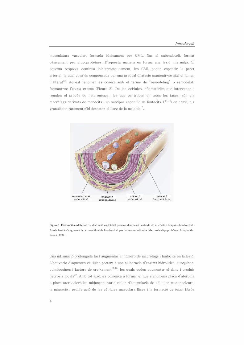
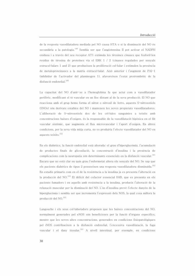
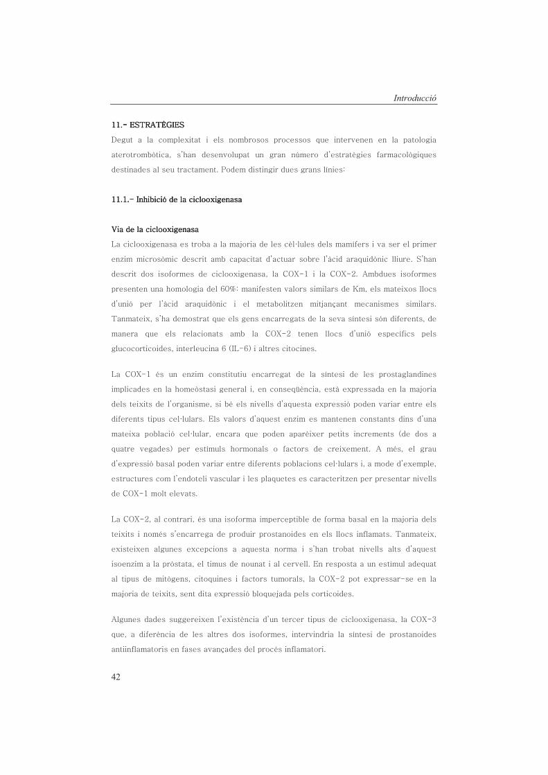
Figura 1. Disfunció endotelial. La disfunció endotelial promou d’adhesió i entrada de leucòcits a l’espai subendotelial.
A més també s’augmenta la permeabilitat de l’endoteli al pas de mecromolècules tals com les lipoproteïnes. Adaptat de
Ross R. 1999.

Introducció
5
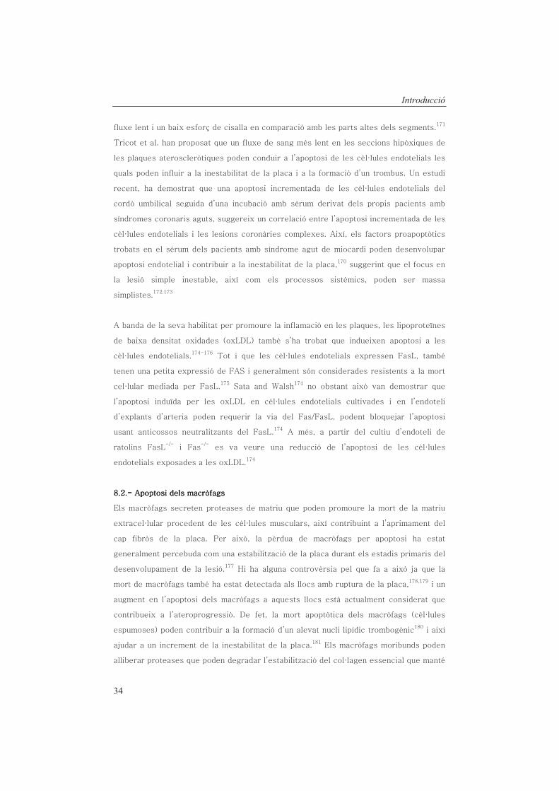
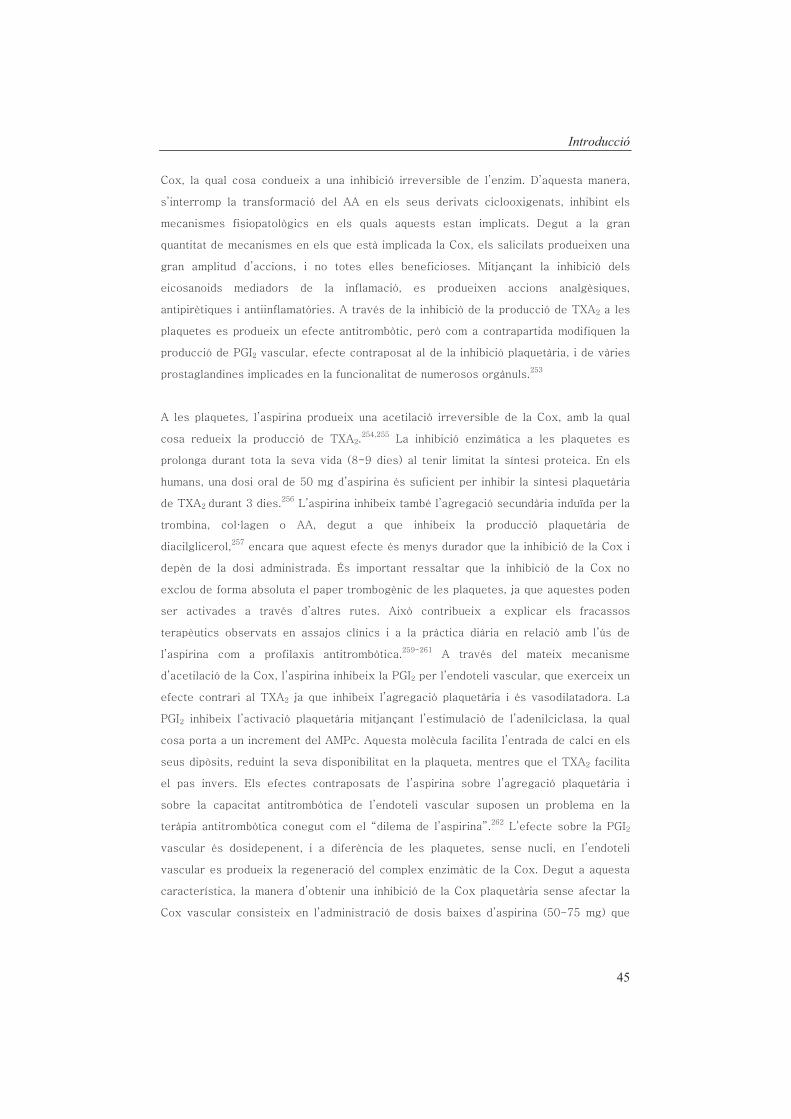
Figura 2. Formació de l’estria grassa. L’acúmul de macròfags i CML poden ocasionar un engruiximent de la paret arterial
conegut com a estria grassa. Aquest fet juntament amb la disfunció endotelial subjacent poden afavorir l’adhesió de les
plaquetes, fet que contribuirà a la progressió de la lesió. Adaptat de Ross R. 1999.

Introducció
6
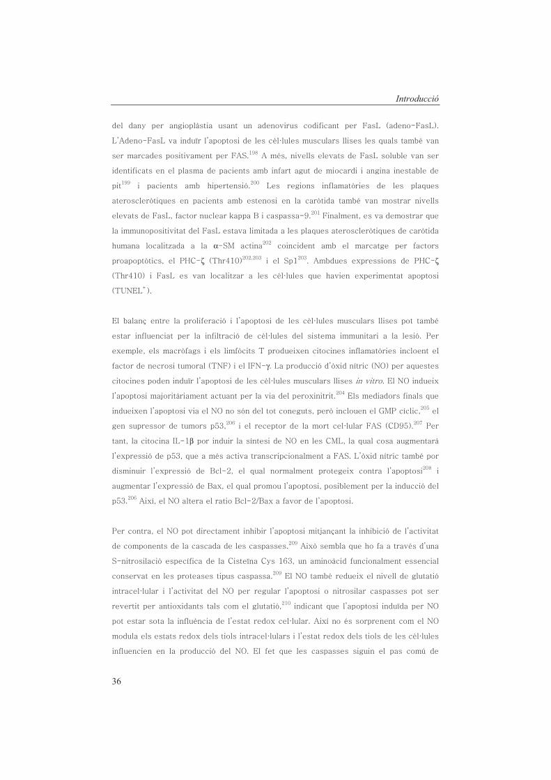
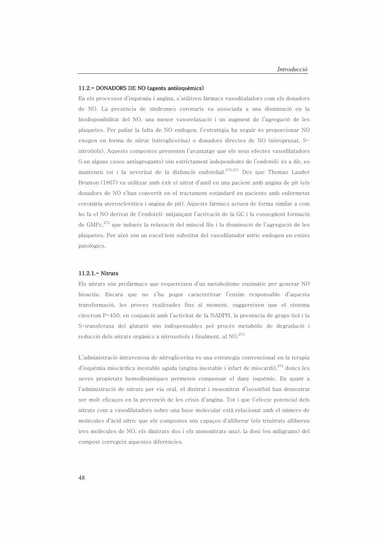
Figura 3. Formació d’una lesió ateroscleròtica avançada. En una agressió continuada en el vas i sota diferents estímuls
es produeix un augment del nombre de macròfags i cèl·lules espumoses que poden acabar morint i constituïr el nucli
necròtic, ric en colesterol i altament protrombòtic. Al voltant d’aquest nucli, les CML sintetitzen una coberta fibrosa que
dóna estabilitat mecànica a la placa ateroscleròtica. Adaptat de Ross R. 1999.

Introducció
7
Introducció
8
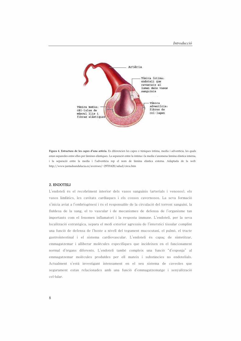
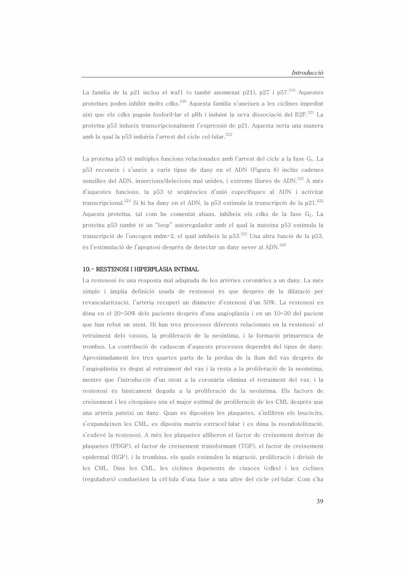
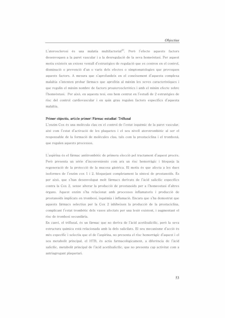
Figura 4. Estructura de les capes d’una artèria. Es diferencien les capes o túniques íntima, media i adventícia, les quals
estan separedes entre elles per làmines elàstiques. La separació entre la íntima i la media s’anomena làmina elàstica interna,
i la separació entre la media i l’adventícia rep el nom de làmina elàstica externa. Adaptada de la web:
http://www.juntadeandalucia.es/averroes/~29701428/salud/circu.htm
Introducció
9
Introducció
10
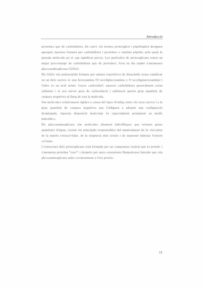
Introducció
11
Introducció
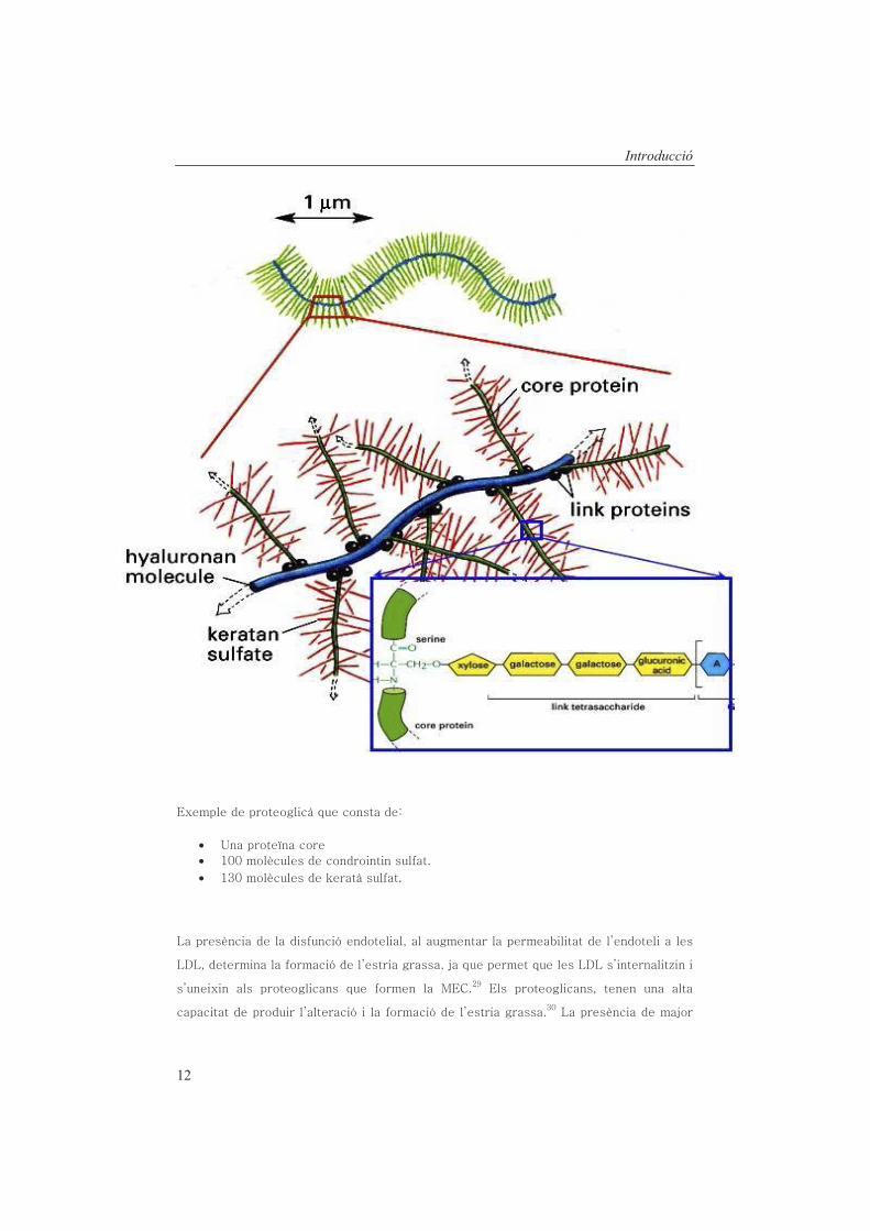
12
••• .
Introducció
13

Introducció
14
Introducció
15
Introducció
16
Introducció
17

Introducció
18
Introducció
19
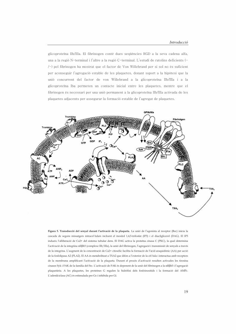
Figura 5. Transducció del senyal durant l’activació de la plaqueta. La unió de l’agonista al receptor (Rec) inicia la
cascada de segons missatgers intracel·lulars incloent el inositol 1,4,5-trifosfat (IP3) i el diacilglicerol (DAG). El IP3
indueix l’alliberació de Ca2+ del sistema tubular dens. El DAG activa la proteïna cinasa C (PKC), la qual determina
l’activació de la integrina αIIβb3 (complexe IIb/IIIa), la unió del fibrinogen, l’agregació i transmissió de senyals a través
de la integrina. L’augment de la concentració de Ca2+ citosòlic facilita la formació de l’àcid araquidònic (AA) per acció
de la fosfolipasa A2 (PLA2). El AA és metabolitzat a TXA2 que difon a l’exterior de la cèl·lula i interactua amb receptors
de la membrana amplificant l’activació de la plaqueta. Durant el procés d’activació resulten activades les tirosina
cinases Syk i FAK de la familia del Src. L’activació de FAK és depenent de la unió del fibrinogen a la αIIβb3 i l’agregació
plaquetària. A les plaquetes, les proteïnes G regulen la hidròlisi dels fosfoinositids i la formació del AMPc.
L’adenilciclasa (AC) és estimulada per Gs i inhibida per Gi.

Introducció
20
Introducció
21
Introducció
22
Introducció
23
Introducció
24

Introducció
25

Introducció
26
Figura 6. Factors derivats de l’endoteli. Vies de la prostaciclina, òxid nítric i endotelina. Entre aquests factors també hi
ha el tromboxà que actuaria sinèrgicament amb l’endotelina. Adaptada de la web:
http://www.antioxidantes.com.ar/Art247.htm.

Introducció
27
Introducció
28

Introducció
29
Introducció
30

Introducció
31

Introducció
32

Introducció
33
Figura 7. Apoptosi cel·lular. Amb la lletra A es representa la via intrínseca de l’apoptosi, la qual es dóna mitjançant la
despolarització de la membrana interna de la mitocondria alliberant el citocrom C que permetrà la formació de
l’apoptosoma. D’altra banda amb la lletra B es representa la via extrínseca de l’apoptosi que s’inicia en el receptor FAS
que transmet l’estímul a la caspassa 8. Adaptada de la web: http://profs.sci.univr.it/~crimi/recent97-00.html
Caspase-8
Introducció
34

Introducció
35
γ γ
γ
Introducció
36
α
ζ ζ
γ
β

Introducció
37

Introducció
38
Figura 8. Representació d’un dany al ADN i les seves posibles respostes moleculars que afecten al cicle cel·lular.
Introducció
39

Introducció
40
β

Introducció
41
β β
Introducció
42

Introducció
43
Figura 9. Biosíntesi dels derivats de l’àcid araquidònic produïts per ciclooxigenació.

Introducció
44
Introducció
45

Introducció
46
κ

Introducció
47
Introducció
48

Introducció
49

Introducció
50

Objectius
51

Objectius
52
Objectius
53

Objectius
54
Mètodes
55

Mètodes
56
Mètodes
57

Mètodes
58
Mètodes
59
Mètodes
60
Mètodes
61

Mètodes
62
Mètodes
63

Mètodes
64

Mètodes
65
Mètodes
66
Mètodes
67

Mètodes
68
Mètodes
69
Mètodes
70
µ
µ
µ
µ µ
µ
µ

Mètodes
71
Mètodes
72

Mètodes
73
Mètodes
74

Mètodes
75
Mètodes
76

Discussió
77

Discussió
78

Discussió
79
Discussió
80
Discussió
81
Discussió
82

Discussió
83

Discussió
84

Conclusions
85

Conclusions
86
Conclusions
87

Conclusions
88

Bibliografia
89

Bibliografia
90
Bibliografia
91

Bibliografia
92

Bibliografia
93
Bibliografia
94

Bibliografia
95
Bibliografia
96

Bibliografia
97

Bibliografia
98

Bibliografia
99
Bibliografia
100
Bibliografia
101
Bibliografia
102

Bibliografia
103
β
β
Bibliografia
104
κ
κ
Bibliografia
105
Bibliografia
106

Bibliografia
107

Bibliografia
108
Publicacions
109

Publicacions
110

Publicacions
111

Publicacions
112
Publicacions
113
Publicacions
114
Publicacions
115

Publicacions
116
Publicacions
117

Publicacions
118

ORIGINAL ARTICLE
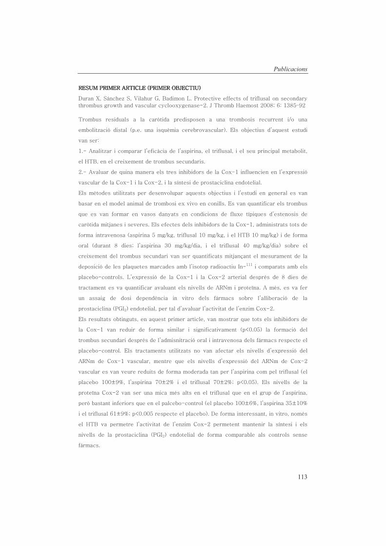
Protective effects of triflusal on secondary thrombus growthand vascular cyclooxygenase-2
X . DURAN,* S . S ANCHEZ ,* G . V I LAHUR*� and L . BADIMON*��*Cardiovascular Research Center, CSIC-ICCC, Hospital de la Santa Creu i Sant Pau, Barcelona; �CIBEROBN-Instituto Salud Carlos III, Barcelona;
and �Cardiovascular Research Chair, UAB, Barcelona, Spain
To cite this article: Duran X, Sanchez S, Vilahur G, Badimon L. Protective effects of triflusal on secondary thrombus growth and vascular
cyclooxygenase-2. J Thromb Haemost 2008; 6: 1385–92.
Summary. Background: Carotid residual mural thrombus
predisposes to recurrent thrombosis and/or distal embolization
(i.e. cerebrovascular ischemia). Objectives:Our aims were (i) to
analyze and compare the efficacy of aspirin, triflusal, and its
main metabolite 2-hydroxy-4-trifluorometylbenzoic acid
(HTB) on secondary thrombus growth; and (ii) evaluate to
what extent the three Cox-1 inhibitors influenced vascular Cox-
1/Cox-2 expression and endothelial prostacyclin synthesis.
Methods: In a rabbit model of ex vivo thrombosis, a fresh
mural thrombus was formed on damaged vessels at flow
conditions typical of mild and severe carotid stenoses. The
effects of Cox-1 inhibitors administered both intravenously
(i.v.) (aspirin 5 mg kg)1, triflusal 10 mg kg)1, and HTB
10 mg kg)1) and orally (p.o.) (8 days; aspirin
30 mg kg)1 day)1, and triflusal 40 mg kg)1 day)1) on second-
ary thrombus growth were assessed by In-111deposited platelets
and compared with a placebo control. Arterial Cox-1/Cox-2
expression after 8-day treatment was evaluated at mRNA and
protein levels. Additionally, a drug-related dose-dependent
in vitro assay was performed for endothelial PGI2 release
measurement (Cox-2 activity). Results: All Cox inhibitors
similarly and significantly (P < 0.05) reduced secondary
thrombus formation after i.v. and p.o. administration versus
placebo control. Treatments exertedno effect onvascularCox-1
mRNA whereas Cox-2 mRNA was moderately reduced by
aspirin and triflusal (placebo 100% ± 9%, aspirin
70% ± 2% and triflusal 70% ± 2%; P < 0.05). Cox-2
protein levels were slightly higher in the triflusal versus aspirin
group (placebo 100% ± 6%, aspirin 35% ± 10% and triflu-
sal 61% ± 9%; P < 0.005 versus placebo). Interestingly,
in vitro, HTB solely maintained endothelial PGI2 synthesis
levels similar to the control. Conclusions: At a similar level of
efficacy in inhibiting secondary thrombosis, triflusal seems to
better preserve Cox-2 expression than aspirin and itsmetabolite
HTB was able to protect endothelial prostacyclin production.
Keywords: cyclooxygenase inhibitors, prostacyclin, secondary
thrombus.
Introduction
Approximately, 25% of the cerebrovascular events (CVE)
derive from an atherothrombotic vessel occlusion within the
carotid system which might then embolize and cause cerebral
infarction. The outcome of a thrombotic event is dependent
on rheological factors, derived from the hemodynamic
compromise (i.e. vessel occlusion) and local risk factors.
Within the latter, the degree of vascular damage at the time of
plaque disruption and the presence of a residual thrombus
may partly determine the thrombogenic outcome [1,2]. Indeed,
the surface of the residual thrombus becomes a very thromb-
ogenic triggering substrate as a result of fibrin bound
thrombin in the original fragmented thrombus [3,4]. There-
fore, antiplatelet therapy is the cornerstone for prevention of
cerebral ischemia.
The efficacy of aspirin therapy in the prevention and
treatment of various cerebrovascular diseases is well established
and supported by a wealth of clinical data [5–8]. However,
emerging data regarding the use of aspirin for primary
prevention (e.g. gastrointestinal and bleeding complications)
or the aspirin �resistance� phenomenon restricts the efficacy of
this compound in various settings [9]. Therefore, introduction
of agents with efficacy at least similar to that of aspirin, yet with
potentially reduced adverse events, may represent a useful
addition to the therapeutic options available. In this regard,
several large-scale clinical trials [10,11] and a meta-analysis [12]
have assessed the comparable efficacy of triflusal (fluorinated
derivative of salicylic acid) relative to aspirin in reducing
ischemic cerebral events of vascular origin yet with a potentially
reduced risk of treatment-related bleeding hemorrhagic
complications [11].
Aspirin, depending on the dose, inhibits cyclooxygenase
(Cox) isoforms 1 and 2. Cox-1 [13] mediates tromboxane A2
Correspondence: Lina Badimon, Cardiovascular Research Center,
C/Sant Antoni Mª Claret 167, 08025 Barcelona, Spain.
Tel.: +34 93 556 58 80; fax: +34 93 556 55 59.
E-mail: [email protected]
Received 7 February 2008, accepted 13 May 2008
Journal of Thrombosis and Haemostasis, 6: 1385–1392 DOI: 10.1111/j.1538-7836.2008.03036.x
� 2008 International Society on Thrombosis and Haemostasis

(TXA2) platelet production leading to platelet aggregation
and vasoconstriction. Cox-2 catalyzes endothelial prostacy-
clin (PGI2) synthesis [14], which counteracts TXA2 and
triggers vasodilation and platelet inhibition and thus renders
vascular protection. Triflusal and its main metabolite 2-
hydroxy-4-trifluorometylbenzoic acid (HTB) [11,15] also
inhibit the platelet Cox-1 isoform; however, in vitro [16]
and in vivo [17] data have suggested that triflusal has little
inhibitory effect in Cox-2 expression; thus far, the mecha-
nisms by which this occurs is not fully understood.
Moreover, in contrast to aspirin, triflusal has been shown
to inhibit cyclic adenosine monophosphate (cAMP) and
cyclic guanosine monophosphate (cGMP) phosphodiesteras-
es, thus diminishing calcium-dependent platelet aggregation
similarly to dipyridamole [18].
Although both Cox-1 inhibitors have proven efficacy at a
clinical level and have both been shown to inhibit platelet
activation, it is yet unknown whether both drugs are
efficacious and equipotent in blocking the growth of pre-
existing thrombus. Moreover, a better understanding of their
effects on vascular Cox expression is required as it may
provide new insights regarding important clinical aspects and
properties of these antiplatelet agents. Thus, we firstly
evaluated the effect of a pharmacological intervention
targeted at the cyclooxygenase pathway to inhibit the
propagation and growth of secondary thrombi. To address
this issue, we have modeled ex vivo the cell–cell and fluid
phase–protein interaction at the particular microenvironment
of the ruptured plaque with a pre-existing thrombus, and
evaluated secondary thrombus growth under carotid-like flow
conditions in an ex vivo rabbit model of thrombosis. Second,
we ascertained to what extent Cox-1 inhibitors, at the tested
antithrombotic doses, also influenced vascular Cox expression
and activity.
Materials and methods
Evaluation of the Cox-1 inhibitors on secondary thrombus
formation
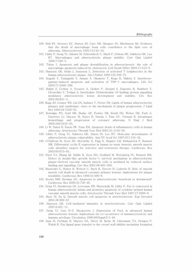
Animal preparation and experimental protocol Eighty-
eight New Zealand White male rabbits obtained from a
single local provider (body weight, 2.8 ± 1.7 kg) were kept
for 5 days in the animal facility without any intervention. As
shown in Fig. 1A, first we evaluated in a subgroup of animals
(Rabbit-In111, n = 5) the amount of primary thrombus
formation by measuring In-111 labeled platelet deposition
triggered by a damaged vessel wall under flow conditions
typical of mildly and severely stenotic carotid arteries using
our ex vivo perfusion chamber as reported elsewhere [1,19–
22]. Next, in order to quantitatively study the growth of
arterial thrombus (i.e. secondary thrombus formation) we
modified our ex vivo perfusion system [1,19–22] (Fig. 1B).
Briefly, blood from Rabbit A (unlabeled platelets) was passed
through the chamber at a flow rate of 10 mL min)1 for 5 min
on de-endothelialized vessel and tunica media with the
intention of creating the fresh thrombus. Thereafter, blood
with autologous 111In-labeled platelets from Rabbit B (with
or without drug treatment) was perfused, under identical flow
conditions, to measure secondary thrombus growth onto the
preformed primary thrombus (acting as the thrombogenic
substrate).
Before performing the perfusion experiments, Rabbits B
were randomly distributed into two types of studies. In the first
study, rabbits were intravenously (i.v.) treated with (i) aspirin
(lysidated acetylsalicylic acid; Synthelabo-Pharma; 5 mg kg)1);
(ii) triflusal (2-acetyloxy-4-trifluorometyl-benzoic acid; Uriach;
10 mg kg)1); (iii) the main triflusal metabolite HTB (2-
hydroxy-4-trifluorometylbenzoic acid; Uriach, 10 mg kg)1);
or (iv) a placebo control. These i.v. doses were considered
equimolar and were selected according to previous studies [23].
In the second study, rabbits were orally treated (p.o.) during
8 days with (i) aspirin (30 mg kg)1 day)1); (ii) triflusal
(40 mg Kg)1 day)1); or (iii) a placebo control. We performed
dose-related finding studies in order to detect the minimal oral
dose required to exert significant antiplatelet effects. Thus, we
evaluated the antithrombotic potential of 10, 20 and
30 mg kg)1 day)1 aspirin and 15, 30, 40 mg kg)1 day)1 triflu-
sal. We observed that 30 mg kg)1 day)1 aspirin and
40 mg kg)1 day)1 triflusal were able to inhibit secondary
thrombus formation (data not included).
All procedures performed in this study followed the Amer-
ican Heart Association guidelines for animal research.
Platelet labeling and ex vivo perfusion experiments The
day before the experiments, blood was drawn from the rabbits
via the ear central artery to perform platelet labeling with 111In-
oxine as described previously [1].
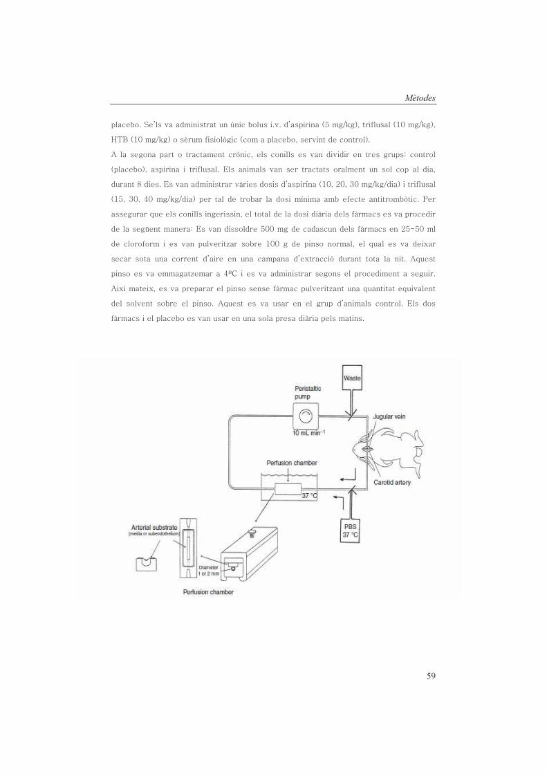
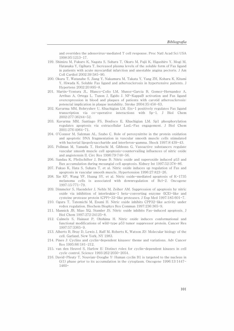
Twenty-four hours after labeling, animals were anesthetized
[intramuscularly (i.m.) ketamine 35 mg kg)1 and xylazine
5 mg kg)1] and the jugular vein and the contra-lateral carotid
artery were catheterized (Fig. 2). Animals were heparinized
with an i.v. bolus of 30 U kg)1 heparin (Liquemine; Roche,
Barcelona, Spain) and activated partial thromboplastin time
Primary thrombus formation withlabeled blood
A
B
5 min perfusion
5 min perfusion 5 min perfusion
- Inductor (damaged vessel wall)
- Inductor: Performed thrombus with blood fromRabbit A- Flow conditions
- Treatment:a) intravenous:
1. Aspirin 5 mg kg–1 (i.v.)2. Triflusal 10 mg kg–1 (i.v.)3. HTB 10 mg kg–1 (i.v.)
1. Aspirin 30 mg kg–1
2. Triflusal 40 mg kg–1
4. Placebo - control
3. Placebo - control
b) oral (8 days):
- Flow conditions:
- Rabbit de-endothelialized vessel
mildly stenotic (212 s–1)
Thrombus growth formed fromlabeled blood from Rabbit B
severely stenotic (1690 s–1)
mildly stenotic (212 s–1)severely stenotic (1690 s–1)- Flow conditions:
mildly stenotic (212 s–1)severely stenotic (1690 s–1)
Fresh thrombus formed fromunlabeled blood from Rabbit A
- Pig tunica media
- Inductor (damaged vessel wall)- Rabbit de-endothelialized vessel- Pig tunica media
Fig. 1. Schematic representation of the perfusion studies.
1386 X. Duran et al
� 2008 International Society on Thrombosis and Haemostasis

(aPTT) levels of 1.88 ± 0.06 were maintained throughout the
experiment. An arterio-venous shunt was established with the
aid of a peristaltic pump (Master-flex, Model 7518-10) at a
fixed rate of 10 mL min)1. Vessel wall segments were mounted
in the chamber and experimental perfusions were performed.
Radioactivity in each vessel segment was measured as counts
per minute with a gamma-well counter (Wizard 1470; Wallac,
Perkin Elmer, Waltham, MA, USA) and transformed in
platelet number per surface unit, as previously described [1]. In
the present study, we used two types of chambers with an
internal diameter of 0.2 and 0.1 cm modeling local flow
conditions typical of mildly and severely stenotic carotid
arteries (shear rates of 212 and 1690 s)1, respectively). Rabbit
subendothelium and pig tunica media were used as models of
wall damage (i.e. the triggering surfaces for primary thrombus
formation).
Thrombus triggering substrates Aortas from rabbits and
pigs were obtained fresh from a slaughterhouse, immediately
cleaned of adventitia and frozen at )80 �C until needed.
Before the experiments, the aortas were thawed in phosphate-
buffered saline (PBS), opened longitudinally and cut into
30 · 10 mm segments. De-endothelialized rabbit vessels (i.e.
subendothelium) were used for modeling mildly damaged
vessels whereas porcine tunica media was used for modeling
severely damaged vessels. Tunica media exposure requires
removing the intima with a thin portion of the subjacent
media as previously described [1,2]. Thus, owing to the
vessel-related difficulties in the intima removal in rabbit
aortas (i.e. thin vessel layers) we used pig aortas for tunica
media obtention.
Laboratory measurements After each perfusion, blood
samples were collected and evaluated for platelet and red
blood cell (RBC) count, hematocrit, fibrinogen levels,
prothrombin time (PT), aPTT, and indium lysis.
Effect of the different Cox-1 inhibitors on systemic vascular
Cox expression and endothelial Cox-2 activity
Vascular Cox protein and gene expression
analysis Immediately after being killed, aortas from orally
treated rabbits (i.e. aspirin and triflusal) were collected, cleaned
and frozen in liquid nitrogen. Aortic vessel wall RNA and
protein were obtained using the Tripure method (Boehringer
Mannheim, Barcelona, Spain) and immediately processed for
reverse transcription polymerase chain reaction (RT-PCR) and
Westernblotting todetectCox-1 andCox-2mRNAandprotein
levels, respectively.
RT-PCR analysis: Next, 1 lg of RNA was reverse tran-
scribed in a 20-lL reaction mixture (0.02 lg lL)1 oligo-dT,
1 mM dNTPs, 20 mM DTT, 200U SuperScript II Reverse
Transcriptase, 50 mM Tris-HCl pH 8.3, 75 mM KCl and 3 mM
MgCl2). Reaction was performed at 42 �C for 55 min and was
stopped by heating at 75 �C for 15 min, RNA residues were
degraded by RNAse (1U) at 37 �C for 20 min. Cox-1/Cox2
primers were as follows: 5¢-TCAATGCCACCTTCATCCGG-
3¢ (sense) and 5¢-ATCCAGCACCTGGTACTTGA-3¢ (anti-sense) for Cox-1 (466bp); 5¢-TCAGCCACGAGCAAATCCT-
3¢ (sense) and 5¢-GTGATCTGGATGTCAGCACG-3¢ (anti-sense) for Cox-2 (282 bp); and 5¢-GTCACCAGGGCTGC-
TTTTAA-3¢ (sense) and 5¢-ACGGAAGGCCATGCCAGT-
GA-3¢ (antisense) for GAPDH (652 bp). Amplification was
Peristalticpump
Waste
Jugular vein
10 mL min–1
Perfusion chamber
Perfusion chamber
Carotid artery
37 °C
Diameter1 or 2 mm
PBS37 °C
Arterial substrate(media or subendothelium)
Fig. 2. Representation of the extracorporeal perfusion system.
Triflusal and thrombus growth 1387
� 2008 International Society on Thrombosis and Haemostasis

carried out by 30 (Cox-2 and GAPDH) or 35 (Cox-1) cycles of
95 �C 45 s, 56 �C 45 s and 72 �C 1 min and 30 s, followed by a
final extension of 72 �C for 2 min. PCRproducts were resolved
by electrophoresis in 2% agarose gels with etidium bromure.
Western Blot analysis: Protein analysis was performed
according to standard procedures [mini-PROTEAN II Dual
Slab Cell (BioRad,Hercules, CA,USA)]. Blots were incubated
with monoclonal antibodies against Cox-2 (Oxford Biomedi-
cal, Oxford, MI, USA) and Cox-1 (Cayman, Ann Arbor, MI,
USA) and the signal was detected with Super Signal (Pierce,
USA). Positive controls for Cox-1 and Cox-2 detection
(Cayman) were run with the samples.
Measurement of endothelial Cox-2 activity
Cell culture: Porcine aortic endothelial cells (PAEC) primary
cultures were prepared from fresh pig aortas by a modification
of the collagenase method as described previously [24].
Thereafter, PAECs were seeded in pretreated gelatine (1%)
plates and incubated at 37 �C in a humidified atmosphere 5%
CO2. PAEC grew in a monolayer in medium M199 (Gibco
Laboratories) supplemented with 5% fetal calf serum (FCS),
2 mmol L)1 L-glutamine, 100 U mL)1 penicillin G, and
100 lg mL)1 streptomycin. Cell viability was determined by
trypan blue exclusion and PAECs were counted in the
Neubauer Chamber. PAECs were studied at passage 3–4.
Cellular treatment and PGI2 determinations: Near-confluent
PAECs were placed in serum-free media for 24 h to remove the
effects of serum. Subsequently, cells were incubated for 30 min
with M199 media in the absence or the presence of increasing
concentrations (37.5, 75, 300 lmols L)1) of aspirin, triflusal
and HTB. Then, Phorbol 12-Myristate 13-Acetate (PMA;
Sigma-Aldrich, Madrid, Spain; 100 nmols L)1) was added to
all plates except for the negative control and maintained for 4,
12, and 24 h. After these different incubation periods, the
media was aspirated and kept at )80 �C until the time of assay
for PGI2. Conversely, PAECs were processed for protein and
mRNA obtention according to the Tripure method.
Release of endothelial PGI2 was measured as 6-keto-
prostaglandin F1a (6-keto-PGF1 a; its stable hydrolysis
product), by radioimmunoassay according to the manu-
facturer�s instructions (Cayman Chemical Co.).
RT-PCR and immunoblot analysis: mRNA was analyzed by
real-time PCR 7000 (ABIPRISM, Applied Biosystems, Foster
City, CA, USA) as previously described in our laboratory [25].
TaqMan fluorescent real-time PCR primers and probes (6-
FAM-MGB) for pig Cox-2 detection were designed by use of
Primer Express software (Applied Biosystems) and were as
follows: Cox-2 pig probe (5¢-ATGCCATGGAGCTGTATC-
CTGCCCTT-3¢- MGB) (250 nM), Cox-2 pig forward primer
(5¢-TTAGAAGCGTCTATGGTGACATT-3¢) (300 nM) and
Cox-2 pig reverse primer (5¢-TCTGGGCGAGGCTTTTC-
TAC-3¢) (300 nM). The threshold cycle (Ct) values were
determined and normalized to the housekeeping gene
18SrRNA. Western blot analysis was performed as mentioned
above. Blots were incubated with monoclonal antibodies
against Cox-2 (Oxford Biomedical).
Statistical analysis
Statistical comparison of data was carried out using STAT-
VIEW II (Abacus Concepts Inc., Cary, NC, USA). Between-
group analysis was made using one-way ANOVA, followed by
Fisher PLSD test to assess specific group differences. All values
are presented as mean ± SEM and P < 0.05 was considered
significant.
Results
Effect of the Cox inhibitors on thrombus progression
Primary thrombus formation: a highly thrombogenic
substrate Before evaluating the effects of the different Cox-
1 inhibitors on secondary thrombus formation, we sought to
assess in this rabbit model of ex vivo thrombosis, the
thrombogenicity of a preformed thrombus. For this purpose,
labeled blood fromRabbits-In111 (n = 5)was directly perfused
for 5 min at both 212 and 1690 s)1 over subendothelium and
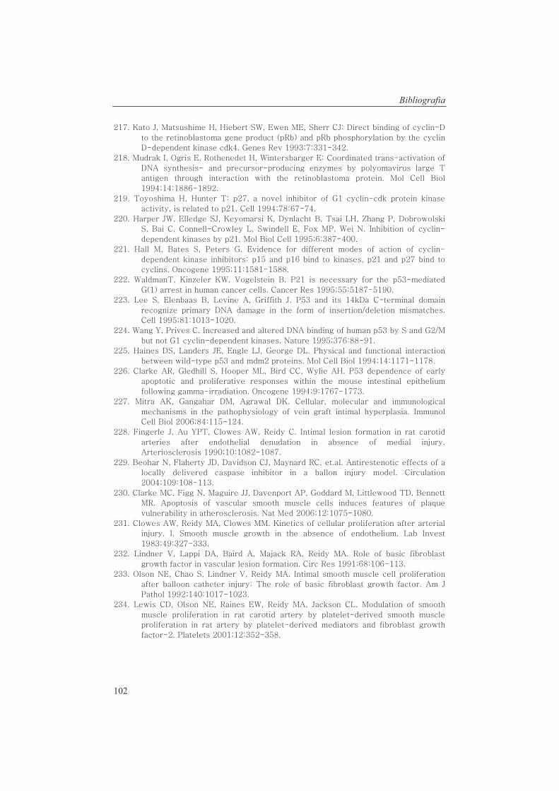
tunica media. At 212 s)1, a mean of 11.6 ± 3.7 and 29.8
± 6.8 · 106 platelets cm)2 were deposited on subendothelium
and tunica media, respectively, whereas at 1690 s)1
subendothelium and tunica media induced a mural thrombus
of about 157.6 ± 25.4 and 563.9 ± 22.7 · 106 platelets cm)2,
respectively (Fig. 3A). Then, in order to evaluate the
thrombogenicity of the resulting primary thrombus, unlabeled
blood from Rabbit-A was perfused over both vascular
substrates for 5 min under the same flow conditions.
Immediately after, this unlabeled freshly formed thrombus
was perfused with blood from Rabbit-B with 111In-labeled
platelets for additional 5 min.Theamountofplatelets deposited
on the preformed thrombus was 23.1 ± 3.8 and 63.8 ±
7.1 · 106 platelets cm)2 at 212 s)1 and 172 ± 43 and 515 ±
104 · 106 platelets cm)2 at 1690 s)1 on subendothelium and
tunica media, respectively. In summary, exposure of a
preformed thrombus to flowing blood at 212 s)1 increased by
2-fold the total amount of deposited platelets when compared
with platelet-free vascular substrates (P < 0.05) while at
1690 s)1 the primary thrombus exhibited similar
thrombogenicity than the triggering substrate (P = NS).
Effect of acute treatment with aspirin, triflusal and HTB on
thrombus progression Cox-1 inhibitors (aspirin, triflusal,
and its active metabolite HTB) similarly and significantly
(P < 0.01) reduced platelet deposition on a fresh primary
thrombus triggered by both substrates (subendothelium and
tunica media) at shear rates typical of mildly stenotic vessels
(Fig. 3B–C). Thus, platelet deposition on subendothelium-
induced primary thrombus was reduced by about 68% by
aspirin (n = 6) and by about 66% by both triflusal and HTB
(n = 4 and n = 5; respectively) when compared with placebo-
control animals (n = 11) (Fig. 3B). Similarly, platelet
deposition on a fresh thrombus formed over tunica media
(Fig. 3C) was reduced by around 62%, 48% and 66% using
aspirin (n = 6), triflusal (n = 7) and HTB (n = 3)-treated
1388 X. Duran et al
� 2008 International Society on Thrombosis and Haemostasis
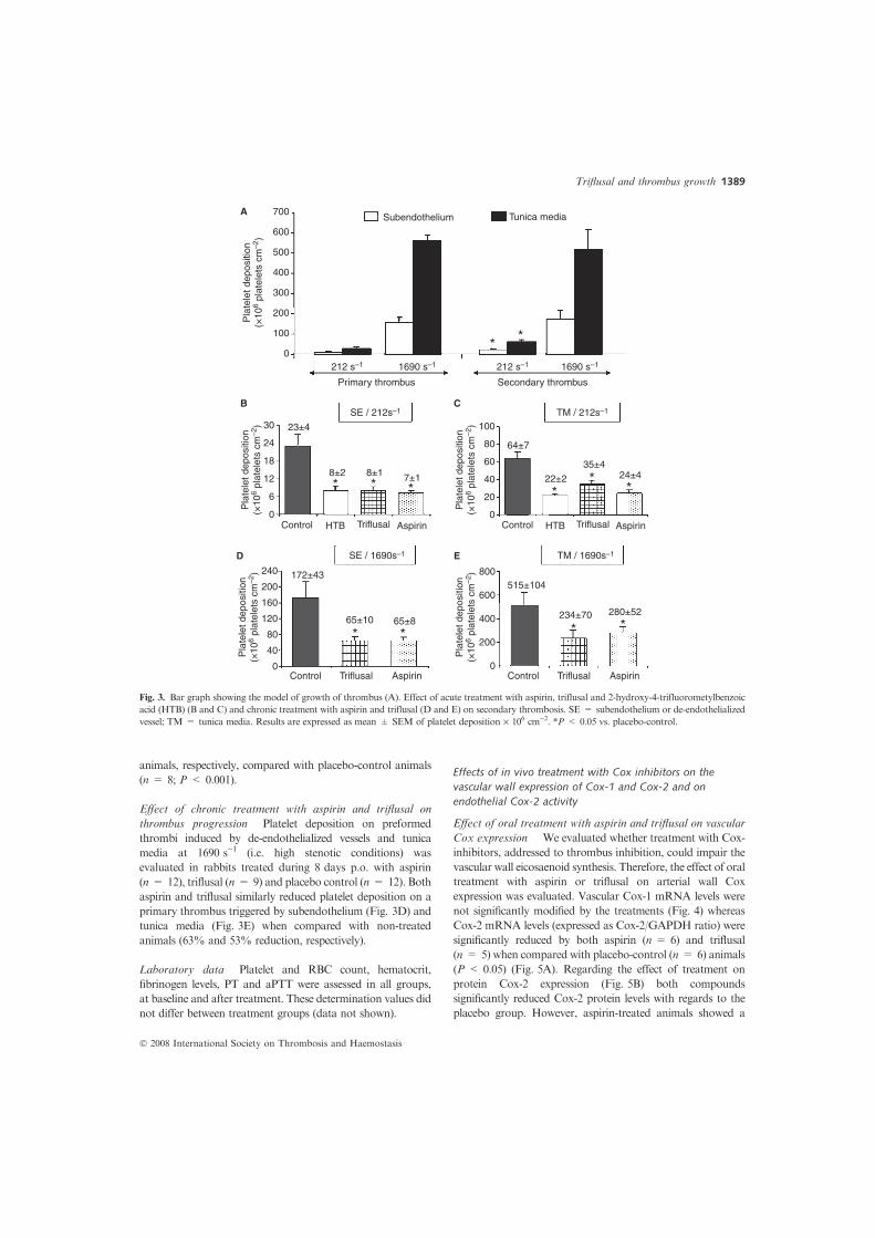
animals, respectively, compared with placebo-control animals
(n = 8; P < 0.001).
Effect of chronic treatment with aspirin and triflusal on
thrombus progression Platelet deposition on preformed
thrombi induced by de-endothelialized vessels and tunica
media at 1690 s)1 (i.e. high stenotic conditions) was
evaluated in rabbits treated during 8 days p.o. with aspirin
(n = 12), triflusal (n = 9) and placebo control (n = 12). Both
aspirin and triflusal similarly reduced platelet deposition on a
primary thrombus triggered by subendothelium (Fig. 3D) and
tunica media (Fig. 3E) when compared with non-treated
animals (63% and 53% reduction, respectively).
Laboratory data Platelet and RBC count, hematocrit,
fibrinogen levels, PT and aPTT were assessed in all groups,
at baseline and after treatment. These determination values did
not differ between treatment groups (data not shown).
Effects of in vivo treatment with Cox inhibitors on the
vascular wall expression of Cox-1 and Cox-2 and on
endothelial Cox-2 activity
Effect of oral treatment with aspirin and triflusal on vascular
Cox expression We evaluated whether treatment with Cox-
inhibitors, addressed to thrombus inhibition, could impair the
vascular wall eicosaenoid synthesis. Therefore, the effect of oral
treatment with aspirin or triflusal on arterial wall Cox
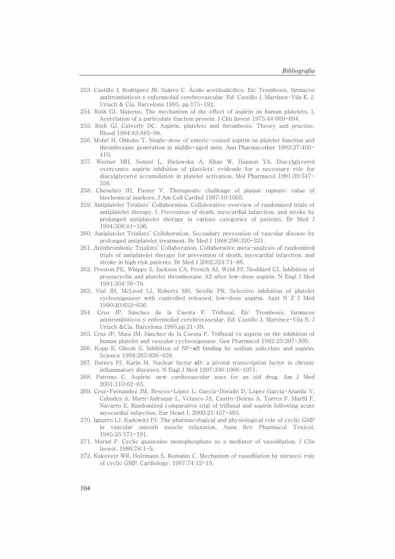
expression was evaluated. Vascular Cox-1 mRNA levels were
not significantly modified by the treatments (Fig. 4) whereas
Cox-2 mRNA levels (expressed as Cox-2/GAPDH ratio) were
significantly reduced by both aspirin (n = 6) and triflusal
(n = 5) when compared with placebo-control (n = 6) animals
(P < 0.05) (Fig. 5A). Regarding the effect of treatment on
protein Cox-2 expression (Fig. 5B) both compounds
significantly reduced Cox-2 protein levels with regards to the
placebo group. However, aspirin-treated animals showed a
700A
B C
D E
Subendothelium
212 s–1 1690 s–1
Primary thrombus
Control HTB Triflusal Aspirin
Control Triflusal Aspirin Control Triflusal Aspirin
Control HTB Triflusal Aspirin
SE / 212s–1 TM / 212s–1
SE / 1690s–1 TM / 1690s–1
Secondary thrombus
212 s–1
* *
1690 s–1
Pla
tele
t dep
ositi
on(×
106
plat
elet
s cm
–2)
Pla
tele
t dep
ositi
on(×
106
plat
elet
s cm
–2)
Pla
tele
t dep
ositi
on(×
106
plat
elet
s cm
–2)
Pla
tele
t dep
ositi
on(×
106
plat
elet
s cm
–2)
Pla
tele
t dep
ositi
on(×
106
plat
elet
s cm
–2)
Tunica media
600
500
400
300
200
600
800
400
200
0
100
0
30
240
200
160
120
80
40
0
23±4
8±2
172±43
65±10 65±8
515±104
234±70 280±52
8±1 7±1
64±7
22±2
35±424±4
**
**
* * **
**
24
18
12
6
0
100
80
60
40
20
0
Fig. 3. Bar graph showing the model of growth of thrombus (A). Effect of acute treatment with aspirin, triflusal and 2-hydroxy-4-trifluorometylbenzoic
acid (HTB) (B and C) and chronic treatment with aspirin and triflusal (D and E) on secondary thrombosis. SE = subendothelium or de-endothelialized
vessel; TM = tunica media. Results are expressed as mean ± SEM of platelet deposition · 106 cm)2. *P < 0.05 vs. placebo-control.
Triflusal and thrombus growth 1389
� 2008 International Society on Thrombosis and Haemostasis
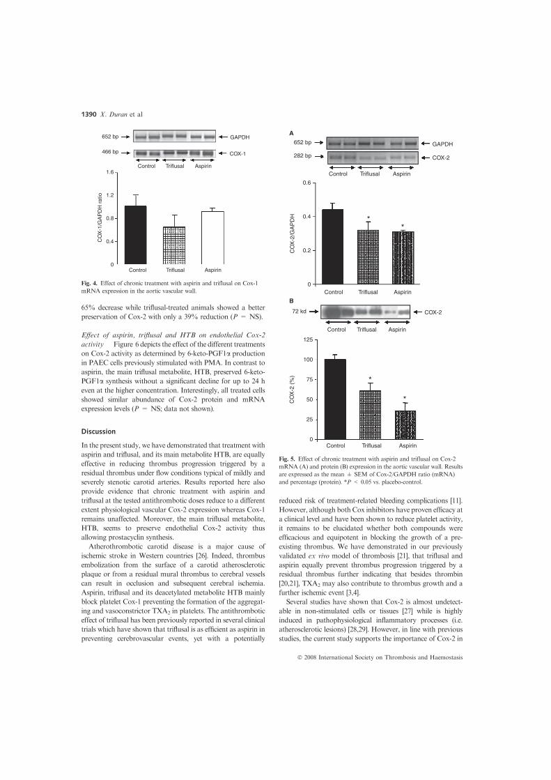
65% decrease while triflusal-treated animals showed a better
preservation of Cox-2 with only a 39% reduction (P = NS).
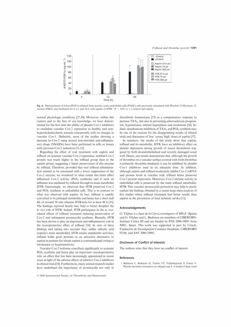
Effect of aspirin, triflusal and HTB on endothelial Cox-2
activity Figure 6 depicts the effect of the different treatments
on Cox-2 activity as determined by 6-keto-PGF1a production
in PAEC cells previously stimulated with PMA. In contrast to
aspirin, the main triflusal metabolite, HTB, preserved 6-keto-
PGF1a synthesis without a significant decline for up to 24 h
even at the higher concentration. Interestingly, all treated cells
showed similar abundance of Cox-2 protein and mRNA
expression levels (P = NS; data not shown).
Discussion
In the present study, we have demonstrated that treatment with
aspirin and triflusal, and its main metabolite HTB, are equally
effective in reducing thrombus progression triggered by a
residual thrombus under flow conditions typical of mildly and
severely stenotic carotid arteries. Results reported here also
provide evidence that chronic treatment with aspirin and
triflusal at the tested antithrombotic doses reduce to a different
extent physiological vascular Cox-2 expression whereas Cox-1
remains unaffected. Moreover, the main triflusal metabolite,
HTB, seems to preserve endothelial Cox-2 activity thus
allowing prostacyclin synthesis.
Atherothrombotic carotid disease is a major cause of
ischemic stroke in Western countries [26]. Indeed, thrombus
embolization from the surface of a carotid atherosclerotic
plaque or from a residual mural thrombus to cerebral vessels
can result in occlusion and subsequent cerebral ischemia.
Aspirin, triflusal and its deacetylated metabolite HTB mainly
block platelet Cox-1 preventing the formation of the aggregat-
ing and vasoconstrictor TXA2 in platelets. The antithrombotic
effect of triflusal has been previously reported in several clinical
trials which have shown that triflusal is as efficient as aspirin in
preventing cerebrovascular events, yet with a potentially
reduced risk of treatment-related bleeding complications [11].
However, although both Cox inhibitors have proven efficacy at
a clinical level and have been shown to reduce platelet activity,
it remains to be elucidated whether both compounds were
efficacious and equipotent in blocking the growth of a pre-
existing thrombus. We have demonstrated in our previously
validated ex vivo model of thrombosis [21], that triflusal and
aspirin equally prevent thrombus progression triggered by a
residual thrombus further indicating that besides thrombin
[20,21], TXA2 may also contribute to thrombus growth and a
further ischemic event [3,4].
Several studies have shown that Cox-2 is almost undetect-
able in non-stimulated cells or tissues [27] while is highly
induced in pathophysiological inflammatory processes (i.e.
atherosclerotic lesions) [28,29]. However, in line with previous
studies, the current study supports the importance of Cox-2 in
652 bp GAPDH
COX-1466 bp
1.6
1.2
0.8
CO
X-1
/GA
PD
H r
atio
0.4
0
Control Triflusal Aspirin
Control Triflusal Aspirin
Fig. 4. Effect of chronic treatment with aspirin and triflusal on Cox-1
mRNA expression in the aortic vascular wall.
GAPDH
COX-2
652 bp
A
B
282 bp
Control Triflusal
0.6
**
0.4
CO
X-2
/GA
PD
H
0.2
0
Aspirin
Control Triflusal Aspirin
Control
125
100
75
50
25
0
72 kd
Triflusal Aspirin
Control Triflusal Aspirin
*
*
COX-2
CO
X-2
(%
)
Fig. 5. Effect of chronic treatment with aspirin and triflusal on Cox-2
mRNA (A) and protein (B) expression in the aortic vascular wall. Results
are expressed as the mean ± SEM of Cox-2/GAPDH ratio (mRNA)
and percentage (protein). *P < 0.05 vs. placebo-control.
1390 X. Duran et al
� 2008 International Society on Thrombosis and Haemostasis
normal physiologic conditions [27,30]. Moreover, within this
context and to the best of our knowledge, we have demon-
strated for the first time the ability of platelet Cox-1 inhibitors
to modulate vascular Cox-2 expression in healthy and non-
hypercholesterolemic animals concurrently with no changes in
vascular Cox-1. Definitely, most of the studies showing a
decrease in Cox-2 using several non-steroidal anti-inflamma-
tory drugs (NSAIDs) have been performed in cells or tissues
with previous Cox-2 induction [31,32].
Regarding the effect of oral treatment with aspirin and
triflusal on systemic vascular Cox-2 expression, residual Cox-2
protein was much higher in the triflusal group than in the
aspirin group, suggesting a better preservation of this enzyme
by triflusal. Therefore, provided that oral triflusal administra-
tion seemed to be associated with a lower suppression of the
Cox-2 enzyme, we wondered to what extent this latter effect
influenced Cox-2 activity (PGI2 synthesis) and if such an
influence was mediated by triflusal through its main metabolite
HTB. Interestingly, we observed that HTB preserved Cox-2
and PGI2 synthesis in endothelial cells. This is in contrast to
what was observed with aspirin. In fact, triflusal is rapidly
converted to its principal metabolite and hence has a short half
life of around 30 min whereas HTB lasts for at least 48 h [33].
The findings reported herein may help to better decipher the
in vivo role of HTB. Indeed, HTB participates in the in vivo-
related effects of triflusal treatment inducing preservation of
Cox-2 and subsequent prostacyclin synthesis. Recently, HTB
has been shown to play an important anti-inflammatory role in
the neuroprotective effect of triflusal [18]. In view of these
findings and taking into account that, unlike salicylic acid
(aspirin�s main metabolite), HTB retains antiplatelet activities,
triflusal holds great promise as an attractive alternative to
aspirin in patients for whom aspirin is contraindicated owing to
intolerance or hypersensitivity.
Vascular Cox-2 isoforms contribute significantly to systemic
PGI2 synthesis and hence play an important vasculoprotective
role; an effect that has been increasingly appreciated in recent
years in light of the adverse effects of selective Cox-2 inhibitors
in clinical trials [34]. Furthermore,manyanimal research studies
have underlined the importance of prostacyclin not only in
thrombotic homeostasis [35] as a compensatory response to
increase TXA2, but also in preventing atherosclerosis progress-
ion, hypertension, intimal hyperplasia and reestenosis [36]. In-
deed, simultaneous inhibition of TXA2 and PGI2 synthesismay
be one of the reasons for the disappointing results of clinical
trials and discussion of �low� versus �high� doses of aspirin [37].
In summary, the results of this study show that aspirin,
triflusal and its metabolite, HTB, have an inhibitory effect on
platelet deposition during growth of mural thrombosis trig-
gered by both de-endothelialized and severely damaged vessel
wall. Hence, our results demonstrate that, although the growth
of thrombus on a vascular surface covered with fresh thrombus
is primarily thrombin mediated, it can be inhibited by platelet
Cox-1 inhibitors used in an adequate dose. In addition,
although aspirin and triflusal moderately inhibit Cox-2 mRNA
and protein levels in vascular wall, triflusal better preserves
Cox-2 protein expression. Moreover, Cox-2 enzyme activity in
endothelial cells is preserved by the main triflusal metabolite,
HTB. This vascular prostacyclin protection may help to partly
explain the findings obtained in a recent large meta-analysis of
five studies where triflusal treatment had better results than
aspirin in the prevention of fatal ischemic stroke [12].
Acknowledgements
G. Vilahur is a Juan de la Cierva investigator ofMEyC (Spain)
and G. Vilahur and L. Badimon are members of CIBEROBN-
Institute Carlos III and are funded by PNS 2006-10091 from
MEC, Spain. This work was supported in part by Uriach,
Fundacion de Investigacion Catalana Occidente, CIBEROBN
03/06, and SAF 2006/10091.
Disclosure of Conflict of Interests
The authors state that they have no conflict of interest.
References
1 Badimon L, Badimon JJ, Turitto VT, Vallabhajosula S, Fuster V.
Platelet thrombus formation on collagen type I. Amodel of deep vessel
140
120
1006-
KE
TO P
GF
1α (n
g m
L–1 )
80
60
40
20
04 12 24
–Control+Control
Aspirin 37.5 µM
Aspirin 75 µM
Aspirin 300 µM
HTB 37.5 µM
HTB 75 µM
HTB 300 µM
Time (h)
* * * *
**
*
*
* * *
*
Fig. 6. Measurements of 6-keto-PGF1a released from porcine aortic endothelial cells (PAEC) cells previously stimulated with Phorbol 12-Myristate 13-
Acetate (PMA) and incubated for 4, 12, and 24 h with aspirin or HTB. *P < 0.05 vs. ()) control and aspirin.
Triflusal and thrombus growth 1391
� 2008 International Society on Thrombosis and Haemostasis
injury. Influence of blood rheology, von Willebrand factor, and blood
coagulation. Circulation 1988; 78: 1431–42.
2 Badimon L, Badimon JJ. Mechanisms of arterial thrombosis in non-
parallel streamlines: platelet thrombi grow on the apex of stenotic
severely injured vessel wall. Experimental study in the pig model. J Clin
Invest 1989; 84: 1134–44.
3 Lassila R, Badimon JJ, Vallabhajosula S, Badimon L. Dynamic
monitoring of platelet deposition on severely damaged vessel wall in
flowing blood. Effects of different stenoses on thrombus growth.
Arteriosclerosis 1990; 10: 306–15.
4 Weitz JI, Hudoba M, Massel D, Maraganore J, Hirsh J. Clot-bound
thrombin is protected from inhibition by heparin-antithrombin III but
is susceptible to inactivation by antithrombin III-independent inhibi-
tors. J Clin Invest 1990; 86: 385–91.
5 Weinberger J. Adverse effects and drug interactions of antithrombotic
agents used in prevention of ischaemic stroke.Drugs 2005; 65: 461–71.
6 Albers GW, Amarenco P, Easton JD, Sacco RL, Teal P. Antithrom-
botic and thrombolytic therapy for ischemic stroke: the SeventhACCP
Conference on Antithrombotic and Thrombolytic Therapy. Chest
2004; 3(Suppl.): 483S–512S.
7 ISIS-2 (Second International Study of Infarct Survival) Collaborative
Group. Randomised trial of intravenous streptokinase, oral aspirin,
both, or neither among 17,187 cases of suspected acute myocardial
infarction: ISIS-2. Lancet 1988; 2: 349–60.
8 The Stroke Prevention in Reversible Ischemia Trial (SPIRIT) Study
Group. A randomized trial of anticoagulants versus aspirin after
cerebral ischemia of presumed arterial origin. Ann Neurol 1997; 42:
857–65.
9 Diener HC, Bogousslavsky J, Brass LM, Cimminiello C, Csiba L,
Kaste M, Leys D, Matias-Guiu J, Rupprecht HJ. Aspirin and clopi-
dogrel compared with clopidogrel alone after recent ischaemic stroke
or transient ischaemic attack in high-risk patients (MATCH): rando-
mised, double-blind, placebo-controlled trial. Lancet 2004; 364: 331–7.
10 Culebras A, Rotta-Escalante R, Vila J, Dominguez R, Abiusi G,
Famulari A, Rey R, Bauso-Tosselli L, Gori H, Ferrari J, Reich E.
Triflusal vs aspirin for prevention of cerebral infarction: a randomized
stroke study. Neurology 2004; 62: 1073–80.
11 Matias-Guiu J, Ferro JM, Alvarez-Sabin J, Torres F, Jimenez MD,
Lago A, Melo T. Comparison of triflusal and aspirin for prevention of
vascular events in patients after cerebral infarction: the TACIP Study:
a randomized, double-blind, multicenter trial. Stroke 2003; 34: 840–8.
12 Costa J, Ferro JM,Matias-Guiu J, Alvarez-Sabin J, Torres F. Triflusal
for preventing serious vascular events in people at high risk. Cochrane
Database Syst Rev 2005; CD004296.
13 Weiss HJ, Aledort LM. Impaired platelet-connective-tissue reaction in
man after aspirin ingestion. Lancet 1967; 2: 495–7.
14 McAdam BF, Catella-Lawson F, Mardini IA, Kapoor S, Lawson JA,
FitzGerald GA. Systemic biosynthesis of prostacyclin by cyclooxy-
genase (COX)-2: the human pharmacology of a selective inhibitor of
COX-2. Proc Natl Acad Sci U S A 1999; 96: 272–7.
15 McNeely W, Goa KL. Triflusal. Drugs 1998; 55: 823–33; discussion
834–825.
16 Cruz-Fernandez J. Antiplatelet drugs in the treatment of acute coro-
nary syndromes: focus on ciclyogenase inhibitors. Eur Heart J Suppls
2001; 3(Suppl. I): I23–30.
17 De la Cruz JP, Pavia J, Garcia-Arnes J, Sanchez de la Cuesta F. Effects
of triflusal and acetylsalicylic acid on platelet aggregation in whole
blood of diabetic patients. Eur J Haematol 1988; 40: 232–6.
18 Gonzalez-Correa JA, Arrebola MM, Urena IM, Ruiz-Villafranca D,
Munoz-Marin J, Guerrero A, Sanchez de la Cuesta F, De La Cruz JP.
Protective effect of triflusal and its main metabolite HTB in an in vitro
model of anoxia-reoxygenation in rat brain slices: comparison with
acetylsalicylic and salicylic acids. Naunyn Schmiedebergs Arch
Pharmacol 2005; 371: 81–8.
19 Badimon L, Turitto V, Rosemark JA, Badimon JJ, Fuster V.
Characterization of a tubular flow chamber for studying platelet
interaction with biologic and prosthetic materials: deposition of
indium 111-labeled platelets on collagen, subendothelium, and ex-
panded polytetrafluoroethylene. J Lab Clin Med 1987; 110: 706–18.
20 Meyer BJ, Badimon JJ, Mailhac A, Fernandez-Ortiz A, Chesebro JH,
FusterV,BadimonL. Inhibition of growth of thrombus on freshmural
thrombus. Targeting optimal therapy. Circulation 1994; 90: 2432–8.
21 Meyer BJ, Badimon JJ, Chesebro JH, Fallon JT, Fuster V, Badimon
L. Dissolution of mural thrombus by specific thrombin inhibition with
r-hirudin: comparison with heparin and aspirin. Circulation 1998; 97:
681–5.
22 Badimon JJ, Lettino M, Toschi V, Fuster V, Berrozpe M, Chesebro
JH, Badimon L. Local inhibition of tissue factor reduces the
thrombogenicity of disrupted human atherosclerotic plaques: effects of
tissue factor pathway inhibitor on plaque thrombogenicity under flow
conditions. Circulation 1999; 99: 1780–7.
23 Garcia Rafanell J, Forn J. Intravenous arachidonate-induced respira-
tory distress: comparative study of three in vivo models for the measure-
ment of antithrombotic activity. J Pharmacol Methods 1982; 7: 231–8.
24 Rodriguez C, Alcudia JF, Martinez-Gonzalez J, Raposo B, Navarro
MA, Badimon L Lysyl oxidase (LOX) down-regulation by TNFalpha:
a new mechanism underlying TNFalpha-induced endothelial dys-
function. Atherosclerosis 2008; 196: 558–64.
25 Llorente-Cortes V, Otero-VinasM, Sanchez S, Rodriguez C, Badimon
L. Low-density lipoprotein upregulates low-density lipoprotein
receptor-related protein expression in vascular smooth muscle cells:
possible involvement of sterol regulatory element binding protein-2-
dependent mechanism. Circulation 2002; 106: 3104–10.
26 Rosamond W, Flegal K, Furie K, Go A, Greenlund K, Haase N,
Hailpern SM, HoM, Howard V, Kissela B, Kittner S, Lloyd-Jones D,
McDermott M, Meigs J, Moy C, Nichol G, O’Donnell C, Roger V,
Sorlie P, Steinberger J et al. Heart disease and stroke statistics–2008
update: a report from the American Heart Association Statistics
Committee and Stroke Statistics Subcommittee. Circulation 2008; 117:
e25–146.
27 Catella-Lawson F, Crofford LJ. Cyclooxygenase inhibition and
thrombogenicity. Am J Med 2001; 110(Suppl 3A): 28S–32S.
28 Schonbeck U, Sukhova GK, Graber P, Coulter S, Libby P.
Augmented expression of cyclooxygenase-2 in human atherosclerotic
lesions. Am J Pathol 1999; 155: 1281–91.
29 Stemme V, Swedenborg J, Claesson H, Hansson GK. Expression of
cyclo-oxygenase-2 in human atherosclerotic carotid arteries. Eur J
Vasc Endovasc Surg 2000; 20: 146–52.
30 FitzGerald GA. Cardiovascular pharmacology of nonselective non-
steroidal anti-inflammatory drugs and coxibs: clinical considerations.
Am J Cardiol 2002; 89: 26D–32D.
31 Xu XM, Sansores-Garcia L, Chen XM, Matijevic-Aleksic N, Du M,
WuKK. Suppression of inducible cyclooxygenase 2 gene transcription
by aspirin and sodium salicylate. Proc Natl Acad Sci U S A 1999; 96:
5292–7.
32 Fernandez de Arriba A, Cavalcanti F,Miralles A, BayonY, AlonsoA,
Merlos M, Garcia-Rafanell J, Forn J. Inhibition of cyclooxygenase-2
expression by 4-trifluoromethyl derivatives of salicylate, triflusal, and
its deacetylated metabolite, 2-hydroxy-4-trifluoromethylbenzoic acid.
Mol Pharmacol 1999; 55: 753–60.
33 Murdoch D, Plosker GL. Triflusal: a review of its use in cerebral
infarction and myocardial infarction, and as thromboprophylaxis in
atrial fibrillation. Drugs 2006; 66: 671–92.
34 Grosser T, Fries S, FitzGerald GA. Biological basis for the cardio-
vascular consequences of COX-2 inhibition: therapeutic challenges and
opportunities. J Clin Invest 2006; 116: 4–15.
35 Cheng Y, Austin SC, Rocca B, Koller BH, Coffman TM, Grosser T,
Lawson JA, FitzGeraldGA. Role of prostacyclin in the cardiovascular
response to thromboxane A2. Science 2002; 296: 539–41.
36 Fetalvero KM, Martin KA, Hwa J. Cardioprotective prostacyclin
signaling in vascular smooth muscle. Prostaglandins Other Lipid
Mediat 2007; 82: 109–18.
37 Marcus AJ. Aspirin as an antithrombotic medication. N Engl J Med
1983; 309: 1515–7.
1392 X. Duran et al
� 2008 International Society on Thrombosis and Haemostasis

Atherosclerosis 205 (2009) 101–106
Contents lists available at ScienceDirect
Atherosclerosis
journa l homepage: www.e lsev ier .com/ locate /a therosc leros is
Exogenous in vivo NO-donor treatment preserves p53 levels and protectsvascular cells from apoptosis
Xavier Duran a,1, Gemma Vilahur a,b,1, Lina Badimon a,b,c,∗
a Cardiovascular Research Center, CSIC-ICCC, Barcelona, Spainb CIBEROBN CB06/03, Instituto de Salud Carlos III, Spainc Cátedra Investigación Cardiovascular (UAB), Catalana-Occidente, Hospital de la Santa Creu i Sant Pau, Barcelona, Spain
a r t i c l e i n f o
Article history:Received 24 October 2007Received in revised form 27 August 2008Accepted 18 November 2008Available online 25 November 2008
Keywords:Nitric oxide donorsApoptosisVascular smooth muscle cells
a b s t r a c t
Objective: Nitric oxide (NO) is critical in cardiovascular protection. However, NO production is impairedin atherosclerosis resulting in thrombosis, vasoconstriction, and restenosis. Exogenous NO-donors (NOd)have proven protection against ischemia and thrombosis. However, their effect on vascular cell functionremains unknown. Our objective was to determine the effect of NOd on vascular smooth muscle cell(VSMC) survival.Methods: Pigs (N = 16) were randomly distributed in the following treatment groups: (I) LA419(nitratethiol; 10-day p.o. 0.9 mg/kg bid); (II) LA816 (nitrosothiol; 2 h i.v. infusion, 6.6 nmol/(kg min));(III) nitroglycerine (GTN, 2 h i.v. infusion, 2.5 mg/kg); and (IV) placebo-control. After sacrifice, pig aorticarch explants were either frozen or incubated with 20% homologous pig serum for different time periods(18 h, 2 and 10 days). Bcl2/bax ratio, phosphorylated-p53, cyclin-D2, casp-3, casp-8, and bax mRNA levelsand protein expression were evaluated by real-time PCR and western blot, respectively.Results: Within the first 2 days explants from NOd-treated animals had an increased pro-survival Bcl2/baxratio compared with placebo-control (5× higher in LA816 and GTN i.v.-treated animals and 10× higherin 10-day orally treated LA419 animals; P < 0.05). Phosphorylated-P53-protein levels were consistentlyincreased by all NOd-treatments vs. placebo-control (P < 0.05) and p53 mRNA levels showed an increase inplacebo-controls at day 10 while they did not rise in NOd-treated animals (P < 0.05). Cyclin-D2, casp-3, andbax mRNA content were 6× lower in NOd-treated animals than in control explants at day-10 (P < 0.001).No changes were detected on caspase-8.Conclusions: Taken together, our findings suggest that exogenous in vivo NO treatment seems to preserveVSMC from mitochondrial-dependent apoptosis and drive cells to quiescence through p53 increase.
© 2008 Elsevier Ireland Ltd. All rights reserved.
1. Introduction
Migration of vascular smooth muscle cells (VSMCs) from thetunica media into the intima and subsequent proliferation (i.e., inti-mal hyperplasia) are important components of the pathogenesis ofatherosclerosis and a principal underlying cause of restenosis afterangioplasty and arteriovenous bypass grafts [1,2]. VSMC migra-tion and proliferation mainly derives from a complex interactionof multiple growth factors (e.g., mitogenic factors), an imbalanceof cell-cycle regulator molecules (e.g., the tumor suppressor pro-tein p53 and the Bcl2 family genes) [3] and a reduction in nitric
∗ Corresponding author at: Cardiovascular Research Center, C/Sant Antoni Ma
Claret 167, 08025 Barcelona, Spain. Tel.: +34 93 556 59 00; fax: +34 93 556 55 59.E-mail addresses: [email protected], [email protected]
(L. Badimon).1 Both authors have equally contributed to the work.
oxide (NO) synthesis [4]. In fact, our group has previously described[5] that p53 maintains VSMC quiescence in the healthy vascularwall whereas exposure to serum-related mitogens (i.e., fetal calfserum), induces a time-dependent suppression of p53 and subse-quent VSMC migration. Conversely, and depending on the natureand intensity of the stimuli and the cellular context, expressionof p53 may lead to growth arrest (i.e., quiescence), adaptation, orapoptosis [6]. Indeed, activated p53, through its downstream effec-tors (e.g., cyclins) may regulate cell cycle G1/S transition leadingcells to quiescence. P53 activation may also enhance bax expres-sion (a pro-apoptotic Bcl2 homologous protein) committing cellsto death through caspase-dependent and independent pathways[7].
Vascular injury and atherosclerosis are associated with NO-impairment. NO is a key molecule in terms of cardiovascularprotection since inhibits platelet aggregation, the synthesis ofextracellular matrix, leukocyte interaction with the vessel wall,and promotes vasodilation [8]. Moreover, in the past years several
0021-9150/$ – see front matter © 2008 Elsevier Ireland Ltd. All rights reserved.doi:10.1016/j.atherosclerosis.2008.11.016

102 X. Duran et al. / Atherosclerosis 205 (2009) 101–106
studies in cultured VSMCs exogenously supplied with NO have beenconducted in order to ascertain NO’s role in cell proliferation [9]. Onone hand, physiological NO concentrations have been proposed topromote cell cycle arrest through p53 activation and accumulation[10,11]. On the other hand, high cytotoxic NO levels have shownto switch from NO-mediated proliferative arrest to NO-mediatedapoptotic cell death [8,12]. Nevertheless, reduction in NO produc-tion, already detected on the primary disease step of endothelialdysfunction or after vessel mechanical injury promotes VSMC pro-liferation [13].
In summary, previous in vitro studies have suggested thatatherosclerotic VSMC outgrowth partly results from mitogen-related p53 down-regulation [5] and/or the absence of cell-growthinhibitory mediators such as NO. However, the modulatory effectsof in vivo NO administration on VSMCs remains to be elucidated.Hence, we conducted this study in order to examine the effect of invivo donation of different NO releasing compounds on explantedVSMC survival in culture in the context of our overall interest tobetter characterize NO-donor biological effects. In fact, due to theclinical implications of the decrease of NO bioavailability, there isgreat interest in developing NO-donor compounds able to restorephysiological NO levels and subsequently re-establish NO-relatedcardiovascular protective roles.
2. Materials and methods
2.1. Animals and drug administration
Pigs (N = 16; ≈36 kg) were housed for 1 week before any exper-imental procedure. In order to evaluate the effect of both oraland intravenous exogenous NO-donation on VSMC migration andproliferation, pigs were randomly distributed in the following treat-ment groups: (I) 10-day oral administration of LA419 (a neutralsugar organic nitrate with a protected thiol group in its molecu-lar structure) [14–16]; (II) 2 h intravenous administration of LA816(a non-natural amino acid that is S-nitrosylated) [17]; and (III) 2 hintravenous administration of nitroglycerine (glyceryl trinitrate;GTN). All animals were sacrificed just after the treatment periodhad concluded. LA419 and LA816 are novel platelet-selective NOdonors currently under clinical research whereas GTN is a commonNO-donor compound currently used in the clinical setting. All com-pounds were administered at therapeutic doses. Thus, LA419 wasgiven at 0.9 mg/kg bid [14,16] whereas LA816 and GTN were admin-istered intravenously at 6.6 nmol/(kg min) [17] and 2.5 mg/kg [18],respectively. A placebo-control (non-treated) group was also evalu-ated for comparative purposes. All procedures in this study conformwith the Guide for the Care and Use of Laboratory Animals.
2.2. Preparation of pig aortic arch explants
All animals were sedated with Azaperone and euthanized withan overdose of pentobarbital. The aortic arch of each animal wasexposed, isolated from the surrounding tissues, washed in PBSand immersed in a sterile dissecting medium consisting of DMEM(GIBCO) with penicillin-streptomycin (100 units/ml and 100 �g/ml,respectively). The blood vessel was cleaned from the adventitiaand connective tissue in a tissue culture hood. Uniform 1–2 mm2
explants were prepared and either directly frozen (time = 0; noincubation) or placed in tissue plates (10 explants per petri dish)for further culture.
2.3. Evaluation of serum-related effects on VSMC outgrowth andvascular tissue survival
Before proceeding to tissue explant culture and based upon ourprevious publication on the effect of fetal calf serum (FCS)-related
mitogens on p53 loss, we analyzed the effect of homologous PS inVSMC outgrowth [5]. Indeed, the use of PS not only closely resem-bles the in vivo scenario (i.e., pig tissue vs. pig serum) thus avoidinginterspecies related-stress signals but may also reduce mitogenconcentration in the culture media. For this purpose, after sacri-fice pig aortic arch explants from non-treated animals were directlyfrozen (time = 0, without incubation period) or incubated with cul-ture medium supplemented with either 20% (v/v) of FCS or PS.Medium was replaced every 2 days and explanted cultures weremaintained at 37 ◦C in a humidified atmosphere of 5%CO2/95% airfor 18 h, 2 days, and 10 days. At each time-point, explants weremicroscopically examined for the presence of outgrowing VSMCcells and counted by a blinded observer (cells/field). Some explantspieces removed, washed with cold PBS, and frozen in liquid N2for further mRNA and protein analysis of p53 and cleaved casp-3expression.
FCS was obtained from Biological Industries, Ltd. PS wasobtained in our lab as follows. Blood from adult non-treated healthypigs was drawn, spontaneously coagulated for 1 h, centrifuged(2400 rpm, 20 min, 10 ◦C), inactivated at 56 ◦C for 30 min, and fil-tered under sterile conditions.
2.4. Aortic arch explants from exogenous NO-donor treatedanimals
Based on the results obtained above, explants were directlyfrozen (time = 0) upon aortic arch dissection or incubated with PS(20%, v/v)-supplemented medium for 18 h, 2 days, and 10 days asalready described.
2.5. Real time polymerase chain reaction
Frozen aortic explants were pulverized and homogenized inTripure for RNA and protein isolation according to the manu-facturer’s instructions. P53, Bcl2, bax, and casp-3, mRNA levelswere analyzed by real time PCR-7000 Sequence Detection Systemof ABIPRISM (Applied Biosystems). TaqMan fluorescent Real-Time PCR primers and probes (6-FAM-MGB) were designed byuse of Primer Express 2.0 software from Applied Biosystemsand were as follows; p53: probe (5′-tccgtcccagcaagttgccactg-3′)(250 nM), forward primer (5′-ccatcctcaccatcatcacact-3′) (300 nM)and reverse primer (5′-gcacaaacacgcacctcaaa-3′) (300 nM); cyclinD2: probe (5′-ttcgccatgtacccgc-3′) (250 nM), forward primer(5′-ctctgtgcgccactgacttc-3′) (300 nM) and reverse primer (5′-ttcgccatgtacccgc-3′) (300 nM); Bcl-2: probe (5′-tcgtggccttcttt-3′)(250 nM), forward primer (5′-acggtggtggagagactcttca-3′) (900 nM),reverse primer (5′-tgacgctctccacacacatgac-3′) (300 nM); bax:probe (5′-agctccatgttactgtcca-3′) (250 nM), forward primer (5′-ctc-aagcgcattggagatgaa-3′) (300 nM), reverse primer (5′-cacggct-gcgatcatcct-3′) (300 nM); caspase-8: probe (5′-gcgctggctctcctctt-3′) (250 nM), forward primer (5′-tgggaaagcatatgagctattcaa-3′)(300 nM), reverse primer (5′-ttccggagtcatctgtgagtga-3′) (300 nM);casp-3: probe (5′-agcatccacatctgtacca-3′) (250 nM), forwardprimer (5′-ccggaatggcatgtcgat-3′) (300 nM), reverse primer (5′-tgaaggtctccctgagattttgc-3′) (900 nM). The specificity and theoptimal primer and probe concentrations were previously tested.The threshold cycle (Ct) values were determined and normalizedto the housekeeping gene 18SrRNA in order to adjust for equalamounts of RNA
2.6. Western blots
Protein concentrations were determined by the BCA proteinassay (Pierce). Equal amounts of proteins (50 �g) were elec-trophoresed according to Laemmli in SDS-polyacrylamide gel forblotting against cleaved casp-3, p53, and phosphorylated-p53. After
X. Duran et al. / Atherosclerosis 205 (2009) 101–106 103
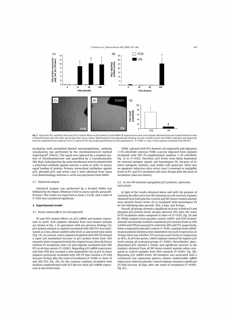
Fig. 1. Pig serum (PS)- and fetal calf serum (FCS)-related effects on p53 protein (A) and mRNA (B) expression in aortic arch explants obtained from non-treated animals at time0 (directly frozen) and 18 h, 48 h, and 10 days after tissue culture. Representative microphotograph showing vascular smooth muscle cell (VSMC) migration and outgrowthfrom the explanted tissue cultured in FCS (C) and in PS (D). Bar-graph quantification of cell-outgrowth (E). *P < 0.001 vs. time = 0 and explants incubated with 20% FCS.
incubation with peroxidase-labeled immunoglobulins, antibodyvisualization was performed by the chemiluminescent methodSuperSignal® (Pierce). The signal was captured by a standard sys-tem of chemiluminescent and quantified by a transilluminator(Bio-Rad). Subsequently, the same membranes were incubated witha polyclonal antibody against porcine �-actin in order to ensureequal loading of protein. Primary monoclonal antibodies againstp53, phospho-p53, and active casp-3 were obtained from SantaCruz Biotechnology whereas �-actin was purchased from DAKO.
2.7. Statistical analysis
Statistical analysis was performed by a Kruskal Wallis testfollowed by the Mann–Whitney-U test to assess specific group dif-ferences. The results are expressed as mean ± S.E.M., and a value ofP < 0.05 was considered significant.
3. Experimental results
3.1. Serum-related effects on cell outgrowth
PS and FCS-related effects on p53 mRNA and protein expres-sion in aortic arch explants obtained from non-treated animalsare shown in Fig. 1. In agreement with our previous findings [5],p53 protein amount in explants incubated with 20% FCS was main-tained at a low, almost undetectable level, at any tested time-point(Fig. 1A). In contrast, aortic explants incubated with 20% PS showeda rapid and maintained increase in p53 protein levels from 18 honwards when compared to both the original tissue (directly frozenwithout PS incubation, time = 0) and explants incubated with 20%FCS at all time-points (P < 0.001). Regarding p53 mRNA expression,real time PCR also revealed a time-dependent rise in p53 in thoseexplants previously incubated with 20% PS that reached a 25-foldincrease 10 days after the onset of incubation (P < 0.001 vs. time = 0and 20% FCS; Fig. 1B). On the contrary, explants incubated withmedium supplemented with FCS did not show p53 mRNA expres-sion at any tested time.
VSMC cultured with FCS showed cell outgrowth and migration(≈70 cells/field) whereas VSMC scarcely migrated from explantsincubated with 20% PS-supplemented medium (≈10 cells/field;Fig. 1C–E; P < 0.05). Therefore, p53 levels seem likely dependenton external mitogen signals and homologous PS, because of itslower mitogenic stimulus, may render cells quiescent. There wasno apoptotic induction since active casp-3 remained at negligiblelevels in PS- and FCS-incubated cells even 10 days after the onset ofincubation (data not shown).
3.2. In vivo NO-donation upregulates p53 synthesis, expression,and activity
In light of the results obtained above and with the purpose ofisolating the effect of in vivo NO-donation on cell-survival, explantsobtained from both placebo-control and NO-donor treated animalswere directly frozen (time = 0) or incubated with homologous PSfor the following time-periods: 18 h, 2 days, and 10 days.
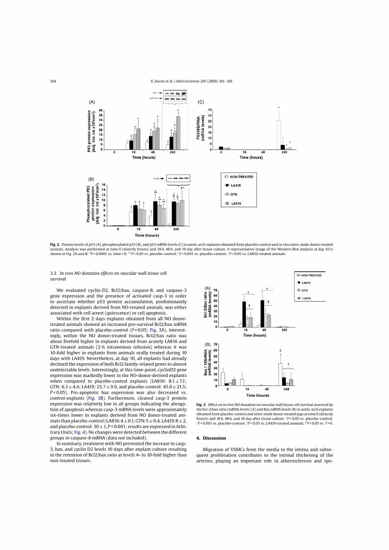
Overall, all groups showed a significant increase in both p53 andphospho-p53 protein levels already detected 18 h after the onsetof PS incubation when compared to time = 0 (P < 0.05; Fig. 2A andB). While explants from placebo-control, LA419- and GTN-treated-animals had already reached a maximum p53 protein levels at 18 h(LA419 and GTN increased p53 content by 30% and 57%, respectively,when compared to placebo-control; P < 0.05), explants from LA816-treated animals showed a time-dependent rise in p53 expression (at10 days there was a further 37% increase in p53 levels in comparisonto 18 h). At all time points, LA816 explants showed the highest p53levels among all evaluated groups (P < 0.001). Nevertheless, phos-phorylated p53 showed a similar and significant increase in theexplants obtained from all NO donor-treated animals when com-pared to control explants from 48 h onwards (P < 0.001; Fig. 2B).Regarding p53 mRNA levels, NO-donation was associated with acontinuous low expression pattern (almost undetectable mRNAexpression) whereas placebo-control explants showed a significant25-fold increase 10 days after the onset of incubation (P < 0.001;Fig. 2C).
104 X. Duran et al. / Atherosclerosis 205 (2009) 101–106
Fig. 2. Protein levels of p53 (A), phosphorylated p53 (B), and p53 mRNA levels (C) in aortic arch explants obtained from placebo-control and in vivo nitric oxide donor-treatedanimals. Analysis was performed at time 0 (directly frozen) and 18 h, 48 h, and 10 day after tissue culture. A representative image of the Western Blot analysis at day 10 isshown in Fig. 2A and B. *P < 0.0001 vs. time = 0; **P < 0.05 vs. placebo-control; †P < 0.001 vs. placebo-control; ‡P < 0.05 vs. LA816-treated animals.
3.3. In vivo NO donation effects on vascular wall tissue cellsurvival
We evaluated cyclin-D2, Bcl2/bax, caspase-8, and caspase-3gene expression and the presence of activated casp-3 in orderto ascertain whether p53 protein accumulation, predominantlydetected in explants derived from NO-treated animals, was eitherassociated with cell arrest (quiescence) or cell apoptosis.
Within the first 2 days explants obtained from all NO donor-treated animals showed an increased pro-survival Bcl2/bax mRNAratio compared with placebo-control (P < 0.05; Fig. 3A). Interest-ingly, within the NO donor-treated tissues, Bcl2/bax ratio wasabout fivefold higher in explants derived from acutely LA816 andGTN-treated animals (2-h intravenous infusion) whereas it was10-fold higher in explants from animals orally treated during 10days with LA419. Nevertheless, at day 10, all explants had alreadydeclined the expression of both Bcl2 family-related genes to almostundetectable levels. Interestingly, at this time-point, cyclinD2 geneexpression was markedly lower in the NO-donor-derived explantswhen compared to placebo-control explants (LA816: 8.1 ± 7.1;GTN: 6.3 ± 4.4; LA419: 25.7 ± 9.9, and placebo-control: 45.0 ± 21.5;P < 0.05). Pro-apoptotic bax expression was also decreased vs.control-explants (Fig. 3B). Furthermore, cleaved casp-3 proteinexpression was relatively low in all groups indicating the abroga-tion of apoptosis whereas casp-3 mRNA levels were approximatelysix-times lower in explants derived from NO donor-treated ani-mals than placebo-control (LA816:4 ± 0.1, GTN:5 ± 0.4, LA419:8 ± 2,and placebo-control: 30 ± 1, P < 0.001; results are expressed in Arbi-trary Units; Fig. 4). No changes were detected between the differentgroups in caspase-8 mRNA (data not included).
In summary, treatment with NO prevented the increase in casp-3, bax, and cyclin D2 levels 10 days after explant culture resultingin the retention of Bcl2/bax ratio at levels 4- to 10-fold higher thannon-treated tissues.
Fig. 3. Effect on in vivo NO donation on vascular wall tissue cell survival assessed bythe bcl-2/bax ratio (mRNA levels) (A) and Bax mRNA levels (B) in aortic arch explantsobtained from placebo-control and nitric oxide donor-treated pigs at time 0 (directlyfrozen) and 18 h, 48 h, and 10 day after tissue culture. *P < 0.05 vs. placebo-control;†P < 0.001 vs. placebo-control; ‡P < 0.05 vs. LA419-treated animals; ıP < 0.05 vs. T = 0.
4. Discussion
Migration of VSMCs from the media to the intima and subse-quent proliferation contributes to the intimal thickening of thearteries, playing an important role in atherosclerosis and spe-

X. Duran et al. / Atherosclerosis 205 (2009) 101–106 105
Fig. 4. Caspase-3 protein expression (A) and mRNA levels (B) in aortic arch explantsobtained from placebo-control and nitric oxide donor-treated pigs at time 0 (directlyfrozen) and 18 h, 48 h, and 10 day after tissue culture. *P < 0.05 vs. time = 0; †P < 0.05vs. placebo-control animals.
cially in restenosis after percutaneous transluminal angioplasty[1]. Therefore, agents that restrict VSMC growth may contributetowards a potentially efficient preventive strategy for coronaryheart disease. Herein, we report that three different NO-releasingcompounds (two novel platelet-selective NO-donors – LA419 andLA816 – and GTN) are capable of modulating VSMC survival whilelikely limiting proliferation and migration, effects that are beyondtheir previously reported antithrombotic and anti-ischemic prop-erties [14–17].
NO has several biochemical activities including mediatingvasodilation, directly scavenging superoxide, attenuating leuko-cyte adhesion and activation, inhibiting platelet aggregation, andmaintaining endothelial integrity [19]. Moreover, NO has shownto exert antiproliferative properties for VSMCs in vitro and in vivosuggesting an important role of NO in atherogenesis [20]. Indeed,upon vascular damage the subsequent disruption in NO produc-tion enhances VSMC proliferation. Concretely, VSMCs undergoa “phenotypic modulation” and migrate from the media to theintima where they begin to proliferate and produce collagen fur-ther straining vascular architecture (i.e., vascular remodelling) [21].In this regard, here we have shown that in vivo treatment withthree differently structured NO-releasing compounds, when givenat therapeutical doses for antiplatelet and anti-ischemic effects,exhibit a significant increase in both p53 content and activity. Acti-vation of the tumour suppressor p53 is best known for its ability toblock VSMC proliferation during the formation of atheroscleroticlesions either by transiently arresting the cell cycle or inducingapoptosis [22,23]. NO has previously shown to produce a dualeffect in VSMC apoptosis depending on the concentration used.It has been reported that physiological amounts of NO have ananti-apoptotic effect [24] while relatively higher concentrationspromote apoptosis [25]. Our results provide evidence that exoge-nous donation of NO at the doses used, both administered acutely(i.e., GTN or LA816) and orally (i.e., LA419), promote vascular cellsquiescence rather than apoptosis, an effect that is maintained forat least 10 days after the withdrawal of NO donation. As such,
NO treatment was associated with a clear reduction in the p53downstream effector cyclin-D2. Indeed, cyclin D1/D2 by associ-ating to Cdc4/6 is essentially involved in the transition from onecell phase to another. Thus, our results demonstrate the ability ofexogenous NO-donation to block cell cycle progression likely driv-ing cells to quiescence [26]. These findings were further supportedby the detected down-regulation in the p53 apoptotic target Bax,key mediator of the mitochondrial-dependent apoptotic pathway.In fact, we did not observe any alterations of caspase-8 expressionlikely excluding NO-effects on the intrinsic apoptotic pathway [27].Furthermore, exogenous NO donation is also associated with anincreased bcl-2/bax ratio further attenuating the pro-apoptotic sig-nals and subsequently favouring a pro-survival state [28]. In fact,active caspase-3 (a well-known irreversible effector of the apop-totic signal) [29] was barely apparent in vascular explants derivedfrom NO-treated animals within the periods analyzed herein. Insummary, administration of LA419, LA816, and GTN at therapeuticaldoses preserves VSMC from apoptosis. Moreover, taking into con-sideration our previous findings, showing that p53 suppression inVSMC is associated with a marked cell out-growth, we hypothesizethat NO treatment may limit VSMC proliferation and migration thusrendering an overall protective mechanism against atherogenesisand vascular remodelling. Further studies are needed to properlyaddress these issues.
We have previously reported in pigs and healthy human subjectsthe powerful antithrombotic effects associated with the adminis-tration of LA816 and LA419 (two novel platelet-selective NO-donorcompounds) [14–17] further supporting their use far beyond theiranti-ischemic properties. Vascular remodelling after balloon dilata-tion is also mediated by an increased thrombus formation. Indeed,upon vascular damage platelets interact with the vessel wall lead-ing to thrombus formation. Once activated platelets release localconcentrations of platelet-derived mitogens that further promoteVSMC proliferation [30] and long-term vascular remodeling. Inthis regard, since we have previously demonstrated the powerfulantiplatelet effects associated to LA419 and LA816 administrationunder the same conditions used to carry out this study, NO donationwith these NO donors not only may reduce thrombus formation butmay also influence VMSC behavior indirectly through their effectson platelets.
It deserves to be acknowledged that our study was carried out inhealthy animals. Certainly, further studies in relevant disease mod-els (NO-deficiency, etc.) seem required to provide further relevantproof-of-concept preclinical data.
In conclusion, proliferation of VSMC has drawn a lot of interestdue to its importance during atherogenesis and vascular remod-elling. In our study we have elucidated the contribution of threedifferent NO donor compounds (LA419, LA816, and GTN) to VSMCsurvival. We have shown that exogenous in vivo NO treatmentpreserves VSMC from apoptosis while renders cells quiescencethus likely limiting proliferation and further migration. Addingthis atheroprotective effect to the previously described powerfulantithrombotic and anti-ischemic properties, NO-donors show apotential valuable therapeutic profile for prevention and treatmentof coronary heart disease and vascular remodelling.
Funding
This work has been supported by the funds provided by CIBER-OBN CB06/03, PN-SAF 2006/10091, and FIC-Catalana Occidente.
Conflict of interest
No conflict of interest to disclose.

106 X. Duran et al. / Atherosclerosis 205 (2009) 101–106
Acknowledgements
We thank Lacer S.A, Spain, for providing the compounds andauthors thank P. Catalina and Ma.A. Cánovas for their technical sup-port. GV is a Juan de la Cierva Investigator of the MEyC of Spain.
References
[1] Chesebro J, Lam J, Badimon L, Fuster V. Restenosis after arterial angioplasty: ahemorrheologic response to injury. Am J Cardiol 1987;60:10B–6B.
[2] Angelini GD, Newby AC. The future of saphenous vein as a coronary arterybypass conduit. Eur Heart J 1989;10:273–80.
[3] Vousden KH. Activation of the p53 tumor suppressor protein. Biochim BiophysActa 2002;1602:47–59.
[4] Villalobo A. Nitric oxide and cell proliferation. FEBS 2006;273:2329–44.[5] Rodriguez-Campos A, Ruiz-Enriquez P, Faraudo S, Badimon L. Mitogen-
induced p53 downregulation precedes vascular smooth muscle cell migrationfrom healthy tunica media and proliferation. Arterioscler Thromb Vasc Biol2001;2:214–9.
[6] Brüne B, von Knethen A, Sandau K. Transcription factors p53 and HIF-1alpha astargets of nitric oxide. Cell Signal 2001;13:525–33.
[7] Auchon D, Jiang X, Morgan D, et al. Three-dimensional structure of the apop-tosome: implications for assembly, procaspase-9 binding, and activation. MolCell 2002;9:423–32.
[8] Walford G, Loscalzo J. Nitric oxide in vascular biology. J Thromb Haemost2003;1:2112–8.
[9] Jeremy JY, Rowe D, Emsley A, Newby A. Nitric oxide and the proliferation ofvascular smooth muscle cells. Cardiovasc Res 1999;43:580–94.
[10] Sarkar R, Gordon D, Stanley J, Webb R. Cell cycle effects of nitric oxide on vascularsmooth muscle cells. Am J Physiol 1997;272:H1810–8.
[11] Tanner F, Meier P, Greutert H, et al. Nitric oxide modulates expression of cellcycle regulatory proteins: a cytostatic strategy for inhibition of human vascularsmooth muscle cell proliferation. Circulation 2000;101:1982–9.
[12] Messmer U, Brüne B. Nitric oxide-induced apoptosis: p53-dependent and p53-independent signaling pathways. Biochem J 1996;319:299–305.
[13] Jeremy JY, Rowe D, Emsley AM, Newby AC. Nitric oxide and the proliferation ofvascular smooth muscle cells. Cardiovasc Res 1999;43:580–94.
[14] Vilahur G, Segales E, Casani L, Badimon L. A novel anti-ischemic nitric oxidedonor inhibits thrombosis without modifying haemodynamic parameters.Thromb Haemost 2004;91:1035–43.
[15] Urooj Zafar M, Vilahur G, Choi BG, et al. A novel anti-ischemic nitric oxide donor(LA419) reduces thrombogenesis in healthy human subjects. J Thromb Haemost2007.
[16] Vilahur G, Pena E, Padro T, Badimon L. Protein disulphide isomerase-mediatedLA419-NO release provides additional antithrombotic effects to the blockade ofthe ADP receptor. Thromb Haemost 2007;97:650–7.
[17] Vilahur G, Segales E, Salas E, Badimon L. Effects of a novel platelet nitric oxidedonor (LA816), aspirin, clopidogrel, and combined therapy in inhibiting flow-and lesion-dependent thrombosis in the porcine ex vivo model. Circulation2004;110:1686–93.
[18] Manabe T, Yamamoto A, Satoh K, Ichihara K. Tolerance to nitroglycerin inducedby isosorbide-5-mononitrate infusion in vivo. Biol Pharm Bull 2001;24:1370–2.
[19] Loscalzo J. Nitric oxide insufficiency, platelet activation, and arterial thrombosis.Circ Res 2001;88:756–62.
[20] Garg UC, Hassid A. Nitric oxide-generating vasodilators and 8-bromo-cyclicguanosine monophosphate inhibit mitogenesis and proliferation of culturedrat vascular smooth muscle cells. J Clin Invest 1989;83:1774–7.
[21] Geng YJ, Libby P. Progression of atheroma: a struggle between death and pro-creation. Arterioscler Thromb Vasc Biol 2002;22:1370–80.
[22] Ihling C, Menzel G, Wellens E, Monting JS, Schaefer HE, Zeiher AM. Topo-graphical association between the cyclin-dependent kinases inhibitor P21, p53accumulation, and cellular proliferation in human atherosclerotic tissue. Arte-rioscler Thromb Vasc Biol 1997;17:2218–24.
[23] Bennett MR, Littlewood TD, Schwartz SM, Weissberg PL. Increased sensitivityof human vascular smooth muscle cells from atherosclerotic plaques to p53-mediated apoptosis. Circ Res 1997;81:591–9.
[24] Dimmeler S, Haendeler J, Nehls M, Zeiher AM. Suppression of apoptosis by nitricoxide via inhibition of interleukin-1beta-converting enzyme (ICE)-like and cys-teine protease protein (CPP)-32-like proteases. J Exp Med 1997;185:601–7.
[25] Pollman MJ, Yamada T, Horiuchi M, Gibbons GH. Vasoactive substances regulatevascular smooth muscle cell apoptosis. Countervailing influences of nitric oxideand angiotensin II. Circ Res 1996;79:748–56.
[26] Sancar A, Lindsey-Boltz LA, Unsal-Kacmaz K, Linn S. Molecular mechanisms ofmammalian DNA repair and the DNA damage checkpoints. Annu Rev Biochem2004;73:39–85.
[27] Hengartner MO. The biochemistry of apoptosis. Nature 2000;407:770–6.[28] Xie K, Huang S, Wang Y, et al. Bcl-2 protects cells from cytokine-induced nitric-
oxide-dependent apoptosis. Cancer Immunol Immunother 1996;43:109–15.[29] Green DR, Reed JC. Mitochondria and apoptosis. Science 1998;281:1309–12.[30] Janssens S, Flaherty D, Nong Z, et al. Human endothelial nitric oxide synthase
gene transfer inhibits vascular smooth muscle cell proliferation and neointimaformation after balloon injury in rats. Circulation 1998;97:1274–81.
133

134

135

136

Annex
137

Annex
138

ORIGINAL ARTICLE
Short-term myocardial ischemia induces cardiac modifiedC-reactive protein expression and proinflammatory gene(cyclo-oxygenase-2, monocyte chemoattractant protein-1, andtissue factor) upregulation in peripheral blood mononuclearcells
G. V I LAHUR ,*� R . HERNANDEZ-VERA ,*� B . MOL INS , *� L . CASAN I , *� X . DURAN,* T . PADRO* and
L . BAD IMON*��*Cardiovascular Research Centre, CSIC-ICCC, Hospital de la Santa Creu i Sant Pau, Barcelona; �CIBEROBN-Instituto Salud Carlos III, Barcelona;
and �Cardiovascular Research Chair, UAB, Barcelona, Spain
To cite this article: Vilahur G, Hernandez-Vera R, Molins B, Casanı L, Duran X, Padro T, Badimon L. Short-term myocardial ischemia induces cardiac
modified C-reactive protein expression and proinflammatory gene (cyclo-oxygenase-2, monocyte chemoattractant protein-1, and tissue factor)
upregulation in peripheral blood mononuclear cells. J Thromb Haemost 2009; 7: 485�93.
Summary. Background: Prompt coronary thrombus resolu-
tion, reducing time of ischemia, improves cardiac recovery. The
factors triggered by ischemia that contribute to the clinical
outcome are not fully known. We hypothesize that unabated
inflammation due to cardiac ischemia may be a contributing
factor. Aims: As a proof-of-concept, we evaluated the effect of
short-term myocardial ischemia on the local and systemic
inflammatory response.Methods:Pigs underwent either 90-min
mid-left anterior descending (LAD) coronary artery balloon
occlusion (infarct size 25% ± 1% left ventricle; 29% heart
function deterioration) or a sham-operation procedure. Peri-
infarcted and non-ischemic cardiac tissue was obtained for
histopathologic, molecular and immunohistochemical analysis
of inflammatory markers [interleukin-6 (IL-6), tumor necrosis
factor-a (TNF-a), modified C-reactive protein (mCRP), and
human alveolar macrophage-56 (HAM-56)]. Blood (femoral
vein) was withdrawn prior to myocardial infarction (MI)
induction (t = 0) and at 30 and 90 min to evaluate: (i) systemic
cytokine levels (IL-6, TNF-a, CRP); (ii) proinflammatory gene
and protein expression in peripheral blood mononuclear cells
(PBMCs) of tissue factor (TF), cyclo-oxygenase-2 (Cox-2),
monocyte chemoattractant protein-1 (MCP-1), and CRP; and
(iii) platelet activation (assessed by perfusion studies and RhoA
activation).Results: Short-term ischemia triggered cardiac IL-6
and TNF-a expression, recruitment of inflammatory cells, and
mCRP expression in infiltrated macrophages (P < 0.05 vs.
t = 0 and sham). PBMC mRNA and protein expression of
MCP-1, Cox-2 and TF was significantly increased by ischemia,
whereas no differences were detected in CRP. Ischemia
increased cardiac troponin-I, IL-6 and TNF-a systemic levels,
and was associated with higher platelet deposition and RhoA
activation (P < 0.001 vs. t = 0 and sham).Conclusion: Short-
term myocardial ischemia, even without atherosclerosis,
induces an inflammatory phenotype by inducing local recruit-
ment of macrophages and systemic activation of mononuclear
cells, and renders platelets more susceptible to activation.
Keywords: inflammation, ischemia, mCRP, mononuclear cells,
myocardial infarction, platelets.
Occlusive intracoronary thrombosis leads to myocardial
ischemia and to cardiac dysfunction. Several studies have
shown that serum cardiac troponin levels are associated with
myocardial tissue injury and are powerful predictors of both
short-term and long-term adverse outcomes in patients with
ST-segment elevation myocardial infarction (STEMI) [1].
Thus, early detection of acute myocardial infarction (AMI)
and prompt coronary thrombus resolution (i.e. fibrinolytic
therapy or mechanical reperfusion) may reduce the extent
of necrosis and facilitate myocardial function recovery. The
factors contributing to the impact of symptom-onset-to-
reperfusion time on clinical outcomes and even mortality are
not fully known. It is our hypothesis that unabated inflamma-
tion due to ischemia may be a contributing factor.
Although there is overwhelming evidence that an inflamma-
tory outburst is associated with acute coronary syndromes, the
causes and nature are still largely unknown. Indeed, it is widely
recognized that the atherosclerotic process and its thrombotic
complications constitute a source of inflammatory stimuli [2,3].
Correspondence: Lina Badimon, Cardiovascular Research Centre,
c/Sant Antoni MªClaret 167, 08025 Barcelona, Spain.
Tel.: +34 935 565880; fax: +34 935 565559.
E-mail: [email protected]
Received 4 August 2008, accepted 20 November 2008
Journal of Thrombosis and Haemostasis, 7: 485�493 DOI: 10.1111/j.1538-7836.2008.03244.x
� 2008 International Society on Thrombosis and Haemostasis

However, several reports have postulated the existence of
alternative inflammatory sources [4]. In this regard, reperfusion
of the ischemic heart has been shown to play a prominent role
in the inflammatory response, either by recruiting inflamma-
tory cells to the site of injury [5,6] or through the overproduc-
tion of reactive oxygen radicals, further enhancing the local
inflammatory milieu [7]. In addition, myocardial necrotic tissue
has also been suggested to contribute to the release of
proinflammatory cytokines, including interleukin-6 (IL-6),
interleukin-1, and tumor necrosis factor-a (TNF-a), which in
turn induce hepatic production of native C-reactive protein
(nCRP), a key systemic inflammatory marker [8]. Indeed,
serum levels of the acute-phase reactant nCRP have been
linked to myocardial necrosis in STEMI patients [4], indicating
the ability of myocyte damage to disseminate the local
inflammatory response to distal organs through C-reactive
protein (CRP). However, AMI patients generally present an
atherosclerosis burden concurrently with a combination of risk
factors, which makes it difficult to determine the individual
contribution of each proinflammatory stimulus to the overall
inflammatory phenotype. Hence, the precise site, stimulus and
mechanisms that cause a systemic inflammatory response after
AMI are not completely understood and remain controversial.
Furthermore, to the best of our knowledge, whether ischemia
itself, without underlying coronary vascular disease, may
acutely cause a local and systemic inflammatory response has
not been formerly addressed in a human-like animal model of
coronary occlusion. Thus, the aim of our study was to evaluate,
in a pig model of closed-chest total coronary occlusion, the
effect of ischemia per se on local and systemic inflammatory
responses. Here we report, as a proof-of-concept, that short-
term myocardial ischemia suffices to induce activation of
myocardium-recruited and circulating peripheral mononuclear
cells and to render platelets more susceptible to activation and
deposition.
Materials and methods
Experimental procedures
AMI induction Crossbred commercial pigs (Landrace-
Largewhite; n = 19) weighing 35�40 kg were acclimated for
1 week before any experimental procedure. Twelve hours
before the experimental coronary occlusion, a loading dose of
clopidogrel (150 mg) was administered to avoid thrombotic
complications due to catheter manipulation. AMI (n = 15)
was experimentally induced by complete balloon occlusion in
the left anterior descending (LAD) coronary artery as
previously described [9] (see Supporting Information). The
study protocol was approved by an institutional animal
research committee and conformed to the Guide for the Care
and Use of Laboratory Animals published by the US National
Institutes of Health. A sham-operated group of animals
(n = 4) underwent the same experimental procedure
(antithrombotic therapy, anesthesia, catheterization, etc.) but
without balloon occlusion.
Animal monitoring The hemodynamic parameters (heart
rate, mean systemic arterial pressure, mixed venous oxygen
saturation) were recorded for all animals throughout the study.
We used two-dimensional echocardiograms (Phillips iE33,
Amsterdam, the Netherlands) to assess global heart function
(i.e. left ventricle ejection fraction) in all animals at baseline
(T0) and 90 min after the onset of ischemia (T90).
Histopathologic evaluation of the infarcted myocardium
After 90 min, animals from both groups (sham-operated,
n = 4; animals subjected to ischemia, n = 7) were killed;
hearts were perfused with cold phosphate-buffered saline
(PBS), and subjected to a 1-h freeze cycle at ) 20 �C in
order to stiffen the tissue for further slicing. Hearts from
both ischemic and non-ischemic animals were sliced into 5-
mm-thick slices from the apex towards the atrioventricular
groove. Consecutive slices were alternately collected for
triphenyl tetrazolium chloride staining for infarct size
measurement (% left ventricle), as previously described [9],
and molecular and histologic examination of the perinecrotic
area (3 mm surrounding the necrotic core) and the
topographically corresponding area in non-ischemic animals.
For these purposes, samples were either directly frozen in
liquid nitrogen for mRNA and protein analysis of several
inflammatory markers (described below), or embedded in
optimal cutting temperature compound (OCT) and frozen.
Four 5-lm sections were obtained from each of these latter
samples, and mounted on gelatinized slides for: (i) conven-
tional hematoxylin�eosin staining for nuclear staining and
inflammatory infiltration visualization; (ii) modified CRP
(mCRP) detection (porcine mCRP antibody kindly provided
by L. A. Potempa); and (iii) macrophage staining [human
alveolar macrophage-56 (HAM-56) antibody, Dako]. Images
were captured with an Olympus Vanox AHBT3 microscope
and digitalized with a Sony 3CCD.
Blood sampling
Once the animal was fully anesthetized and the vital signs were
stable, the femoral vein was accessed percutaneously with an
18-gauge needle and a 6F arterial sheath for blood withdraw-
als. Blood was drawn at three different time points: T0
(baseline), which was 20 min after heparin bolus administra-
tion and just prior to the start of LAD coronary artery
occlusion; T30 (30 min after onset of ischemia); and T90
(90 min after start of ischemia). The first 3 mL of blood were
discarded, and blood was withdrawn gently to minimize shear-
induced platelet activation. Blood from sham-operated animals
was drawn at the same time points.
Cardiac troponin-I (cTnI) levels Blood was induced to clot
(20 min at 37 �C) to obtain serum (4 �C, 15 min, 1800 g)
that was stored at) 80 �Cuntil cTnI analysis. The quantitative
determination of pig cTnI in serum was performed by using an
ADVIA Centaur Automated Chemiluminescence System
486 G. Vilahur et al
� 2008 International Society on Thrombosis and Haemostasis
(Bayer Corporation) in a medical analysis laboratory. The
sensitivity threshold for cTnI was 0.01 ng mL)1. Values above
0.1 ng mL)1 were considered to be positive for cTnI.
Hematologic, coagulation and biochemical parameters Blood
was collected in sodium citrate (0.16 mol L)1), gently mixed,
and immediately centrifuged at 2800 · g to obtain plasma for
subsequent hematologic determinations (System 9000, Serono-
Baker Diagnostics, Allentown, Philadelphia, PA, USA),
coagulometry [fibrinogen concentration, standard prothrom-
bin time (PT), activated partial thromboplastin time (APTT);
ST4, Diagnostica Stago] and routine biochemical analysis
(glucose, lipids, and liver and kidney parameters).
Circulating cytokines and nCRP measurement: Cytokines
(IL-6 and TNF-a) and nCRP were analyzed in plasma and
serum, respectively, using commercially available enzyme-
linked immunosorbent assay kits (porcine IL-6 and porcine
TNF-a from Quantikine Porcine Kits from R&D Systems;
porcine nCRP from Immunological Consultant Laboratories).
According to the manufacturers, the minimum detection limits
for IL-6, TNF-a and nCRP are 10 pg mL)1, 3.7 pg mL)1, and
20 ng mL)1, respectively.
Peripheral blood mononuclear cell (PBMC) isolation: Twenty
milliliters of blood were collected into EDTA tubes at the three
tested timepoints (T0,T30, andT90) fromall animals.All blood
samples were immediately subjected to Ficoll-paque Plus
(Amersham Biosciences, Piscataway, NJ, USA) density
gradient centrifugation to isolate PBMCs. Samples were
counted and then directly frozen in liquid nitrogen and stored
at ) 80 �C until mRNA and protein evaluation.
Real-time polymerase chain reaction (PCR) analysis
Perinecrotic and non-ischemic myocardial tissue, and PBMC
mRNA and protein, were extracted with Tripure Reagent
(Roche Molecular Biochemicals, Indianapolis, IN, USA)
according to the manufacturer�s instructions. Tissue factor
(TF), cyclo-oxygenase (Cox)-2, CRP and monocyte chemo-
attractant protein-1 (MCP-1/CCL2) mRNA levels were ana-
lyzed by real-time PCR (7000 Sequence Detection System of
ABIPRISM; Applied Biosystems) in both PBMCs and the
perinecrotic myocardium. In addition, IL-6 and TNF-a gene
expression was also evaluated in the cardiac tissue. TaqMan
fluorescent real-time PCR primers and probes (6-FAM-MGB)
were designed with the use of PRIMER EXPRESS 2.0 software
from Applied Biosystems, and real-time PCR was performed
(see Supporting Information). The threshold cycle (Ct) values
were determined and normalized to the housekeeping gene 18S
rRNA in order to adjust for equal amounts of RNA.
Immunoblotting
The protein phase recovered from the Tripure Reagent was
quantified by using the bicinchonic acid method (Pierce
Biotechnology, Rockford, IL, USA). Samples (30 lg per lane)were subjected to sodium dodecylsulfate polyacrylamide gel
electrophoresis and blotted onto nitrocellulose membranes.
Blots were blocked for 2 h at room temperature, and then
incubated overnight at 4 �C with antibodies known to cross-
react with pig proteins, including anti-TF (American Diag-
nostica, Greenwich, CT, USA), anti-Cox-2 (Santa Cruz
Biotechnology, Santa Cruz, CA, USA), anti-MCP-1/CCL2
(Cell Signaling), and anti-mCRP (monocloncal mCRP; kindly
provided by L. A. Potempa). Thereafter, membranes were
incubated with horseradish peroxidase-conjugated secondary
antibodies and detected with a SuperSignal chemiluminescence
system (Pierce). Protein expression was determined using a
computerized software package (QUANTITY-ONE; Bio-Rad),
and b-actin was used as a protein loading control.
Platelet activation assays
Platelet adhesion Type I collagen-coated slides were prepared
and placed in a parallel plate chamber, described in detail
elsewhere [10]. Briefly, the flow chamber was assembled and
filled with Tyrode�s buffer. A peristaltic pump was used to
perfuse the buffer through the chamber. After 1 min of buffer
preperfusion, blood was introduced into the chamber at a
constant shear rate of 1500 s)1 for 5 min. At the end of blood
perfusion, buffer was again circulated for 1 min through the
chamber under identical flow conditions. The entire systemwas
kept at 37 �C. After perfusions, collagen-coated slides were
carefully removed from the system, rinsed with PBS, and fixed
with paraformaldehyde for 15 min. The fixed slides were then
washed with PBS and mounted on glass slides with Glycerol
Mounting Medium (Dako Cytomation, Copenhagen,
Denmark). Five fields along the adhesion surface were
systematically acquired for total platelet deposition (field:
750 · 750 lm). Images were analyzed with a commercial
software package (METAMORPH; Universal Imaging Corp.,
West Chester, PA, USA). The surface covered by platelets was
expressed as the area covered by platelets per field analyzed
(lm2/field). Further information regarding platelet adhesion
analysis is provided in the supporting information.
Western blot analysis of platelet-activated RhoA Blood was
collected in acid citrate dextrose solution for platelet RhoA
antigen determination (anti-RhoA; Santa Cruz Biotech-
nology). Platelets were isolated and subfractioned for both
total and cytosolic/membrane RhoA fraction evaluation, as
previously described [11].
Statistical analysis
Continuous variables are expressed as mean ± standard error
of themean. The differences between different time periods and
animal groups were evaluated using a one-way ANOVA followed
by Scheffe�s comparison. A P-value < 0.05 was considered to
be significant. All statistical analyses were performed with the
statistical software package STATVIEW.
Ischemia effects on local and systemic inflammation 487
� 2008 International Society on Thrombosis and Haemostasis
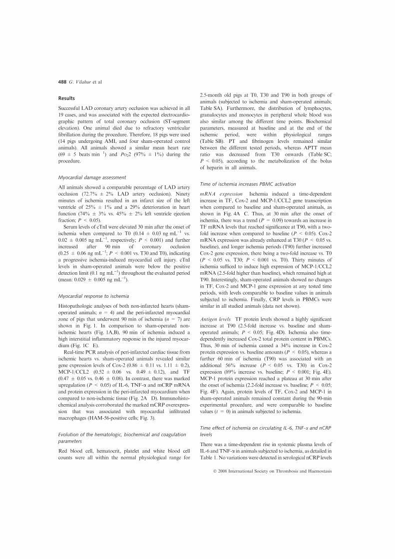
Results
Successful LAD coronary artery occlusion was achieved in all
19 cases, and was associated with the expected electrocardio-
graphic pattern of total coronary occlusion (ST-segment
elevation). One animal died due to refractory ventricular
fibrillation during the procedure. Therefore, 18 pigs were used
(14 pigs undergoing AMI, and four sham-operated control
animals). All animals showed a similar mean heart rate
(69 ± 5 beats min�1) and PO22 (97% ± 1%) during the
procedure.
Myocardial damage assessment
All animals showed a comparable percentage of LAD artery
occlusion (72.7% ± 2% LAD artery occlusion). Ninety
minutes of ischemia resulted in an infarct size of the left
ventricle of 25% ± 1% and a 29% deterioration in heart
function (74% ± 3% vs. 45% ± 2% left ventricle ejection
fraction; P < 0.05).
Serum levels of cTnI were elevated 30 min after the onset of
ischemia when compared to T0 (0.14 ± 0.03 ng mL)1 vs.
0.02 ± 0.005 ng mL)1, respectively; P < 0.001) and further
increased after 90 min of coronary occlusion
(0.25 ± 0.06 ng mL)1; P < 0.001 vs. T30 and T0), indicating
a progressive ischemia-induced myocardial cell injury. cTnI
levels in sham-operated animals were below the positive
detection limit (0.1 ng mL)1) throughout the evaluated period
(mean: 0.029 ± 0.005 ng mL)1).
Myocardial response to ischemia
Histopathologic analyses of both non-infarcted hearts (sham-
operated animals; n = 4) and the peri-infarcted myocardial
zone of pigs that underwent 90 min of ischemia (n = 7) are
shown in Fig. 1. In comparison to sham-operated non-
ischemic hearts (Fig. 1A,B), 90 min of ischemia induced a
high interstitial inflammatory response in the injured myocar-
dium (Fig. 1C�E).
Real-time PCR analysis of peri-infarcted cardiac tissue from
ischemic hearts vs. sham-operated animals revealed similar
gene expression levels of Cox-2 (0.86 ± 0.11 vs. 1.11 ± 0.2),
MCP-1/CCL2 (0.52 ± 0.06 vs. 0.49 ± 0.12), and TF
(0.47 ± 0.05 vs. 0.46 ± 0.08). In contrast, there was marked
upregulation (P < 0.05) of IL-6, TNF-a and mCRP mRNA
and protein expression in the peri-infarcted myocardium when
compared to non-ischemic tissue (Fig. 2A�D). Immunohisto-
chemical analysis corroborated the marked mCRP overexpres-
sion that was associated with myocardial infiltrated
macrophages (HAM-56-positive cells; Fig. 3).
Evolution of the hematologic, biochemical and coagulation
parameters
Red blood cell, hematocrit, platelet and white blood cell
counts were all within the normal physiological range for
2.5-month old pigs at T0, T30 and T90 in both groups of
animals (subjected to ischemia and sham-operated animals;
Table SA). Furthermore, the distribution of lymphocytes,
granulocytes and monocytes in peripheral whole blood was
also similar among the different time points. Biochemical
parameters, measured at baseline and at the end of the
ischemic period, were within physiological ranges
(Table SB). PT and fibrinogen levels remained similar
between the different tested periods, whereas APTT mean
ratio was decreased from T30 onwards (Table SC;
P < 0.05), according to the metabolization of the bolus
of heparin in all animals.
Time of ischemia increases PBMC activation
mRNA expression Ischemia induced a time-dependent
increase in TF, Cox-2 and MCP-1/CCL2 gene transcription
when compared to baseline and sham-operated animals, as
shown in Fig. 4A�C. Thus, at 30 min after the onset of
ischemia, there was a trend (P = 0.09) towards an increase in
TF mRNA levels that reached significance at T90, with a two-
fold increase when compared to baseline (P < 0.05). Cox-2
mRNA expression was already enhanced at T30 (P < 0.05 vs.
baseline), and longer ischemia periods (T90) further increased
Cox-2 gene expression, there being a two-fold increase vs. T0
(P < 0.05 vs. T30; P < 0.001 vs. T0). Thirty minutes of
ischemia sufficed to induce high expression of MCP-1/CCL2
mRNA (2.5-fold higher than baseline), which remained high at
T90. Interestingly, sham-operated animals showed no changes
in TF, Cox-2 and MCP-1 gene expression at any tested time
periods, with levels comparable to baseline values in animals
subjected to ischemia. Finally, CRP levels in PBMCs were
similar in all studied animals (data not shown).
Antigen levels TF protein levels showed a highly significant
increase at T90 (2.5-fold increase vs. baseline and sham-
operated animals; P < 0.05; Fig. 4D). Ischemia also time-
dependently increased Cox-2 total protein content in PBMCs.
Thus, 30 min of ischemia caused a 34% increase in Cox-2
protein expression vs. baseline amounts (P < 0.05), whereas a
further 60 min of ischemia (T90) was associated with an
additional 56% increase (P < 0.05 vs. T30) in Cox-2
expression (89% increase vs. baseline; P < 0.001; Fig. 4E).
MCP-1 protein expression reached a plateau at 30 min after
the onset of ischemia (2.2-fold increase vs. baseline; P < 0.05;
Fig. 4F). Again, protein levels of TF, Cox-2 and MCP-1 in
sham-operated animals remained constant during the 90-min
experimental procedure, and were comparable to baseline
values (t = 0) in animals subjected to ischemia.
Time effect of ischemia on circulating IL-6, TNF-a and nCRP
levels
There was a time-dependent rise in systemic plasma levels of
IL-6 and TNF-a in animals subjected to ischemia, as detailed in
Table 1.No variations were detected in serological nCRP levels
488 G. Vilahur et al
� 2008 International Society on Thrombosis and Haemostasis
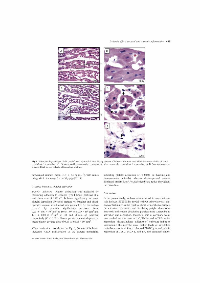
between all animals (mean: 34.4 ± 3.6 ng mL)1), with values
being within the range for healthy pigs [12,13].
Ischemia increases platelet activation
Platelet adhesion Platelet activation was evaluated by
measuring adhesion to collagen type I fibrils perfused at a
wall shear rate of 1500 s)1. Ischemia significantly increased
platelet deposition (five-fold increase vs. baseline and sham-
operated animals at all tested time points; Fig. 5); the surface
covered by platelets significantly increased from
0.23 ± 0.09 · 105 lm2 at T0 to 1.07 ± 0.029 · 105 lm2 and
1.03 ± 0.021 · 105 lm2 at 30 and 90 min of ischemia,
respectively (P < 0.001). Sham-operated animals displayed a
mean platelet-covered area of 0.21 ± 0.026 · 105 lm2.
RhoA activation As shown in Fig. 6, 30 min of ischemia
increased RhoA translocation to the platelet membrane,
indicating platelet activation (P < 0.001 vs. baseline and
sham-operated animals), whereas sham-operated animals
displayed similar RhoA cytosol/membrane ratios throughout
the procedure.
Discussion
In the present study, we have demonstrated, in an experimen-
tally induced STEMI-like model without atherosclerosis, that
myocardial injury as the result of short-term ischemia triggers
the activation of recruited and circulating peripheral mononu-
clear cells and renders circulating platelets more susceptible to
activation and deposition. Indeed, 90 min of coronary occlu-
sion resulted in an increase in IL-6, TNF-a and mCRP cardiac
expression, histopathologic evidence of leukocyte infiltrates
surrounding the necrotic area, higher levels of circulating
proinflammatory cytokines, enhanced PBMC gene and protein
expression of Cox-2, MCP-1, and TF, and increased platelet
240x
A B
900x
C
240x
D
900x
900x
E
Fig. 1. Histopathologic analysis of the peri-infarcted myocardial zone. Ninety minutes of ischemia was associated with inflammatory infiltrate in the
peri-infarcted myocardium (C�E), as assessed by hematoxylin�eosin staining, when compared to non-infarcted myocardium (A, B) from sham-operated
animals. Black arrows indicate inflammatory infiltrate.
Ischemia effects on local and systemic inflammation 489
� 2008 International Society on Thrombosis and Haemostasis

adhesion to collagen-coated surfaces. In addition, we report for
the first time that infiltrated cardiac macrophages express
mCRP. Certainly, our results do not exclude the expression of
mCRP by other cardiac resident or infiltrating cells. Recently,
distinct isoforms of CRP with different activities and functions
have been described; the classically studied serum CRP, the
pentameric or native CRP (nCRP) [14], and the monomeric or
modified CRP (mCRP) found in fibrous tissues of normal and
IL-6
/18s
rR
NA
0.4
0.2
0.0SHAM 90 min Post-AMI
0.6
0.8
1.0
SHAM 90 min Post-AMI
6.05.04.0
TN
F-α
/18s
rR
NA
3.02.01.00.0
7.0
SHAM 90 min Post-AMI
CR
P/1
8s r
RN
A
0.4
0.6
0.8
1.0C
A B
0.2
0.0 SHAM 90 min Post-AMI
5.0*
**
* 4.0
3.0
2.0
1.0
0.0
mC
RP
/β-a
ctin
mCRP (23 kDa)
β-actin (42 kDa)
D
Fig. 2. Interleukin-6 (IL-6), tumor necrosis factor-a (TNF-a) and C-reactive protein (CRP) mRNA analysis in the peri-infarcted myocardium. Coronary
occlusion for 90 min was associated with clear upregulation of IL-6, TNF-a and CRP gene expression levels in the peri-infarcted myocardial zone when
compared to non-ischemic animals (sham-operated animals). Modified CRP protein was also enhanced after the ischemic period (C). AMI, acute
myocardial infarction; mCRP, modified CRP. *P < 0.05 vs. sham-operated animals.
D
900x
mCRP
A
900x
HAM-56
B
900x
900x
C
Fig. 3. C-reactive protein (CRP) immunohistochemical localization in the peri-infarcted myocardium. Modified CRP (mCRP; A, C) and macrophage
[human alveolar macrophage-56 (HAM-56); B, D] content was assessed by immunohistochemical analysis in both sham-operated animals (A, B) and
ischemic animals (C, D). Immunostaining of the peri-infarctedmyocardium indicates mCRP-positive staining in cardiac infiltratedmacrophages (see black
arrows). Perinecrotic myocardium shows loss of integrity because of ischemic injury.
490 G. Vilahur et al
� 2008 International Society on Thrombosis and Haemostasis
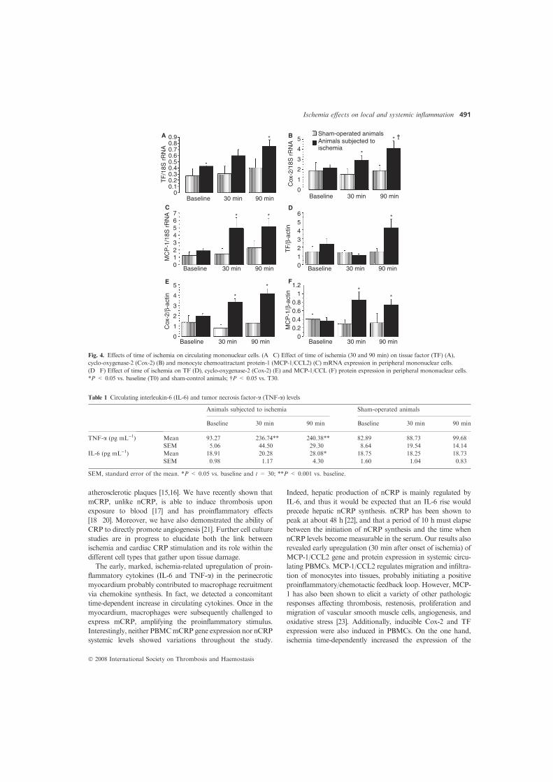
atherosclerotic plaques [15,16]. We have recently shown that
mCRP, unlike nCRP, is able to induce thrombosis upon
exposure to blood [17] and has proinflammatory effects
[18�20]. Moreover, we have also demonstrated the ability of
CRP to directly promote angiogenesis [21]. Further cell culture
studies are in progress to elucidate both the link between
ischemia and cardiac CRP stimulation and its role within the
different cell types that gather upon tissue damage.
The early, marked, ischemia-related upregulation of proin-
flammatory cytokines (IL-6 and TNF-a) in the perinecrotic
myocardium probably contributed to macrophage recruitment
via chemokine synthesis. In fact, we detected a concomitant
time-dependent increase in circulating cytokines. Once in the
myocardium, macrophages were subsequently challenged to
express mCRP, amplifying the proinflammatory stimulus.
Interestingly, neither PBMCmCRP gene expression nor nCRP
systemic levels showed variations throughout the study.
Indeed, hepatic production of nCRP is mainly regulated by
IL-6, and thus it would be expected that an IL-6 rise would
precede hepatic nCRP synthesis. nCRP has been shown to
peak at about 48 h [22], and that a period of 10 h must elapse
between the initiation of nCRP synthesis and the time when
nCRP levels become measurable in the serum. Our results also
revealed early upregulation (30 min after onset of ischemia) of
MCP-1/CCL2 gene and protein expression in systemic circu-
lating PBMCs. MCP-1/CCL2 regulates migration and infiltra-
tion of monocytes into tissues, probably initiating a positive
proinflammatory/chemotactic feedback loop. However, MCP-
1 has also been shown to elicit a variety of other pathologic
responses affecting thrombosis, restenosis, proliferation and
migration of vascular smooth muscle cells, angiogenesis, and
oxidative stress [23]. Additionally, inducible Cox-2 and TF
expression were also induced in PBMCs. On the one hand,
ischemia time-dependently increased the expression of the
Baseline 30 min 90 min0123456
D
TF
/β-a
ctin
0Baseline 30 min 90 min
Sham-operated animalsAnimals subjected toischemia
0.10.20.30.40.50.60.70.80.9 *
* *
*
**
*
*
*
* †
TF
/18S
rR
NA
A
0
1
2
3
4
5B
Baseline
Cox
-2/1
8S r
RN
A
30 min 90 min
Baseline 30 min 90 min01234567
C
MC
P-1
/18S
rR
NA
0
1
2
3
4
5E
Baseline
Cox
-2/β
-act
in
30 min 90 min
F
Baseline 30 min 90 min0
11.2
0.2
0.8
0.40.6
MC
P-1
/β-a
ctin
Fig. 4. Effects of time of ischemia on circulating mononuclear cells. (A�C) Effect of time of ischemia (30 and 90 min) on tissue factor (TF) (A),
cyclo-oxygenase-2 (Cox-2) (B) and monocyte chemoattractant protein-1 (MCP-1/CCL2) (C) mRNA expression in peripheral mononuclear cells.
(D�F) Effect of time of ischemia on TF (D), cyclo-oxygenase-2 (Cox-2) (E) and MCP-1/CCL (F) protein expression in peripheral mononuclear cells.
*P < 0.05 vs. baseline (T0) and sham-control animals; �P < 0.05 vs. T30.
Table 1 Circulating interleukin-6 (IL-6) and tumor necrosis factor-a (TNF-a) levels
Animals subjected to ischemia Sham-operated animals
Baseline 30 min 90 min Baseline 30 min 90 min
TNF-a (pg mL)1) Mean 93.27 236.74** 240.38** 82.89 88.73 99.68
SEM 5.06 44.50 29.30 8.64 19.54 14.14
IL-6 (pg mL)1) Mean 18.91 20.28 28.08* 18.75 18.25 18.73
SEM 0.98 1.17 4.30 1.60 1.04 0.83
SEM, standard error of the mean. *P < 0.05 vs. baseline and t = 30; **P < 0.001 vs. baseline.
Ischemia effects on local and systemic inflammation 491
� 2008 International Society on Thrombosis and Haemostasis
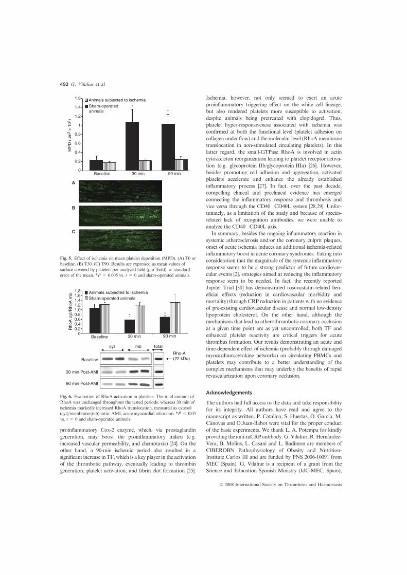
proinflammatory Cox-2 enzyme, which, via prostaglandin
generation, may boost the proinflammatory milieu (e.g.
increased vascular permeability, and chemotaxis) [24]. On the
other hand, a 90-min ischemic period also resulted in a
significant increase in TF,which is a key player in the activation
of the thrombotic pathway, eventually leading to thrombin
generation, platelet activation, and fibrin clot formation [25].
Ischemia, however, not only seemed to exert an acute
proinflammatory triggering effect on the white cell lineage,
but also rendered platelets more susceptible to activation,
despite animals being pretreated with clopidogrel. Thus,
platelet hyper-responsiveness associated with ischemia was
confirmed at both the functional level (platelet adhesion on
collagen under flow) and the molecular level (RhoAmembrane
translocation in non-stimulated circulating platelets). In this
latter regard, the small-GTPase RhoA is involved in actin
cytoskeleton reorganization leading to platelet receptor activa-
tion (e.g. glycoprotein IIb/glycoprotein IIIa) [26]. However,
besides promoting cell adhesion and aggregation, activated
platelets accelerate and enhance the already established
inflammatory process [27]. In fact, over the past decade,
compelling clinical and preclinical evidence has emerged
connecting the inflammatory response and thrombosis and
vice versa through the CD40�CD40L system [28,29]. Unfor-
tunately, as a limitation of the study and because of species-
related lack of recognition antibodies, we were unable to
analyze the CD40�CD40L axis.
In summary, besides the ongoing inflammatory reaction in
systemic atherosclerosis and/or the coronary culprit plaques,
onset of acute ischemia induces an additional ischemia-related
inflammatory boost in acute coronary syndromes. Taking into
consideration that the magnitude of the systemic inflammatory
response seems to be a strong predictor of future cardiovas-
cular events [2], strategies aimed at reducing the inflammatory
response seem to be needed. In fact, the recently reported
Jupiter Trial [30] has demonstrated rosuvastatin-related ben-
eficial effects (reduction in cardiovascular morbidity and
mortality) through CRP reduction in patients with no evidence
of pre-existing cardiovascular disease and normal low-density
lipoprotein cholesterol. On the other hand, although the
mechanisms that lead to atherothrombotic coronary occlusion
at a given time point are as yet uncontrolled, both TF and
enhanced platelet reactivity are critical triggers for acute
thrombus formation. Our results demonstrating an acute and
time-dependent effect of ischemia (probably through damaged
myocardium/cytokine networks) on circulating PBMCs and
platelets may contribute to a better understanding of the
complex mechanisms that may underlay the benefits of rapid
revascularization upon coronary occlusion.
Acknowledgements
The authors had full access to the data and take responsibility
for its integrity. All authors have read and agree to the
manuscript as written. P. Catalina, S. Huertas, O. Garcıa, M.
Canovas and O.Juan-Babot were vital for the proper conduct
of the basic experiments. We thank L. A. Potempa for kindly
providing the anti-mCRP antibody.G. Vilahur, R.Hernandez-
Vera, B. Molins, L. Casanı and L. Badimon are members of
CIBEROBN Pathophysiology of Obesity and Nutrition-
Institute Carlos III and are funded by PNS 2006-10091 from
MEC (Spain). G. Vilahur is a recipient of a grant from the
Science and Education Spanish Ministry (JdC-MEC, Spain),
MP
D (
µm2
× 1
05)
0Baseline 30 min
A
B
C
90 min
0.2
0.4
0.6
0.8
1
1.2
1.4 **
1.6 Animals subjected to ischemiaSham-operatedanimals
Fig. 5. Effect of ischemia on mean platelet deposition (MPD). (A) T0 or
baseline. (B) T30. (C) T90. Results are expressed as mean values of
surface covered by platelets per analyzed field (lm2/field) ± standard
error of the mean. *P < 0.005 vs. t = 0 and sham-operated animals.
Animals subjected to ischemiaSham-operated animals
Baseline
* *
0
Rho
A c
yt/R
hoA
mb
0.20.40.60.81.01.21.41.61.8
Baseline
cyt mb Total
Rho-A(22 kDa)
30 min
30 min Post-AMI
90 min Post-AMI
90 min
Fig. 6. Evaluation of RhoA activation in platelets. The total amount of
RhoA was unchanged throughout the tested periods, whereas 30 min of
ischemia markedly increased RhoA translocation, measured as cytosol
(cyt)/membrane (mb) ratio. AMI, acute myocardial infarction. *P < 0.05
vs. t = 0 and sham-operated animals.
492 G. Vilahur et al
� 2008 International Society on Thrombosis and Haemostasis

R. Hernandez-Vera is a pre-doctoral CSIC fellow (JAE-CSIC,
Spain), and B.Molins is a pre-doctoral fellow of Generalitat de
Catalunya. X. Duran and T. Padro state that they have no
conflict of interest.
Disclosure of Conflict of Interests
The authors state that they have no conflict of interest.
Supporting Information
Additional Supporting Informationmay be found in the online
version of this article:
Methods (Supplemental file).
Table S1. Follow-up of hematologic (A), biochemical (B) and
coagulation (C) parameters in sham-operated animals
(SHAM) and animals subjected to ischemia (acute myocardial
infarction). *P < 0 05 vs. baseline.
Please note:Wiley-Blackwell are not responsible for the content
or functionality of any supporting materials supplied by the
authors. Any queries (other than missing material) should be
directed to the corresponding author for the article.
References
1 Heidenreich PA, Alloggiamento T, Melsop K, McDonald KM, Go
AS, Hlatky MA. The prognostic value of troponin in patients with
non-ST elevation acute coronary syndromes: a meta-analysis. J Am
Coll Cardiol 2001; 38: 478�85.
2 Ross R. Atherosclerosis is an inflammatory disease. Am Heart J 1999;
138: S419�20.
3 Libby P. Inflammation in atherosclerosis. Nature 2002; 420: 868�74.
4 Cusack MR, Marber MS, Lambiase PD, Bucknall CA, Redwood SR.
Systemic inflammation in unstable angina is the result of myocardial
necrosis. J Am Coll Cardiol 2002; 39: 1917�23.
5 Hoffman M, Blum A, Baruch R, Kaplan E, Benjamin M. Leukocytes
and coronary heart disease. Atherosclerosis 2004; 172: 1�6.
6 Kukielka GL, Smith CW, Manning AM, Youker KA, Michael LH,
Entman ML. Induction of interleukin-6 synthesis in the myocardium.
Potential role in postreperfusion inflammatory injury. Circulation
1995; 92: 1866�75.
7 Kakio T, Matsumori A, Ono K, Ito H, Matsushima K, Sasayama S.
Roles and relationship ofmacrophages andmonocyte chemotactic and
activating factor/monocyte chemoattractant protein-1 in the ischemic
and reperfused rat heart. Lab Invest 2000; 80: 1127�36.
8 Libby P, Ridker PM. Novel inflammatory markers of coronary risk:
theory versus practice. Circulation 1999; 100: 1148�50.
9 Ibanez B, Prat-Gonzalez S, Speidl WS, Vilahur G, Pinero A, Cimmino
G, Garcia MJ, Fuster V, Sanz J, Badimon JJ. Early metoprolol
administration before coronary reperfusion results in increased myo-
cardial salvage: analysis of ischemic myocardium at risk using cardiac
magnetic resonance. Circulation 2007; 115: 2909�16.
10 LawrenceMB,McIntire LV, Eskin SG. Effect of flow on polymorpho-
nuclear leukocyte/endothelial cell adhesion. Blood 1987; 70: 1284�90.
11 Bulhak A, Roy J, Hedin U, Sjoquist PO, Pernow J. Cardioprotective
effect of rosuvastatin in vivo is dependent on inhibition of geranyl-
geranyl pyrophosphate and altered RhoA membrane translocation.
Am J Physiol Heart Circ Physiol 2007; 292: H3158�63.
12 Tam CS, Wong M, Tam K, Aouad L, Waters KA. The effect of acute
intermittent hypercapnic hypoxia treatment on IL-6, TNF-alpha, and
CRP levels in piglets. Sleep 2007; 30: 723�7.
13 Martinez-Subiela S, Eckersall PD, Campbell FM, Parra MD, Fuentes
P, Ceron JJ. A time-resolved immunofluorometric assay for porcine
C-reactive protein quantification in whole blood. Luminescence 2007;
22: 171�6.
14 Wu Y, Wang HW, Ji SR, Sui SF. Two-dimensional crystallization of
rabbit C-reactive protein monomeric subunits.Acta Crystallogr D Biol
Crystallogr 2003; 59: 922�6.
15 Diehl EE, Haines GK 3rd, Radosevich JA, Potempa LA. Immuno-
histochemical localization of modified C-reactive protein antigen in
normal vascular tissue. Am J Med Sci 2000; 319: 79�83.
16 Krupinski J, Turu MM, Martinez-Gonzalez J, Carvajal A, Juan-
Babot JO, Iborra E, Slevin M, Rubio F, Badimon L. Endogenous
expression of C-reactive protein is increased in active (ulcerated
noncomplicated) human carotid artery plaques. Stroke 2006; 37:
1200�4.
17 Molins B, Pena E, Vilahur G, Mendieta C, Slevin M, Badimon L.
C-reactive protein isoforms differ in their effects on thrombus growth.
Arterioscler Thromb Vasc Biol 2008; 28: 2239�46.
18 Khreiss T, Jozsef L, Potempa LA, Filep JG. Opposing effects of
C-reactive protein isoforms on shear-induced neutrophil�platelet
adhesion and neutrophil aggregation in whole blood.Circulation 2004;
110: 2713�20.
19 Khreiss T, Jozsef L, Potempa LA, Filep JG. Conformational
rearrangement in C-reactive protein is required for proinflammatory
actions on human endothelial cells. Circulation 2004; 109: 2016�22.
20 Potempa LA, Zeller JM, Fiedel BA, Kinoshita CM, Gewurz H.
Stimulation of human neutrophils, monocytes, and platelets by mod-
ified C-reactive protein (CRP) expressing a neoantigenic specificity.
Inflammation 1988; 12: 391�405.
21 TuruMM, SlevinM,Matou S,West D, Rodriguez C, LuqueA, Grau-
OlivaresM, Badimon L,Martinez-Gonzalez J, Krupinski J. C-reactive
protein exerts angiogenic effects on vascular endothelial cells and
modulates associated signalling pathways and gene expression. BMC
Cell Biol 2008; 9: 47.
22 Vigushin DM, Pepys MB, Hawkins PN. Metabolic and scintigraphic
studies of radioiodinated human C-reactive protein in health and dis-
ease. J Clin Invest 1993; 91: 1351�7.
23 Egashira K. Molecular mechanisms mediating inflammation in vas-
cular disease: special reference to monocyte chemoattractant protein-1.
Hypertension 2003; 41: 834�41.
24 Linton MF, Fazio S. Cyclooxygenase-2 and inflammation in athero-
sclerosis. Curr Opin Pharmacol 2004; 4: 116�23.
25 Badimon L, Badimon JJ, Vilahur G, Segales E, Llorente V. Patho-
genesis of the acute coronary syndromes and therapeutic implications.
Pathophysiol Haemost Thromb 2002; 32: 225�31.
26 Ridley AJ, Hall A. The small GTP-binding protein rho regulates the
assembly of focal adhesions and actin stress fibers in response to
growth factors. Cell 1992; 70: 389�99.
27 GawazM. Role of platelets in coronary thrombosis and reperfusion of
ischemic myocardium. Cardiovasc Res 2004; 61: 498�511.
28 Henn V, Slupsky JR, GrafeM, Anagnostopoulos I, Forster R,Muller-
Berghaus G, Kroczek RA. CD40 ligand on activated platelets triggers
an inflammatory reaction of endothelial cells. Nature 1998; 391:
591�4.
29 Henn V, Steinbach S, Buchner K, Presek P, Kroczek RA. The
inflammatory action of CD40 ligand (CD154) expressed on activated
human platelets is temporally limited by coexpressed CD40. Blood
2001; 98: 1047�54.
30 Ridker PM, Fonseca FA, Genest J, Gotto AM, Kastelein JJ, Khurmi
NS, Koenig W, Libby P, Lorenzatti AJ, Nordestgaard BG, Shepherd
J, Willerson JT, Glynn RJ. Baseline characteristics of participants in
the JUPITER trial, a randomized placebo-controlled primary pre-
vention trial of statin therapy among individuals with low low-density
lipoprotein cholesterol and elevated high-sensitivity C-reactive protein.
Am J Cardiol 2007; 100: 1659�64.
Ischemia effects on local and systemic inflammation 493
� 2008 International Society on Thrombosis and Haemostasis

Atherosclerosis 206 (2009) 95–101
Contents lists available at ScienceDirect
Atherosclerosis
journa l homepage: www.e lsev ier .com/ locate /a therosc leros is
Induction of RISK by HMG-CoA reductase inhibition affords cardioprotectionafter myocardial infarction�
Gemma Vilahur a,b, Laura Casaní a,b, Esther Pena a,b, Xavier Duran a,Oriol Juan-Babot a, Lina Badimon a,b,c,∗
a Cardiovascular Research Center, CSIC-ICCC, Barcelona Spainb Hospital de la Santa Creu i Sant Pau, CIBEROBN-Instituto Salud Carlos III, Barcelona, Spainc UAB, Barcelona, Spain
a r t i c l e i n f o
Article history:Received 23 December 2008Received in revised form 20 January 2009Accepted 5 February 2009Available online 20 February 2009
Keywords:RISK pathwayMyocardial infarctionApoptosisMolecular biologyHeart function
a b s t r a c t
Objectives: Coronary occlusion and revascularization leads to myocardial damage and heart function dete-rioration. Statins can regress atherosclerosis and modulate platelet function, but their effect on post-acutemyocardial infarction (AMI) injury remains to be fully determined. We sought to examine whether rosu-vastatin (R) exerts any effect on the RISK/apoptosis pathway when administered early after coronaryreperfusion.Methods: Pigs were fed 10 days a hypercholesterolemic diet before AMI induction and thereafter for7 days randomly distributed to receive R or placebo (C) with the same diet. At sacrifice, hearts weresliced and alternatively collected for MI size and molecular analysis (gene and protein expression) in theperi-infarcted and remote myocardium. The RISK components (PKC, Erk2, and Akt/PKB) and downstreamtargets (HIF-1� and VEGF), and cell survival/apoptosis markers (Bcl-2, Bax, and Caspase-3) were analyzed.Annexin-V, Mito-Tracker staining, and inflammatory infiltration were also evaluated.Results: R enhanced PKC, Erk2, Akt/PKB and its downstream effectors, and attenuated inflammation andcardiomyocyte apoptosis in the peri-infarcted zone (p < 0.05). No changes were detected in the remotemyocardium. Infarct size was smaller in R than in C pigs (7% absolute reduction; 36% relative reduction;p < 0.05) and was associated with an absolute 12% recovery of LVEF (24% relative restoration; p < 0.05 vs.post-AMI).Conclusions: HMG-CoA inhibition early after reperfusion activates RISK kinases, reduces the extent ofdamaged myocardium, and improves heart function.
© 2009 Published by Elsevier Ireland Ltd.
1. Introduction
Total occlusion of coronary arteries by thrombosis leads tomyocardial infarction inducing necrosis, apoptosis, and repro-gramming of myocyte function. Several landmark clinical trialshave demonstrated that administration of HMG-CoA reductaseinhibitors reduce the risk of coronary events and mortality inpatients with CAD [1–3]. Statins have also shown to retard pro-gression or stimulate regression of human coronary plaques [4,5],improve endothelial function, decrease inflammation, and reducethrombogenicity. All these anti-atherothrombotic effects are dueto systemic cholesterol-lowering mechanisms [1,2]. However, sub-
� This study was partially presented at the European Society of Cardiology 2008receiving the Young Investigation Award in Thrombosis.
∗ Corresponding author at: Cardiovascular Research Center, c/Sant AntoniMaClaret 167, 08025 Barcelona, Spain. Tel.: +34 935565880; fax: +34 935565559.
E-mail address: [email protected] (L. Badimon).
group analysis of large clinical trials [2,6] and clinical studies[7,8] have suggested that lipid levels modification alone cannotexplain all of the clinical benefits associated to statin treatment[9]. It has become increasingly apparent that statins are involvednot only in immunomodulation [10], neuroprotection [11], andcellular senescence [12], but also in cardiovascular protectionon ischemia/reperfusion (I/R) injury [13,14], and oxidative stressreduction [15]. Early reperfusion is a requirement to salvage viablemyocardium, following an acute MI. Reperfusion of ischaemicmyocardium, however, can paradoxically result in myocyte death:a phenomenon termed lethal reperfusion-induced injury [16].Indeed, fairly recent evidence has involved apoptotic cell death,during the phase of reperfusion, as an important contributor to theextent of MI [17]. Hence, targeting anti-apoptotic mechanisms ofcellular protection at the time of reperfusion may offer a potentialapproach to attenuate reperfusion-induced cell death.
The RISK pathway includes a group of pro-survival anti-apoptotic protein kinases such as Akt/PKB, PKC, and the MAPKErk1/2 (p44/p22) that by inhibiting mitochondrial permeability
0021-9150/$ – see front matter © 2009 Published by Elsevier Ireland Ltd.doi:10.1016/j.atherosclerosis.2009.02.009
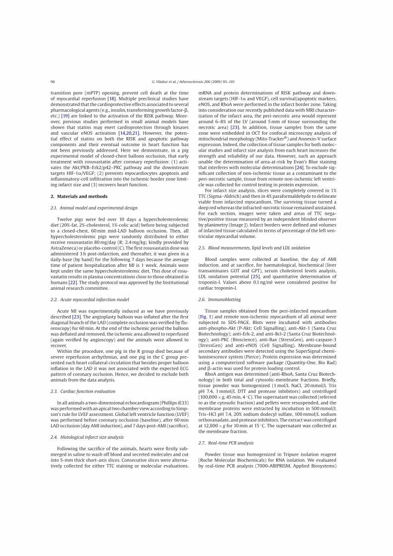
96 G. Vilahur et al. / Atherosclerosis 206 (2009) 95–101
transition pore (mPTP) opening, prevent cell death at the timeof myocardial reperfusion [18]. Multiple preclinical studies havedemonstrated that the cardioprotective effects associated to severalpharmacological agents (e.g., insulin, transforming growth factor-�,etc.) [19] are linked to the activation of the RISK pathway. More-over, previous studies performed in small animal models haveshown that statins may exert cardioprotection through kinasesand vascular eNOS activation [14,20,21]. However, the poten-tial effect of statins on both the RISK and apoptotic pathwaycomponents and their eventual outcome in heart function hasnot been previously addressed. Here we demonstrate, in a pigexperimental model of closed-chest balloon occlusion, that earlytreatment with rosuvastatin after coronary reperfusion: (1) acti-vates the Akt/PKB–Erk2/p42–PKC pathway and the downstreamtargets HIF-1�/VEGF; (2) prevents myocardiocytes apoptosis andinflammatory-cell infiltration into the ischemic border zone limit-ing infarct size and (3) recovers heart function.
2. Materials and methods
2.1. Animal model and experimental design
Twelve pigs were fed over 10 days a hypercholesterolemicdiet (20%-fat, 2%-cholesterol, 1%-colic acid) before being subjectedto a closed-chest, 60 min mid-LAD balloon occlusion. Then, allhypercholesterolemic pigs were randomly distributed to eitherreceive rosuvastatin 80 mg/day (R; 2.4 mg/kg; kindly provided byAstraZeneca) or placebo-control (C). The first rosuvastatin dose wasadministered 3 h post-infarction, and thereafter, it was given in adaily-base (by hand) for the following 7 days because the averagetime of patient hospitalization after MI is 1 week. Animals werekept under the same hypercholesterolemic diet. This dose of rosu-vastatin results in plasma concentrations close to those obtained inhumans [22]. The study protocol was approved by the Institutionalanimal research committee.
2.2. Acute myocardial infarction model
Acute MI was experimentally induced as we have previouslydescribed [23]. The angioplasty balloon was inflated after the firstdiagonal branch of the LAD (complete occlusion was verified by flu-oroscopy) for 60 min. At the end of the ischemic period the balloonwas deflated and removed, the ischemic area allowed to reperfused(again verified by angioscopy) and the animals were allowed torecover.
Within the procedure, one pig in the R group died because ofsevere reperfusion arrhythmias, and one pig in the C group pre-sented such heart collateral circulation that besides proper ballooninflation in the LAD it was not associated with the expected ECGpattern of coronary occlusion. Hence, we decided to exclude bothanimals from the data analysis.
2.3. Cardiac function evaluation
In all animals a two-dimensional echocardiogram (Phillips iE33)was performed with an apical two chamber view according to Simp-son’s rule for LVEF assessment. Global left ventricle function (LVEF)was performed before coronary occlusion (baseline), after 60 minLAD occlusion (day AMI induction), and 7 days post-AMI (sacrifice).
2.4. Histological infarct size analysis
Following the sacrifice of the animals, hearts were firstly sub-merged in saline to wash off blood and secreted molecules and cutinto 5-mm thick short-axis slices. Consecutive slices were alterna-tively collected for either TTC staining or molecular evaluations.
mRNA and protein determinations of RISK pathway and down-stream targets (HIF-1� and VEGF), cell survival/apoptotic markers,eNOS, and RhoA were performed in the infarct border zone. Takinginto consideration our recently published data with MRI character-ization of the infarct area, the peri-necrotic area would representaround 6–8% of the LV (around 5 mm of tissue surrounding thenecrotic area) [23]. In addition, tissue samples from the samezone were embedded in OCT for confocal microscopy analysis ofmitochondrial morphology (Mito-Tracker®) and Annexin-V surfaceexpression. Indeed, the collection of tissue samples for both molec-ular studies and infarct size analysis from each heart increases thestrength and reliability of our data. However, such an approachunable the determination of area-at-risk by Evan’s Blue stainingthat interferes with molecular determinations [24]. To exclude sig-nificant collection of non-ischemic tissue as a contaminant to theperi-necrotic sample, tissue from remote non-ischemic left ventri-cle was collected for control testing in protein expression.
For infarct size analysis, slices were completely covered in 1%TTC (Sigma–Aldrich) and then in 4% paraformaldehyde to delineateviable from infarcted myocardium. The surviving tissue turned adeep red whereas the infracted-necrotic tissue remained unstained.For each section, images were taken and areas of TTC nega-tive/positive tissue measured by an independent blinded observerby planimetry (Image J). Infarct borders were defined and volumesof infarcted tissue calculated in terms of percentage of the left ven-tricular myocardial volume.
2.5. Blood measurements, lipid levels and LDL oxidation
Blood samples were collected at baseline, the day of AMIinduction, and at sacrifice, for haematological, biochemical (livertransaminases GOT and GPT), serum cholesterol levels analysis,LDL oxidation potential [25], and quantitative determination oftroponin-I. Values above 0.1 ng/ml were considered positive forcardiac troponin-I.
2.6. Immunoblotting
Tissue samples obtained from the peri-infarcted myocardium(Fig. 1) and remote non-ischemic myocardium of all animal weresubjected to SDS-PAGE. Blots were incubated with antibodiesanti-phospho-Akt (P-Akt; Cell Signalling), anti-Akt-1 (Santa CruzBiotechnology); anti-Erk-2, and anti-Bcl-2 (Santa Cruz Biotechnol-ogy); anti-PKC (Bioscience), anti-Bax (StressGen), anti-caspase-3(StressGen) and anti-eNOS (Cell Signalling). Membrane-boundsecondary antibodies were detected using the SuperSignal chemi-luminescence system (Pierce). Protein expression was determinedusing a computerized software package (Quantity-One, Bio Rad)and �-actin was used for protein loading control.
RhoA antigen was determined (anti-RhoA, Santa Cruz Biotech-nology) in both total and cytosolic-membrane fractions. Briefly,tissue powder was homogenized (1 mol/L NaCl, 20 mmol/L TrispH 7.4, 1 mmol/L DTT and protease inhibitors) and centrifuged(100,000 × g, 45 min, 4 ◦C). The supernatant was collected (referredto as the cytosolic fraction) and pellets were resuspended, and themembrane proteins were extracted by incubation in 500 mmol/LTris–HCl pH 7.4, 20% sodium dodecyl sulfate, 100 mmol/L sodiumorthovanadate, and protease inhibitors. The extract was centrifugedat 12,000 × g for 10 min at 15 ◦C. The supernatant was collected asthe membrane fraction.
2.7. Real-time PCR analysis
Powder tissue was homogenized in Tripure isolation reagent(Roche Molecular Biochemicals) for RNA isolation. We evaluatedby real-time PCR analysis (7000-ABIPRISM, Applied Biosystems)
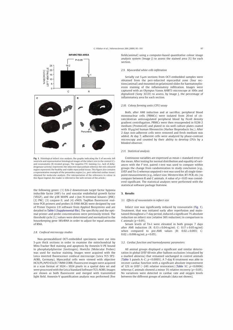
G. Vilahur et al. / Atherosclerosis 206 (2009) 95–101 97
Fig. 1. Histological infarct size analysis. Bar graphs indicating the % of necrotic leftventricle and representative histological images of the infarct size in the control (C)-and rosuvastatin (R)-treated groups. The negative-TTC staining (i.e., lack of dehy-drogenase activity) represents the infarcted myocardium whereas the red-colouredregion represents the healthy and viable myocardial tissue. This figure also containsa representative example of the penumbra region (i.e., peri-infarcted cardiac tissue)obtained for molecular analysis. (For interpretation of the references to colour inthis figure legend, the reader is referred to the web version of the article.)
the following genes: (1) Erk-2 downstream target factor hypoxiainducible factor (HIF)-1� and vascular endothelial growth factor(VEGF), and the p38 MAPK and c-Jun N-terminal kinases (JNK);(2) PKC; (3) caspase-3; and (4) eNOS. TaqMan fluorescent real-time PCR primers and probes (6-FAM-MGB) were designed by useof Primer Express 2.0 software from Applied Biosystems and aredetailed in Table I (Supplemental file). The specificity and the opti-mal primer and probe concentrations were previously tested. Thethreshold cycle (Ct) values were determined and normalized to thehousekeeping gene 18SrRNA in order to adjust for equal amountsof RNA.
2.8. Confocal microscopy studies
Non-permeabilized OCT-embedded specimens were cut into5-�m thick sections in order to examine the mitochondrial byMito-Tracker Red staining and apoptosis by Annexin-V-PE boundto phosphatydylserine (Invitrogen). Hoetchs (Molecular Probes)was used for nuclear staining. Images were acquired with theLeica inverted fluorescence confocal microscope (Leica TCS SP2-AOBS, Germany). Myocardial cells were viewed with objectiveHCX/PL/APO 63x/0.7 IMM CORR. Fluorescent images were acquiredin a scan format of 1024 × 1024 pixels in a spatial data set andwere processed with the Leica Standard Software TCS-AOBS. Imagesare shown as both fluorescent and merged with transmittedlight field. Annexin-V quantification analysis was performed (five
fields/animal) using a computer-based quantitative colour imageanalysis system (Image J) to assess the stained area (%) for eachsection.
2.9. Myocardial white cells infiltration
Serially cut 5 �m sections from OCT-embedded samples wereobtained from the peri-infarcted myocardial zone (four sec-tions/animal) and mounted on gelatinized slides for haematoxylin-eosin staining of the inflammatory infiltration. Images werecaptured with an Olympus Vanox AHBT3 microscope at 100x anddigitalized (Sony 3CCD) to assess, by Image J, the percentage ofinflammatory area for each section.
2.10. Colony forming units (CFU) assay
Both, after AMI induction and at sacrifice, peripheral bloodmononuclear cells (PBMCs) were isolated from 20 ml of cit-rate/dextran anticoagulated peripheral blood by Ficoll densitygradient centrifugation. PBMCs were then resuspended in EGM-2medium (PromoCell) and plated in six-well culture plates coatedwith 10 �g/ml human fibronectin (Harbor Bioproducts Inc.). After2 days non-adherent cells were removed and fresh medium wasadded. At day 7, adherent cells were analyzed by phase-contrastmicroscopy and counted by their ability to develop CFUs by ablinded observer.
2.11. Statistical analysis
Continuous variables are expressed as mean ± standard error ofthe mean. After testing for normal distribution and equality of vari-ances with the F test, paired t-test was used to compare withingroups the change from randomization to study conclusion (e.g.,LVEF and Tn-I) whereas unpaired t-test was used for all single time-point measurements (e.g., infarct size, Western blot, RT-PCR, etc.) tocompare between R and C animals. A value of p < 0.05 was consid-ered significant. The statistical analyses were performed with thestatistical software package Statview.
3. Results
3.1. Effects of rosuvastatin in infarct size
Infarct size was significantly reduced by rosuvastatin (Fig. 1).Treatment, that was initiated early after reperfusion and main-tained throughout a 7-day period, induced a significant 7% absolutereduction on infarct size (relative 36% reduction) in comparison toC animals (p < 0.05).
Serum levels of Tn-I were elevated in both groups 7 daysafter AMI induction (R: 0.13 ± 0.04 ng/ml; C: 0.17 ± 0.05 ng/ml)when compared to pre-AMI values (R: 0.02 ± 0.003; C:0.02 ± 0.006 ng/ml; p < 0.05).
3.2. Cardiac function and haemodynamic parameters
All animal groups displayed a significant and similar deterio-ration in global LVEF 60 min after balloon occlusion (visualized bya marked akinesia) that remained unchanged in control animals(Table 1 panels A–C; p < 0.0005). A 7-day R treatment was able torecover cardiac function with a significant absolute improvementof 12% in LVEF (∼24% relative restoration) (Table 1C; p < 0.0006)whereas C animals showed a minor 3% relative recovery (p < 0.05).No variations were detected in cardiac rate and oxygen levelsbetween the different groups of animals (data not shown).

98 G. Vilahur et al. / Atherosclerosis 206 (2009) 95–101
3.3. Serum cholesterol levels and liver enzymes
Rosuvastatin treatment did not produce any significant differ-ence in blood cell or platelet counts (data not shown). With regardsto liver transaminases, untreated control animals showed a sig-nificant increase at day 7th in both GOT (75 ± 17 IU vs. 36 ± 6 IU)and GPT (66 ± 5 vs. 48 ± 3 IU) (p < 0.05) with respect to baselinepre-intervention values, whereas no changes were detected aftera 7-day treatment with rosuvastatin (GOT: 45 ± 17 IU vs. 36 ± 3 IU;GPT: 34 ± 7 vs. 41 ± 11 IU). As shown in Table II (Supplemental file)and as expected due to the short treatment period, no variations inplasma lipid levels were detected among R-treated and C animals atthe different tested periods. All animals were maintained through-out the entire experimental period under a hypercholesterolemicdiet (favouring cholesterol absorption over synthesis) in order toexclude any systemic cholesterol lowering effects of rosuvastatin.LDL oxidation, measured as maximal conjugated dienes (MaxDC)was similar in both groups (382.7 ± 47.2 vs. 382.0 ± 82.2; p = N.S.).
3.4. Molecular studies
3.4.1. Immunoblot and real-time PCR analysisRosuvastatin was associated with higher protein expression of
the RISK kinases Akt/PKB (p-Akt/Akt ratio), MAPK Erk-2/p44, and
PKC in the myocardial border zone (Fig. 2 and Fig. I Supplemen-tal file). In addition, R-treated animals showed an up-regulatedexpression (p < 0.05) of PKC and the Erk-2/p44 cardioprotectivedownstream targets HIF-1� and VEGF genes (Fig. 3). No changeswere detected in the other MAPK cJNK and p38 between thedifferent groups (data not shown). Regarding the expression of pro-apoptotic/anti-apoptotic signals, R lead to a higher pro-survival Bcl-2/Bax ratio and conversely, a twofold decrease in cleaved-caspase-3protein expression (Fig. 2 and Fig. I Supplemental file). Caspase-3mRNA expression was also significantly decreased by R treatment(72 ± 8% of control; p < 0.05; Fig. 3). In the remote myocardiumpAkt, Erk-2, PKC, Bcl-2, Bax, and Truncated Casp-3 protein levelswere significantly lower than in the peri-necrotic (penum-bra) area and did not differ between the two groups (data notshown).
Neither eNOS mRNA nor protein levels were affected by a 7-daytreatment with a non-lipid lowering dose of rosuvastatin (99.8 ± 6%of control, p = 0.98; Fig. I Supplemental file).
Total myocardial RhoA protein as well as both the cytosolic (inac-tive) and membrane (active) fractions was determined by imm-unoblotting. As depicted in Fig. 4A the total amount of RhoAwas unchanged. However, R markedly decreased RhoA translo-cation, measured as membrane-to-cytosol ratio (p < 0.001 vs. C;Fig. 4B).
Fig. 2. Protein expression levels in the infarct border zone of several components of the RISK pathway (A) and apoptotic cascade (B) in both control (C) and rosuvastatin-treatedanimals (R). *p < 0.05 vs. control animals (C).

G. Vilahur et al. / Atherosclerosis 206 (2009) 95–101 99
Table 1Echocardiography analysis of heart function (left ventricle ejection fraction).
BSL Post-AMI SacrificeA
RMean 77.5 54.5* 66.6†
S.E.M. 3.2 4.2 3.3
CMean 75.4 59.5* 61.4**
S.E.M. 4.5 3.4 3.8
BSL vs. Post-AMI Post-AMI vs. SAC
BR
Mean 23.0* −12.21†
S.E.M. 1.4 1.6
CMean 15.9* −1.9**
S.E.M. 2.1 0.4
BSL vs. post-AMI (% change) Post-AMI vs. SAC (% change)
CR
Mean −30.1* 23.7†
S.E.M. 2.5 4.5
CMean −21.0* 3.1**
S.E.M. 2.2 0.5
* p < 0.0005 vs. baseline.† p < 0.0006 vs. post-AMI.
** p < 0.05 vs. post-AMI.
3.4.2. Mito-Tracker® and Annexin-V detection by confocalanalysis
When compared to R-treated animals cardiac tissue from Canimal showed both an evident “ballooning/swelling” of the mito-chondria (Fig. 5-I) and a marked exposition of phosphatidylserine(PS) on the outer leaflet of the cell surface. Annexin positive area wasreduced an average 87% in rosuvastatin-treated animals (1.5 ± 0.4%in C animals and 0.2 ± 0.06% in R-treated animals; p < 0.05; Fig. 5-II).
3.4.3. Leukocyte infiltrationHistopathologic analysis of the peri-infarcted myocardial zone
showed a significant reduction in inflammatory-cell infiltrates(23.8 ± 4.6% vs. 9.3 ± 2.3%; p < 0.05; Fig. II Supplemental file).
3.5. Effect of rosuvastatin on CFUs development
PBMCs obtained from C and R-treated animals showed a compa-rable ability to form CFUs at both tested periods, just at AMI induc-tion (14 ± 7 and 31 ± 8 CFUs/1 × 106 cells in C group and the group tobe treated with R, respectively) and at sacrifice (46 ± 13 and 56 ± 11CFUs/1 × 106 cells in C and R-treated animals, respectively). Indeed,the increase in CFU during the 7 days post-AMI were similar in C(32 ± 6 CFUs/1 × 106 cells) and R group (25 ± 3 CFUs/1 × 106 cells).
4. Discussion
The results of the present study show that inhibition of HMG-CoA reductase by oral administration of rosuvastatin in the earlyhours after MI and for 7 days thereafter is associated with: (1) amarked activation of the cardioprotective RISK signalling transduc-tion pathway; (2) increase in pro-survival anti-apoptotic markers;(3) reduction in the extent of infarct size by 7% (a relative reduc-tion of 36%); and (4) heart function restoration by 12% (a relativerecovery of 24%). Moreover, all these effects were observed with novariations in plasma lipid levels suggesting that rosuvastatin car-dioprotection was likely derived from blocking the isoprenylationand subsequent membrane translocation of members of the RhoGTPase family, for instance RhoA [26].
It is well established that the final MI size is a major deter-minant of mortality and post-MI remodeling. Thus, investigationof strategies for the amelioration and preservation of myocardialcell viability is a matter of great interest [16,27]. Recent experi-mental studies have demonstrated the cardioprotective actions ofboth lipophilic [13] and hydrophilic [28] statins in the setting ofMI beyond cholesterol effects. The mechanisms by which statinsinduce myocardial salvage have been independently associatedto either activation of the PI3K/Akt/eNOS pathway, to antioxi-dant and anti-inflammatory actions, or to stem cell mobilization[13,21,28,29]. However, a system biological approach analysing
Fig. 3. Gene expression in the peri-infarcted myocardium assessed by real-time PCR in both control (C)- and rosuvastatin (R)-treated animals. *p < 0.05 vs. controlanimals (C).

100 G. Vilahur et al. / Atherosclerosis 206 (2009) 95–101
Fig. 4. Evaluation of RhoA activation in the peri-infarcted myocardium determinedby western blot analysis A. Total RhoA protein expression. (B) Cytosolic (inactive)and membrane (active) RhoA fractions. *p < 0.001 vs. C.
HMG-CoA/myocardial RhoA effects, activation of the RISK signallingpathway, myocyte damage reduction and restoration of heart func-tion, as demonstrated in the present study, has not been addressedpreviously. Indeed, the attenuation of inflammatory-cell recruit-ment to the peri-infarcted area detected in rosuvastatin-treatedanimals might have also contributed to rescue the infarcted heart.
The primary beneficial effects derived from HMG-CoA reductaseinhibitors is the inhibition of cholesterol synthesis and subse-quent plasma lipid lowering. However, in order to exclude suchlipid-lowering beneficial effects we maintained the animals in ahypercholesterolemic diet throughout all the study. This modelallowed us to mainly attribute rosuvastatin cardioprotective effectsto the prevention of isoprenylation of small G-protein functions. Infact, we observed a pronounced inhibition of RhoA translocationto the membrane (i.e., inactivation). In its turn, RhoA inhibition bystatins has shown to activated protein kinase B (i.e., Akt) [30]. Ourresults not only support these previous findings, but also attributeto statin treatment a marked up-regulation of PKC (downstreamtarget of Akt) and Erk-2/p44 MAPK and its downstream cardio-protective targets HIF-1� and VEGF (Fig. III Supplemental file).Furthermore, such statin-derived cardioprotective effects seemedunrelated to both JNK and p38 MAPKs. Our data also suggeststhat activation of the RISK pathway may lead to the triggeringof several anti-apoptotic signals such as Bcl-2 increase and Baxreduction, avoiding mitochondria depolarization and further itsrelease of mitochondrial-related mediators (e.g., cytochrome-c).Rosuvastatin’s halt of the apoptotic process through mitochondriapreservation was histologically evidenced by the nearly normalmitochondrial staining observed in rosuvastatin-treated animals.This overall increase in pro-survival signals eventually translatedin less phosphatidylserine exposure, reduced truncated caspase-3 detection, lower infarct size, and improved heart function
Fig. 5. Mitochondrial shape analysis and Annexin-V staining by confocal microscopy. (I) Cardiac tissue from control (C) animals (A and B) showed an evident “ballooning”of the mitochondria as compared to the nearly normal mitochondrial staining observed in rosuvastatin (R)-treated animals (C and D). (II) Heart sections from C (A–C) and R(D–F)-treated animals. R treatment exhibited a marked reduction in Annexin-V signal (green) when compared to C animals. (For interpretation of the references to colour inthis figure legend, the reader is referred to the web version of the article.)
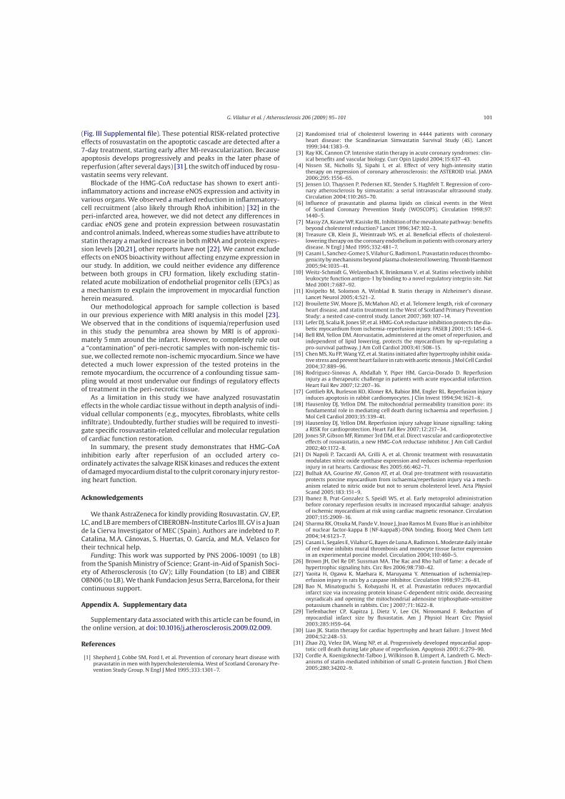
G. Vilahur et al. / Atherosclerosis 206 (2009) 95–101 101
(Fig. III Supplemental file). These potential RISK-related protectiveeffects of rosuvastatin on the apoptotic cascade are detected after a7-day treatment, starting early after MI-revascularization. Becauseapoptosis develops progressively and peaks in the later phase ofreperfusion (after several days) [31], the switch off induced by rosu-vastatin seems very relevant.
Blockade of the HMG-CoA reductase has shown to exert anti-inflammatory actions and increase eNOS expression and activity invarious organs. We observed a marked reduction in inflammatory-cell recruitment (also likely through RhoA inhibition) [32] in theperi-infarcted area, however, we did not detect any differences incardiac eNOS gene and protein expression between rosuvastatinand control animals. Indeed, whereas some studies have attribute tostatin therapy a marked increase in both mRNA and protein expres-sion levels [20,21], other reports have not [22]. We cannot excludeeffects on eNOS bioactivity without affecting enzyme expression inour study. In addition, we could neither evidence any differencebetween both groups in CFU formation, likely excluding statin-related acute mobilization of endothelial progenitor cells (EPCs) asa mechanism to explain the improvement in myocardial functionherein measured.
Our methodological approach for sample collection is basedin our previous experience with MRI analysis in this model [23].We observed that in the conditions of isquemia/reperfusion usedin this study the penumbra area shown by MRI is of approxi-mately 5 mm around the infarct. However, to completely rule outa “contamination” of peri-necrotic samples with non-ischemic tis-sue, we collected remote non-ischemic myocardium. Since we havedetected a much lower expression of the tested proteins in theremote myocardium, the occurrence of a confounding tissue sam-pling would at most undervalue our findings of regulatory effectsof treatment in the peri-necrotic tissue.
As a limitation in this study we have analyzed rosuvastatineffects in the whole cardiac tissue without in depth analysis of indi-vidual cellular components (e.g., myocytes, fibroblasts, white cellsinfiltrate). Undoubtedly, further studies will be required to investi-gate specific rosuvastatin-related cellular and molecular regulationof cardiac function restoration.
In summary, the present study demonstrates that HMG-CoAinhibition early after reperfusion of an occluded artery co-ordinately activates the salvage RISK kinases and reduces the extentof damaged myocardium distal to the culprit coronary injury restor-ing heart function.
Acknowledgements
We thank AstraZeneca for kindly providing Rosuvastatin. GV, EP,LC, and LB are members of CIBEROBN-Institute Carlos III. GV is a Juande la Cierva Investigator of MEC (Spain). Authors are indebted to P.Catalina, M.A. Cánovas, S. Huertas, O. García, and M.A. Velasco fortheir technical help.
Funding: This work was supported by PNS 2006-10091 (to LB)from the Spanish Ministry of Science; Grant-in-Aid of Spanish Soci-ety of Atherosclerosis (to GV); Lilly Foundation (to LB) and CIBEROBN06 (to LB). We thank Fundacion Jesus Serra, Barcelona, for theircontinuous support.
Appendix A. Supplementary data
Supplementary data associated with this article can be found, inthe online version, at doi:10.1016/j.atherosclerosis.2009.02.009.
References
[1] Shepherd J, Cobbe SM, Ford I, et al. Prevention of coronary heart disease withpravastatin in men with hypercholesterolemia. West of Scotland Coronary Pre-vention Study Group. N Engl J Med 1995;333:1301–7.
[2] Randomised trial of cholesterol lowering in 4444 patients with coronaryheart disease: the Scandinavian Simvastatin Survival Study (4S). Lancet1999;344:1383–9.
[3] Ray KK, Cannon CP. Intensive statin therapy in acute coronary syndromes: clin-ical benefits and vascular biology. Curr Opin Lipidol 2004;15:637–43.
[4] Nissen SE, Nicholls SJ, Sipahi I, et al. Effect of very high-intensity statintherapy on regression of coronary atherosclerosis: the ASTEROID trial. JAMA2006;295:1556–65.
[5] Jensen LO, Thayssen P, Pedersen KE, Stender S, Haghfelt T. Regression of coro-nary atherosclerosis by simvastatin: a serial intravascular ultrasound study.Circulation 2004;110:265–70.
[6] Influence of pravastatin and plasma lipids on clinical events in the Westof Scotland Coronary Prevention Study (WOSCOPS). Circulation 1998;97:1440–5.
[7] Massy ZA, Keane WF, Kasiske BL. Inhibition of the mevalonate pathway: benefitsbeyond cholesterol reduction? Lancet 1996;347:102–3.
[8] Treasure CB, Klein JL, Weintraub WS, et al. Beneficial effects of cholesterol-lowering therapy on the coronary endothelium in patients with coronary arterydisease. N Engl J Med 1995;332:481–7.
[9] Casani L, Sanchez-Gomez S, Vilahur G, Badimon L. Pravastatin reduces thrombo-genicity by mechanisms beyond plasma cholesterol lowering. Thromb Haemost2005;94:1035–41.
[10] Weitz-Schmidt G, Welzenbach K, Brinkmann V, et al. Statins selectively inhibitleukocyte function antigen-1 by binding to a novel regulatory integrin site. NatMed 2001;7:687–92.
[11] Kivipelto M, Solomon A, Winblad B. Statin therapy in Alzheimer’s disease.Lancet Neurol 2005;4:521–2.
[12] Brouilette SW, Moore JS, McMahon AD, et al. Telomere length, risk of coronaryheart disease, and statin treatment in the West of Scotland Primary PreventionStudy: a nested case-control study. Lancet 2007;369:107–14.
[13] Lefer DJ, Scalia R, Jones SP, et al. HMG-CoA reductase inhibition protects the dia-betic myocardium from ischemia-reperfusion injury. FASEB J 2001;15:1454–6.
[14] Bell RM, Yellon DM. Atorvastatin, administered at the onset of reperfusion, andindependent of lipid lowering, protects the myocardium by up-regulating apro-survival pathway. J Am Coll Cardiol 2003;41:508–15.
[15] Chen MS, Xu FP, Wang YZ, et al. Statins initiated after hypertrophy inhibit oxida-tive stress and prevent heart failure in rats with aortic stenosis. J Mol Cell Cardiol2004;37:889–96.
[16] Rodriguez-Sinovas A, Abdallah Y, Piper HM, Garcia-Dorado D. Reperfusioninjury as a therapeutic challenge in patients with acute myocardial infarction.Heart Fail Rev 2007;12:207–16.
[17] Gottlieb RA, Burleson KO, Kloner RA, Babior BM, Engler RL. Reperfusion injuryinduces apoptosis in rabbit cardiomyocytes. J Clin Invest 1994;94:1621–8.
[18] Hausenloy DJ, Yellon DM. The mitochondrial permeability transition pore: itsfundamental role in mediating cell death during ischaemia and reperfusion. JMol Cell Cardiol 2003;35:339–41.
[19] Hausenloy DJ, Yellon DM. Reperfusion injury salvage kinase signalling: takinga RISK for cardioprotection. Heart Fail Rev 2007;12:217–34.
[20] Jones SP, Gibson MF, Rimmer 3rd DM, et al. Direct vascular and cardioprotectiveeffects of rosuvastatin, a new HMG-CoA reductase inhibitor. J Am Coll Cardiol2002;40:1172–8.
[21] Di Napoli P, Taccardi AA, Grilli A, et al. Chronic treatment with rosuvastatinmodulates nitric oxide synthase expression and reduces ischemia-reperfusioninjury in rat hearts. Cardiovasc Res 2005;66:462–71.
[22] Bulhak AA, Gourine AV, Gonon AT, et al. Oral pre-treatment with rosuvastatinprotects porcine myocardium from ischaemia/reperfusion injury via a mech-anism related to nitric oxide but not to serum cholesterol level. Acta PhysiolScand 2005;183:151–9.
[23] Ibanez B, Prat-Gonzalez S, Speidl WS, et al. Early metoprolol administrationbefore coronary reperfusion results in increased myocardial salvage: analysisof ischemic myocardium at risk using cardiac magnetic resonance. Circulation2007;115:2909–16.
[24] Sharma RK, Otsuka M, Pande V, Inoue J, Joao Ramos M. Evans Blue is an inhibitorof nuclear factor-kappa B (NF-kappaB)-DNA binding. Bioorg Med Chem Lett2004;14:6123–7.
[25] Casani L, Segales E, Vilahur G, Bayes de Luna A, Badimon L. Moderate daily intakeof red wine inhibits mural thrombosis and monocyte tissue factor expressionin an experimental porcine model. Circulation 2004;110:460–5.
[26] Brown JH, Del Re DP, Sussman MA. The Rac and Rho hall of fame: a decade ofhypertrophic signaling hits. Circ Res 2006;98:730–42.
[27] Yaoita H, Ogawa K, Maehara K, Maruyama Y. Attenuation of ischemia/rep-erfusion injury in rats by a caspase inhibitor. Circulation 1998;97:276–81.
[28] Bao N, Minatoguchi S, Kobayashi H, et al. Pravastatin reduces myocardialinfarct size via increasing protein kinase C-dependent nitric oxide, decreasingoxyradicals and opening the mitochondrial adenosine triphosphate-sensitivepotassium channels in rabbits. Circ J 2007;71:1622–8.
[29] Tiefenbacher CP, Kapitza J, Dietz V, Lee CH, Niroomand F. Reduction ofmyocardial infarct size by fluvastatin. Am J Physiol Heart Circ Physiol2003;285:H59–64.
[30] Liao JK. Statin therapy for cardiac hypertrophy and heart failure. J Invest Med2004;52:248–53.
[31] Zhao ZQ, Velez DA, Wang NP, et al. Progressively developed myocardial apop-totic cell death during late phase of reperfusion. Apoptosis 2001;6:279–90.
[32] Cordle A, Koenigsknecht-Talboo J, Wilkinson B, Limpert A, Landreth G. Mech-anisms of statin-mediated inhibition of small G-protein function. J Biol Chem2005;280:34202–9.
Top Related