CHRONIC INTERSTITIAL PNEUMONITIS IN DOGS NATURALLY ...
6
Rev. Inst. Med. trop. S. Paulo 45(3):153-158, May-June, 2003 Financial Support: FAPEMIG (CDS 2263/97) and CAPES, UFMG. (1) Departamento de Clínica e Cirurgia Veterinárias, Escola de Veterinária; Universidade Federal de Minas Gerais (UFMG), Av. Antônio Carlos 6627, Campus Pampulha, 31270-901 Belo Horizonte, MG, Brazil. Phone/fax: 55 31 3499-2889. (2) Departamento de Patologia Geral, Instituto de Ciências Biológicas (ICB), Universidade Federal de Minas Gerais (UFMG), Brazil. (3) Departamento de Parasitologia, Instituto de Ciências Biológicas (ICB), Universidade Federal de Minas Gerais (UFMG), Brazil. (4) Departamento de Anatomia Patológica e Medicina Legal, Faculdade de Medicina, Universidade Federal de Minas Gerais Correspondence to: Ricardo Gonçalves, Departamento de Clínica e Cirurgia Veterinárias, Escola de Veterinária/UFMG, Av. Antonio Carlos 6627, Campus Pampulha, 31270-901 Belo Horizonte, MG, Brasil. e-mail: [email protected] CHRONIC INTERSTITIAL PNEUMONITIS IN DOGS NATURALLY INFECTED WITH Leishmania (Leishmania) chagasi: A HISTOPATHOLOGICAL AND MORPHOMETRIC STUDY Ricardo GONÇALVES(1), Washington Luiz TAFURI(4), Maria Norma de MELO(3), Pedro RASO(4) & Wagner Luiz TAFURI(2) SUMMARY Eighteen mongrel dogs of unknown age and naturally infected with Leishmania (Leishmania) chagasi, were obtained from the City Hall of Belo Horizonte, Brazil. Four dogs were used as control. Lung samples were obtained and immediately fixed in formalin. The histopathological picture of all lung tissue sections was a chronic and diffuse interstitial pneumonitis. The thickened inter- alveolar septa were characterized by the cellular exudate (mostly macrophages, lymphocytes and plasmocytes) associated with collagen deposition. Morphometric analysis showed greater septal thickness in the infected animals than in controls. In fact, the morphometric study of collagen stained with ammoniac silver confirmed a larger deposition of collagen in the infected animals. The parasitologic method was carried out during the study of the lesions on the slides. However, we did not observe any correlation between the histopathologic and morphometric data and the clinical status of the animals. We conclude that the pulmonary lesions observed in all naturally infected dogs were correlated with the disease and that the morphometric method used was satisfactory for the analysis of septal thickness and of increased collagen deposition, confirming the presence of fibrosis. KEYWORDS: Chronic Interstitial Pneumonitis; Dogs; Histopathology; Morphometric analysis; Leishmania (Leishmania) chagasi. INTRODUCTION Interstitial pneumonitis in visceral leishmaniasis was initially described in humans in 1959 3 and later confirmed by other authors 4,6,12,19 . In dogs, pulmonary lesions are very similar to those observed in humans, and were previously characterized by some authors as interstitial pneumonitis mainly characterized by thickening of alveolar walls associated with the presence of a chronic cellular exudate mainly consisting of macrophages 1,23 . Later on, similar lesions were described in dogs experimentally infected with Leishmania (Leishmania) chagasi 13 . In Brazil, DUARTE et al . (1986) described the histological pulmonary alterations of dogs naturally infected with Leishmania (Leishmania) chagasi. In a study performed with 41 naturally infected dogs they detected interstitial pneumonitis in 81.5% of the animals and no other kind of pulmonary alteration, such as bronchopneumonia. An immunoenzymatic study with specific antibodies (PAP) showed particulate antigenic material and/or amastigotes of Leishmania in the inter-alveolar septa where interstitial pneumonitis was present 11 . As far as we know, a specific pulmonary symptom occurring during visceral leishmaniasis has not been described. On the other hand, there are many reports of dry coughing as a symptom of visceral leishmaniasis in human beings, but there are no references about the correlation between this symptom and interstitial pneumonitis 7,12,14,16,19 . Some authors have proposed compression of the spleen against the vagus nerve 16 as the cause of this symptom and terminal broncopneumonitis as a terminal stage of the disease, or at least the presence of leishmanias in the alveolar septa 2 . The pathogenesis of the pulmonary alterations occurring in visceral leishmaniasis has not been completely elucidated, especially in persistent lesions where the parasite was not found. Moreover, interstitital pneumonitis is an asymptomatic lesion and few studies have investigated the pathogenesis of this disease in dogs. Thus, the aim of the present study was to characterize the interstitial pneumonitis occurring in canine visceral leishmaniasis on the basis of histopathological and morphometric aspects of the lesions associated with the different clinical forms of the disease in the dog. MATERIAL AND METHODS Animals: Eighteen mongrel dogs of unknown age (nine females and nine males) were obtained from the City of Belo Horizonte (Zoonosis Department), MG, Brazil. All dogs were positive for Leishmania by direct immunofluorescence (RIFI) and complement fixation tests (RFC). This group of naturally infected animals was divided into three groups of six animals each. Tissue touch preparations of liver, spleen and bone
Transcript of CHRONIC INTERSTITIAL PNEUMONITIS IN DOGS NATURALLY ...
Rev. Inst. Med. trop. S. Paulo
45(3):153-158, May-June, 2003
Financial Support: FAPEMIG (CDS 2263/97) and CAPES, UFMG.(1) Departamento de Clínica e Cirurgia Veterinárias, Escola de Veterinária; Universidade Federal de Minas Gerais (UFMG), Av. Antônio Carlos 6627, Campus Pampulha, 31270-901 Belo
Horizonte, MG, Brazil. Phone/fax: 55 31 3499-2889.(2) Departamento de Patologia Geral, Instituto de Ciências Biológicas (ICB), Universidade Federal de Minas Gerais (UFMG), Brazil.(3) Departamento de Parasitologia, Instituto de Ciências Biológicas (ICB), Universidade Federal de Minas Gerais (UFMG), Brazil.(4) Departamento de Anatomia Patológica e Medicina Legal, Faculdade de Medicina, Universidade Federal de Minas GeraisCorrespondence to: Ricardo Gonçalves, Departamento de Clínica e Cirurgia Veterinárias, Escola de Veterinária/UFMG, Av. Antonio Carlos 6627, Campus Pampulha, 31270-901 Belo
Horizonte, MG, Brasil. e-mail: [email protected]
CHRONIC INTERSTITIAL PNEUMONITIS IN DOGS NATURALLY INFECTED WITH Leishmania(Leishmania) chagasi: A HISTOPATHOLOGICAL AND MORPHOMETRIC STUDY
Ricardo GONÇALVES(1), Washington Luiz TAFURI(4), Maria Norma de MELO(3), Pedro RASO(4) & Wagner Luiz TAFURI(2)
SUMMARY
Eighteen mongrel dogs of unknown age and naturally infected with Leishmania (Leishmania) chagasi, were obtained from theCity Hall of Belo Horizonte, Brazil. Four dogs were used as control. Lung samples were obtained and immediately fixed in formalin.The histopathological picture of all lung tissue sections was a chronic and diffuse interstitial pneumonitis. The thickened inter-alveolar septa were characterized by the cellular exudate (mostly macrophages, lymphocytes and plasmocytes) associated with collagendeposition. Morphometric analysis showed greater septal thickness in the infected animals than in controls. In fact, the morphometricstudy of collagen stained with ammoniac silver confirmed a larger deposition of collagen in the infected animals. The parasitologicmethod was carried out during the study of the lesions on the slides. However, we did not observe any correlation between thehistopathologic and morphometric data and the clinical status of the animals. We conclude that the pulmonary lesions observed in allnaturally infected dogs were correlated with the disease and that the morphometric method used was satisfactory for the analysis ofseptal thickness and of increased collagen deposition, confirming the presence of fibrosis.
KEYWORDS: Chronic Interstitial Pneumonitis; Dogs; Histopathology; Morphometric analysis; Leishmania (Leishmania) chagasi.
INTRODUCTION
Interstitial pneumonitis in visceral leishmaniasis was initiallydescribed in humans in 19593 and later confirmed by other authors4,6,12,19.In dogs, pulmonary lesions are very similar to those observed in humans,and were previously characterized by some authors as interstitialpneumonitis mainly characterized by thickening of alveolar wallsassociated with the presence of a chronic cellular exudate mainlyconsisting of macrophages1,23. Later on, similar lesions were describedin dogs experimentally infected with Leishmania (Leishmania) chagasi13.
In Brazil, DUARTE et al. (1986) described the histologicalpulmonary alterations of dogs naturally infected with Leishmania(Leishmania) chagasi. In a study performed with 41 naturally infecteddogs they detected interstitial pneumonitis in 81.5% of the animals andno other kind of pulmonary alteration, such as bronchopneumonia. Animmunoenzymatic study with specific antibodies (PAP) showedparticulate antigenic material and/or amastigotes of Leishmania in theinter-alveolar septa where interstitial pneumonitis was present11.
As far as we know, a specific pulmonary symptom occurring duringvisceral leishmaniasis has not been described. On the other hand, there aremany reports of dry coughing as a symptom of visceral leishmaniasis inhuman beings, but there are no references about the correlation between
this symptom and interstitial pneumonitis7,12,14,16,19. Some authors haveproposed compression of the spleen against the vagus nerve16 as the causeof this symptom and terminal broncopneumonitis as a terminal stage ofthe disease, or at least the presence of leishmanias in the alveolar septa2.
The pathogenesis of the pulmonary alterations occurring in visceralleishmaniasis has not been completely elucidated, especially in persistentlesions where the parasite was not found. Moreover, interstititalpneumonitis is an asymptomatic lesion and few studies have investigatedthe pathogenesis of this disease in dogs. Thus, the aim of the presentstudy was to characterize the interstitial pneumonitis occurring in caninevisceral leishmaniasis on the basis of histopathological and morphometricaspects of the lesions associated with the different clinical forms of thedisease in the dog.
MATERIAL AND METHODS
Animals: Eighteen mongrel dogs of unknown age (nine femalesand nine males) were obtained from the City of Belo Horizonte (ZoonosisDepartment), MG, Brazil. All dogs were positive for Leishmania bydirect immunofluorescence (RIFI) and complement fixation tests (RFC).
This group of naturally infected animals was divided into three groupsof six animals each. Tissue touch preparations of liver, spleen and bone
154
GONÇALVES, R.; TAFURI, W.L.; MELO, M.N.; RASO, P. & TAFURI, W.L. - Chronic interstitial pneumonitis in dogs naturally infected with Leishmania (Leishmania) chagasi: a histopathologicaland morphometric study. Rev. Inst. Med. trop. S. Paulo, 45(3):153-158, 2003.
marrow were also positive for all animals. The control group consistedof four dogs seronegative for Leishmania.
Dogs were clinically classified according to previous studies15 andto our own experience as follows:
Group I: Symptomatic - animals that exhibited the classical signs of thedisease such as cutaneous alterations (alopecia, dry exfoliative dermatitisor ulcers), onychogryphosis, keratoconjunctivitis, cachexia, and anemia.
Group II - Oligosymptomatic - animals exhibiting some clinical signsof the disease and/or lesions such as lymphoid adenopathies, moderateweight loss and/or dull brittle hair accompanied by cutaneous lesions.
Group III - Asymptomatic - apparently healthy animals without signsor typical clinical symptoms of visceral leishmaniasis.
Histopathology: Dogs were sacrificed with a lethal dose of 33%Thiopental. Lungs, spleen, liver and lymph nodes were macroscopicallyevaluated. Four to five lung fragments were randomly collected fromthe pulmonary lobules.
Histological lung sections were stained with Hematoxylin-Eosin(HE). Other special staining consisted of: a) Gomori’s silver stain forcollagen (reticular fibers), b) Grocott for differential fungi and yeastdiagnosis and (c) Good Pasture for the presence of bacteria.
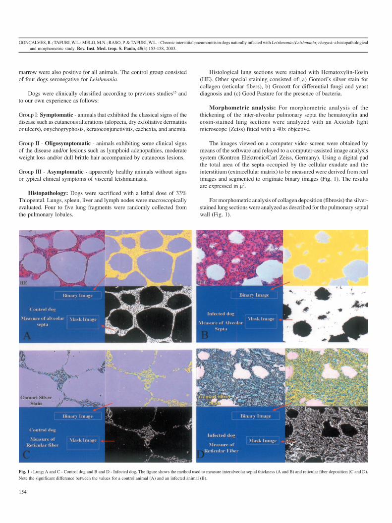
Morphometric analysis: For morphometric analysis of thethickening of the inter-alveolar pulmonary septa the hematoxylin andeosin-stained lung sections were analyzed with an Axiolab lightmicroscope (Zeiss) fitted with a 40x objective.
The images viewed on a computer video screen were obtained bymeans of the software and relayed to a computer-assisted image analysissystem (Kontron Elektronic/Carl Zeiss, Germany). Using a digital padthe total area of the septa occupied by the cellular exudate and theinterstitium (extracellular matrix) to be measured were derived from realimages and segmented to originate binary images (Fig. 1). The resultsare expressed in µ2.
For morphometric analysis of collagen deposition (fibrosis) the silver-stained lung sections were analyzed as described for the pulmonary septalwall (Fig. 1).
Fig. 1 - Lung; A and C - Control dog and B and D - Infected dog. The figure shows the method used to measure interalveolar septal thickness (A and B) and reticular fiber deposition (C and D).
Note the significant difference between the values for a control animal (A) and an infected animal (B).
GONÇALVES, R.; TAFURI, W.L.; MELO, M.N.; RASO, P. & TAFURI, W.L. - Chronic interstitial pneumonitis in dogs naturally infected with Leishmania (Leishmania) chagasi: a histopathologicaland morphometric study. Rev. Inst. Med. trop. S. Paulo, 45(3):153-158, 2003.
155
Statistical analysis: The results obtained in a fully randomizeddesign were log transformed and the means for each group were comparedby the Student t test21. A P value of less than 0.05 was consideredsignificant.
RESULTS
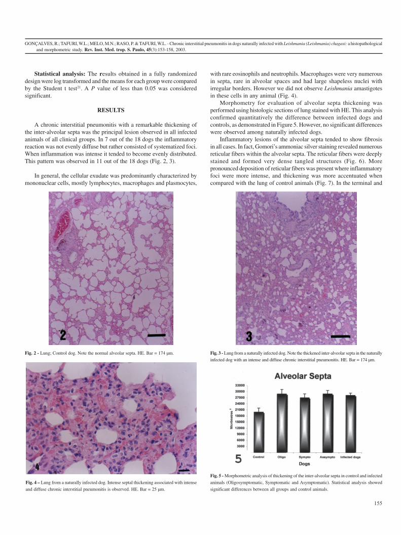
A chronic interstitial pneumonitis with a remarkable thickening ofthe inter-alveolar septa was the principal lesion observed in all infectedanimals of all clinical groups. In 7 out of the 18 dogs the inflammatoryreaction was not evenly diffuse but rather consisted of systematized foci.When inflammation was intense it tended to become evenly distributed.This pattern was observed in 11 out of the 18 dogs (Fig. 2, 3).
In general, the cellular exudate was predominantly characterized bymononuclear cells, mostly lymphocytes, macrophages and plasmocytes,
with rare eosinophils and neutrophils. Macrophages were very numerousin septa, rare in alveolar spaces and had large shapeless nuclei withirregular borders. However we did not observe Leishmania amastigotesin these cells in any animal (Fig. 4).
Morphometry for evaluation of alveolar septa thickening wasperformed using histologic sections of lung stained with HE. This analysisconfirmed quantitatively the difference between infected dogs andcontrols, as demonstrated in Figure 5. However, no significant differenceswere observed among naturally infected dogs.
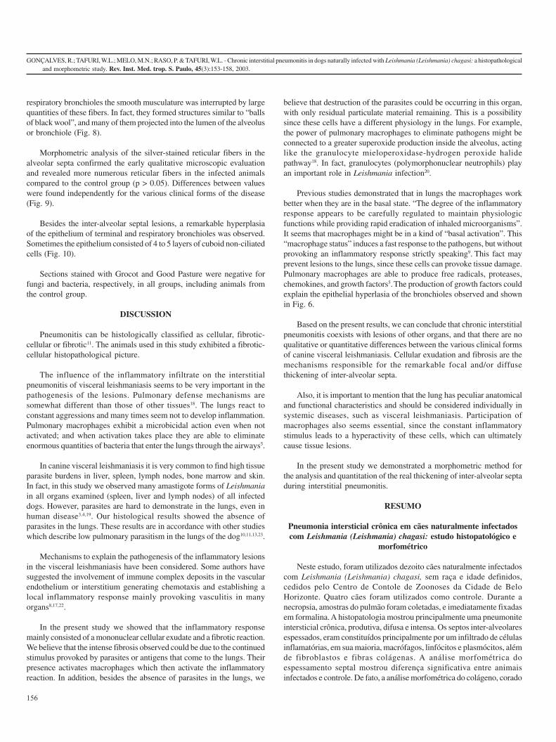
Inflammatory lesions of the alveolar septa tended to show fibrosisin all cases. In fact, Gomori’s ammoniac silver staining revealed numerousreticular fibers within the alveolar septa. The reticular fibers were deeplystained and formed very dense tangled structures (Fig. 6). Morepronounced deposition of reticular fibers was present where inflammatoryfoci were more intense, and thickening was more accentuated whencompared with the lung of control animals (Fig. 7). In the terminal and
Fig. 4 – Lung from a naturally infected dog. Intense septal thickening associated with intense
and diffuse chronic interstitial pneumonitis is observed. HE. Bar = 25 µm.
Fig. 5 - Morphometric analysis of thickening of the inter-alveolar septa in control and infected
animals (Oligosymptomatic, Symptomatic and Asymptomatic). Statistical analysis showed
significant differences between all groups and control animals.
Fig. 3 - Lung from a naturally infected dog. Note the thickened inter-alveolar septa in the naturally
infected dog with an intense and diffuse chronic interstitial pneumonitis. HE. Bar = 174 µm.
Fig. 2 - Lung; Control dog. Note the normal alveolar septa. HE. Bar = 174 µm.
156
GONÇALVES, R.; TAFURI, W.L.; MELO, M.N.; RASO, P. & TAFURI, W.L. - Chronic interstitial pneumonitis in dogs naturally infected with Leishmania (Leishmania) chagasi: a histopathologicaland morphometric study. Rev. Inst. Med. trop. S. Paulo, 45(3):153-158, 2003.
respiratory bronchioles the smooth musculature was interrupted by largequantities of these fibers. In fact, they formed structures similar to “ballsof black wool”, and many of them projected into the lumen of the alveolusor bronchiole (Fig. 8).
Morphometric analysis of the silver-stained reticular fibers in thealveolar septa confirmed the early qualitative microscopic evaluationand revealed more numerous reticular fibers in the infected animalscompared to the control group (p > 0.05). Differences between valueswere found independently for the various clinical forms of the disease(Fig. 9).
Besides the inter-alveolar septal lesions, a remarkable hyperplasiaof the epithelium of terminal and respiratory bronchioles was observed.Sometimes the epithelium consisted of 4 to 5 layers of cuboid non-ciliatedcells (Fig. 10).
Sections stained with Grocot and Good Pasture were negative forfungi and bacteria, respectively, in all groups, including animals fromthe control group.
DISCUSSION
Pneumonitis can be histologically classified as cellular, fibrotic-cellular or fibrotic11. The animals used in this study exhibited a fibrotic-cellular histopathological picture.
The influence of the inflammatory infiltrate on the interstitialpneumonitis of visceral leishmaniasis seems to be very important in thepathogenesis of the lesions. Pulmonary defense mechanisms aresomewhat different than those of other tissues18. The lungs react toconstant aggressions and many times seem not to develop inflammation.Pulmonary macrophages exhibit a microbicidal action even when notactivated; and when activation takes place they are able to eliminateenormous quantities of bacteria that enter the lungs through the airways5.
In canine visceral leishmaniasis it is very common to find high tissueparasite burdens in liver, spleen, lymph nodes, bone marrow and skin.In fact, in this study we observed many amastigote forms of Leishmaniain all organs examined (spleen, liver and lymph nodes) of all infecteddogs. However, parasites are hard to demonstrate in the lungs, even inhuman disease3,4,19. Our histological results showed the absence ofparasites in the lungs. These results are in accordance with other studieswhich describe low pulmonary parasitism in the lungs of the dog10,11,13,23.
Mechanisms to explain the pathogenesis of the inflammatory lesionsin the visceral leishmaniasis have been considered. Some authors havesuggested the involvement of immune complex deposits in the vascularendothelium or interstitium generating chemotaxis and establishing alocal inflammatory response mainly provoking vasculitis in manyorgans8,17,22.
In the present study we showed that the inflammatory responsemainly consisted of a mononuclear cellular exudate and a fibrotic reaction.We believe that the intense fibrosis observed could be due to the continuedstimulus provoked by parasites or antigens that come to the lungs. Theirpresence activates macrophages which then activate the inflammatoryreaction. In addition, besides the absence of parasites in the lungs, we
believe that destruction of the parasites could be occurring in this organ,with only residual particulate material remaining. This is a possibilitysince these cells have a different physiology in the lungs. For example,the power of pulmonary macrophages to eliminate pathogens might beconnected to a greater superoxide production inside the alveolus, actinglike the granulocyte mieloperoxidase-hydrogen peroxide halidepathway18. In fact, granulocytes (polymorphonuclear neutrophils) playan important role in Leishmania infection20.
Previous studies demonstrated that in lungs the macrophages workbetter when they are in the basal state. “The degree of the inflammatoryresponse appears to be carefully regulated to maintain physiologicfunctions while providing rapid eradication of inhaled microorganisms”.It seems that macrophages might be in a kind of “basal activation”. This“macrophage status” induces a fast response to the pathogens, but withoutprovoking an inflammatory response strictly speaking9. This fact mayprevent lesions to the lungs, since these cells can provoke tissue damage.Pulmonary macrophages are able to produce free radicals, proteases,chemokines, and growth factors5. The production of growth factors couldexplain the epithelial hyperlasia of the bronchioles observed and shownin Fig. 6.
Based on the present results, we can conclude that chronic interstitialpneumonitis coexists with lesions of other organs, and that there are noqualitative or quantitative differences between the various clinical formsof canine visceral leishmaniasis. Cellular exudation and fibrosis are themechanisms responsible for the remarkable focal and/or diffusethickening of inter-alveolar septa.
Also, it is important to mention that the lung has peculiar anatomicaland functional characteristics and should be considered individually insystemic diseases, such as visceral leishmaniasis. Participation ofmacrophages also seems essential, since the constant inflammatorystimulus leads to a hyperactivity of these cells, which can ultimatelycause tissue lesions.
In the present study we demonstrated a morphometric method forthe analysis and quantitation of the real thickening of inter-alveolar septaduring interstitial pneumonitis.
RESUMO
Pneumonia intersticial crônica em cães naturalmente infectadoscom Leishmania (Leishmania) chagasi: estudo histopatológico e
morfométrico
Neste estudo, foram utilizados dezoito cães naturalmente infectadoscom Leishmania (Leishmania) chagasi, sem raça e idade definidos,cedidos pelo Centro de Contole de Zoonoses da Cidade de BeloHorizonte. Quatro cães foram utilizados como controle. Durante anecropsia, amostras do pulmão foram coletadas, e imediatamente fixadasem formalina. A histopatologia mostrou principalmente uma pneumoniteintersticial crônica, produtiva, difusa e intensa. Os septos inter-alveolaresespessados, eram constituídos principalmente por um infiltrado de célulasinflamatórias, em sua maioria, macrófagos, linfócitos e plasmócitos, alémde fibroblastos e fibras colágenas. A análise morfométrica doespessamento septal mostrou diferença significativa entre animaisinfectados e controle. De fato, a análise morfométrica do colágeno, corado
GONÇALVES, R.; TAFURI, W.L.; MELO, M.N.; RASO, P. & TAFURI, W.L. - Chronic interstitial pneumonitis in dogs naturally infected with Leishmania (Leishmania) chagasi: a histopathologicaland morphometric study. Rev. Inst. Med. trop. S. Paulo, 45(3):153-158, 2003.
157
Fig. 10 – Lung from a naturally infected dog. Hyperplasia of the epithelium. Presence of
many cells projecting into the lumem. HE. Bar = 25 µm.
Fig. 9 - Morphometric analysis of reticular fibers stained with Gomori’s Ammoniac Silver.
Statistical analysis showed significant differences between all groups and control animals.
Fig. 8 – Lung from a naturally infected dog. Reticular fibers are stained black. Observe the
intense fibrillopoiesis showing dense and coiled fibers (“wool nodules”). Gomori’s Ammoniac
Silver. Bar = 12.5 µm.
Fig. 6 – Lung from a naturally infected dog. Note the intense fibropoiesis in the smooth
muscle of bronchioles, which still exhibit intense hyperplasia of the epithelium. Gomori’s
Ammoniac Silver. Bar = 50 µm.
Fig. 7 – Lung from a control dog. Reticular collagen fibers stained black showing normal
deposition in the alveolar septa. Gomori’s Ammoniac Silver. Bar = 12.5 µm.
158
GONÇALVES, R.; TAFURI, W.L.; MELO, M.N.; RASO, P. & TAFURI, W.L. - Chronic interstitial pneumonitis in dogs naturally infected with Leishmania (Leishmania) chagasi: a histopathologicaland morphometric study. Rev. Inst. Med. trop. S. Paulo, 45(3):153-158, 2003.
pela prata, confirmou um aumento na deposição de colágeno em todosos animais infectados. O exame parasitológico foi ralizado durante aanálise histopatológica, em todos os casos estudados. Contudo nestetrabalho, não observamos qualquer correlação entre os achadoshistopatológicos e morfométricos quando relacionados aos cães emdiferentes formas clínicas. Concluímos que as lesões, encontradas noscães naturalmente infectados, tem correlação com a doença, e que ométodo de análise morfométrica utilizado apresentou resultadossatisfatórios quanto à análise do espessamento septal e ao aumento dadeposição de colágeno, confirmando a fibrose.
REFERENCES
1. ANDERSON, D.C.; BUCKER, R.G.; GLENN, B.L. & MACVEAN, D.W. - Endemiccanine leishmaniasis. Vet. Path., 17: 94-96, 1980.
2. ANDRADE, Z.A. - Anatomia patológica da leishmaniose visceral (calazar). Arq. brasMed. naval, 7: 7346-7472, 1958.
3. ANDRADE, Z.A. - Pneumonite intersticial no calazar. Hospital (Rio de J.), 55: 371-381, 1959.
4. ANDRADE, Z.A. & ANDRADE, S.G. - Alguns novos aspectos da patologia do calazar(estudo morfológico de 13 casos necropsiados). Rev. Inst. Med trop. S. Paulo, 8:259-266, 1966.
5. BITTERMAN, P.B.; SALTZMAN, L.E.; ADELBERG, S.; FERRANS, V.J. & CRYSTAL,R.G. - Alveolar macrophage replication. J. clin. Invest., 74: 460-469, 1984.
6. BOGLIOLO, L. - Sulla Anatomia pathologica della leishmaniose visceral dell’uomo.Arch. ital. Sci. med. colon., 15: 588, 1934.
7. CHAGAS, E. & CHAGAS, A.W. - Notas sobre a epidemiologia da leishmaniose visceralamericana. Hospital (Rio de J.), 13: 147-180, 1938.
8. CIARAMELLA, P.; OLIVA, G., LUNA, R.D. et al. - A retrospective clinical study ofcanine leishmaniasis in 150 dogs naturally infected by Leishmania infantum. Vet.Rec., 22: 539-543, 1997.
9. COONROD, J.D. - Role of leukocytes in lung defenses. Respiration, 55(suppl. 1): 9-13,1989.
10. DUARTE, M.I.S. & CORBETT, C.E.P. - Histopathological and ultrastructural aspects ofinterstitial pneumonitis of experimental visceral leishmaniasis. Trans. roy. Soc. trop.Med. Hyg., 78: 683-688, 1984.
11. DUARTE, M.I.S.; LAURENTI, M.D.; NUNES, V.L.B. et al. - Interstitial pneumonitis incanine visceral leishmaniasis. Rev. Inst. Med. trop. S. Paulo, 28: 431-436, 1986.
12. DUARTE, M.I.S.; MATTA, V.L.R.; CORBETT, C.E.P. et al. - Interstitial pneumonitis inhuman visceral leishmaniasis. Trans. roy. Soc. trop. Med. Hyg., 83: 73-76, 1989.
13. KEENAN, C.M.; HENDRICKS, L.D.; LIGHTNER, L. & JOHNSON, A.J. - Visceralleishmaniasis in the German shepherd dog. II. Pathology. Vet. Path., 21: 80-86,1984..
14. LIPSCOMB, F.E. & BIGSON, M.O.J. - Visceral leishmaniosis (Kala-azar) in an adultcontracted in Malta. Brit. med. J., 1: 492-493, 1944..
15. MANCIANTI, F.; GRAMICCIA, M.; GRANDONI, L. & PIERI, S. - Studies on canineleishmaniasis control. 1. Evolution of infection of different clinical forms of canineleishmaniasis following antimonial treatment. Trans. roy. Soc. trop Med. Hyg., 82:566-567, 1988.
16. NAPIER, L.E. - The principles and practice of tropical medicine. New York, TheMacMillan, 1946. p. 917.
17. PUMAROLA, M.; BREVIK, L.; BADIOLA, J. et al. - Canine leishmaniasis associatedwith systemic vasculitis in two dogs. J. comp. Path., 105: 279-286, 1991.
18. QUIE, P.G. - Lung defense against infection. J. Pediat., 108: 813-816, 1986.
19. RASO, P. & SIQUEIRA, J.T.D. - Subsídio ao conhecimento da anatomia patológica daleishmaniose visceral, com especial referência às lesões pulmonares e cardíacas.Hospital (Rio de J.), 65(6): 145-163, 1964.
20. ROUSSEAU, D.; DEMARTINO, S.; FERRUA, B. et al. - In vivo involvement ofpolymorphonuclear neutrophils in Leishmania infantum infection. BMC Microbiol.,1: 17, 2001.
21. SAMPAIO, I.B.M. - Estatística aplicada à experimentação animal. Belo Horizonte,Fundação de Ensino e Pesquisa em Medicina Veterinária, 1998. p. 221.
22. TAFURI, W.L.; MICHALICK, M.S.M.; DIAS, M. et al. - Estudo ao microscópio ópticoe eletrônico, do rim de cães natural e experimentalmente infectados com Leishmania(Leishmania) chagasi. Rev. Inst. Med. trop. S. Paulo, 31: 139-145, 1989.
23. TRYPHONAS, L.; ZAWIDZKA, Z.; BERNARD, M.A. & JANZEN, E.A. - Visceralleishmaniasis in a dog: clinical, hematological and pathological observations. Canad.J. comp. Med., 41: 1-12, 1977.
Received: 9 January 2003Accepted: 22 April 2003