![Rehabilitació del nen amb disfunció del buidatge vesical · Microsoft PowerPoint - RHB 1 urologica disfuncio buidatge. Acadèmia..ppt [Modo de compatibilidad] Author: tecnicsales](https://static.fdocuments.co/doc/165x107/5b0371687f8b9a0a548c28ed/rehabilitaci-del-nen-amb-disfunci-del-buidatge-powerpoint-rhb-1-urologica-disfuncio.jpg)
Disfunció microcirculatòria sinusoïdal hepàtica en la...
132
Disfunció microcirculatòria sinusoïdal hepàtica en la cirrosi i dany per isquèmia/reperfusió: mecanismes involucrats i noves dianes terapèutiques Sergi Guixé Muntet ADVERTIMENT. La consulta d’aquesta tesi queda condicionada a l’acceptació de les següents condicions d'ús: La difusió d’aquesta tesi per mitjà del servei TDX (www.tdx.cat) i a través del Dipòsit Digital de la UB (diposit.ub.edu) ha estat autoritzada pels titulars dels drets de propietat intel·lectual únicament per a usos privats emmarcats en activitats d’investigació i docència. No s’autoritza la seva reproducció amb finalitats de lucre ni la seva difusió i posada a disposició des d’un lloc aliè al servei TDX ni al Dipòsit Digital de la UB. No s’autoritza la presentació del seu contingut en una finestra o marc aliè a TDX o al Dipòsit Digital de la UB (framing). Aquesta reserva de drets afecta tant al resum de presentació de la tesi com als seus continguts. En la utilització o cita de parts de la tesi és obligat indicar el nom de la persona autora. ADVERTENCIA. La consulta de esta tesis queda condicionada a la aceptación de las siguientes condiciones de uso: La difusión de esta tesis por medio del servicio TDR (www.tdx.cat) y a través del Repositorio Digital de la UB (diposit.ub.edu) ha sido autorizada por los titulares de los derechos de propiedad intelectual únicamente para usos privados enmarcados en actividades de investigación y docencia. No se autoriza su reproducción con finalidades de lucro ni su difusión y puesta a disposición desde un sitio ajeno al servicio TDR o al Repositorio Digital de la UB. No se autoriza la presentación de su contenido en una ventana o marco ajeno a TDR o al Repositorio Digital de la UB (framing). Esta reserva de derechos afecta tanto al resumen de presentación de la tesis como a sus contenidos. En la utilización o cita de partes de la tesis es obligado indicar el nombre de la persona autora. WARNING. On having consulted this thesis you’re accepting the following use conditions: Spreading this thesis by the TDX (www.tdx.cat) service and by the UB Digital Repository (diposit.ub.edu) has been authorized by the titular of the intellectual property rights only for private uses placed in investigation and teaching activities. Reproduction with lucrative aims is not authorized nor its spreading and availability from a site foreign to the TDX service or to the UB Digital Repository. Introducing its content in a window or frame foreign to the TDX service or to the UB Digital Repository is not authorized (framing). Those rights affect to the presentation summary of the thesis as well as to its contents. In the using or citation of parts of the thesis it’s obliged to indicate the name of the author.
Transcript of Disfunció microcirculatòria sinusoïdal hepàtica en la...

Disfunció microcirculatòria sinusoïdal hepàtica
en la cirrosi i dany per isquèmia/reperfusió: mecanismes involucrats i noves dianes terapèutiques
Sergi Guixé Muntet
ADVERTIMENT. La consulta d’aquesta tesi queda condicionada a l’acceptació de les següents condicions d'ús: La difusió d’aquesta tesi per mitjà del servei TDX (www.tdx.cat) i a través del Dipòsit Digital de la UB (diposit.ub.edu) ha estat autoritzada pels titulars dels drets de propietat intel·lectual únicament per a usos privats emmarcats en activitats d’investigació i docència. No s’autoritza la seva reproducció amb finalitats de lucre ni la seva difusió i posada a disposició des d’un lloc aliè al servei TDX ni al Dipòsit Digital de la UB. No s’autoritza la presentació del seu contingut en una finestra o marc aliè a TDX o al Dipòsit Digital de la UB (framing). Aquesta reserva de drets afecta tant al resum de presentació de la tesi com als seus continguts. En la utilització o cita de parts de la tesi és obligat indicar el nom de la persona autora. ADVERTENCIA. La consulta de esta tesis queda condicionada a la aceptación de las siguientes condiciones de uso: La difusión de esta tesis por medio del servicio TDR (www.tdx.cat) y a través del Repositorio Digital de la UB (diposit.ub.edu) ha sido autorizada por los titulares de los derechos de propiedad intelectual únicamente para usos privados enmarcados en actividades de investigación y docencia. No se autoriza su reproducción con finalidades de lucro ni su difusión y puesta a disposición desde un sitio ajeno al servicio TDR o al Repositorio Digital de la UB. No se autoriza la presentación de su contenido en una ventana o marco ajeno a TDR o al Repositorio Digital de la UB (framing). Esta reserva de derechos afecta tanto al resumen de presentación de la tesis como a sus contenidos. En la utilización o cita de partes de la tesis es obligado indicar el nombre de la persona autora. WARNING. On having consulted this thesis you’re accepting the following use conditions: Spreading this thesis by the TDX (www.tdx.cat) service and by the UB Digital Repository (diposit.ub.edu) has been authorized by the titular of the intellectual property rights only for private uses placed in investigation and teaching activities. Reproduction with lucrative aims is not authorized nor its spreading and availability from a site foreign to the TDX service or to the UB Digital Repository. Introducing its content in a window or frame foreign to the TDX service or to the UB Digital Repository is not authorized (framing). Those rights affect to the presentation summary of the thesis as well as to its contents. In the using or citation of parts of the thesis it’s obliged to indicate the name of the author.

1


3
DISFUNCIÓ MICROCIRCULATÒRIA SINUSOÏDAL HEPÀTICA EN LA
CIRROSI I DANY PER ISQUÈMIA/REPERFUSIÓ: MECANISMES
INVOLUCRATS I NOVES DIANES TERAPÈUTIQUES.
Tesi Doctoral presentada per
SERGI GUIXÉ MUNTET
Per a optar al grau de Doctor
Treball realitzat sota la direcció del Dr. Jordi Gracia-Sancho i el Prof. Jaume
Bosch Genover, al Laboratori d’Hemodinàmica Hepàtica i Hipertensió Portal,
Institut d’Investigacions Biomèdiques August Pi i Sunyer (IDIBAPS),
Departament de Medicina, Hospital Clínic de Barcelona.
Dr. Jordi Gracia-Sancho
Prof. Jaume Bosch Genover
Directors
Programa de Doctorat en Medicina
Maig 2017


A la meva família

1
ÍNDEX
INFORME DELS DIRECTORS ........................................................................................................... 3
1. LLISTA D’ABREVIACIONS ............................................................................................................ 7
2. INTRODUCCIÓ.......................................................................................................................... 11
2.1. El sinusoide hepàtic .......................................................................................................... 13
2.2. Regulació del to vascular hepàtic ..................................................................................... 14
2.3. KLF2 .................................................................................................................................. 17
2.3.1. Estatines .................................................................................................................... 17
2.4. Cirrosi ............................................................................................................................... 19
2.4.1. Fisiopatologia de la cirrosi ......................................................................................... 19
2.4.2. Aproximacions farmacològiques ............................................................................... 21
2.4.3. Liraglutida .................................................................................................................. 22
2.5. Isquèmia i reperfusió ........................................................................................................ 22
2.5.1. Isquèmia .................................................................................................................... 23
2.5.2. Reperfusió ................................................................................................................. 24
2.6. Autofàgia .......................................................................................................................... 26
2.6.1. Iniciació de l’autofàgia .............................................................................................. 27
2.6.2. Elongació de l’autofagosoma .................................................................................... 28
2.6.3. Fusió i flux autofàgic .................................................................................................. 28
2.6.4. Autofàgia hepàtica .................................................................................................... 30
3. HIPÒTESIS I OBJECTIUS ............................................................................................................ 31
3.1. Estudi 1 ............................................................................................................................. 34
3.2. Estudi 2 ............................................................................................................................. 34
4. CÒPIA DELS ARTICLES ORIGINALS ........................................................................................... 37
4.1. Estudi 1 ............................................................................................................................. 39
4.2. Estudi 2 ............................................................................................................................. 57
5. RESUM DELS RESULTATS ......................................................................................................... 77
5.1. Estudi 1 ............................................................................................................................. 79
5.2. Estudi 2 ............................................................................................................................. 79
6. DISCUSSIÓ ............................................................................................................................... 81
7. CONCLUSIONS ......................................................................................................................... 89

7.1. Estudi 1 ............................................................................................................................. 91
7.2. Estudi 2 ............................................................................................................................. 91
8. REFERÈNCIES ........................................................................................................................... 93
9. ALTRES PUBLICACIONS .......................................................................................................... 113
10. AGRAÏMENTS ....................................................................................................................... 117

3
INFORME DELS DIRECTORS


INFORME DELS DIRECTORS
5
INFORME DELS DIRECTORS
Barcelona, 28 d’abril de 2017
Jordi Gracia Sancho, Cap de Grup de Recerca a l’Institut D’Investigacions
Biomèdiques August Pi i Sunyer (IDIBAPS), i Jaume Bosch Genover, Professor
en Medicina de la Universitat de Barcelona i Consultor Sènior en Hepatologia a
l’Hospital Clínic de Barcelona,
CERTIFIQUEM:
Que la tesi doctoral titulada “DISFUNCIÓ MICROCIRCULATÒRIA SINUSOÏDAL
HEPÀTICA EN LA CIRROSI I DANY PER ISQUÈMIA/REPERFUSIÓ:
MECANISMES INVOLUCRATS I NOVES DIANES TERAPÈUTIQUES”,
presentada per Sergi Guixé Muntet per a l’obtenció del títol de Doctor per la
Universitat de Barcelona s’ha fet sota la nostra supervisió i compleix tots els
requisits per a ser defensada davant del corresponent comitè avaluador.
Jordi Gracia Sancho Jaume Bosch Genover


7
1. LLISTA D’ABREVIACIONS


LLISTA D’ABREVIACIONS 9
1. LLISTA D’ABREVIACIONS
α-SMA: actina de musculatura llisa alfa
ADMA: dimetilarginina asimètrica
Akt: proteïna quinasa B
AMPK: proteïna-quinasa activada per AMP
Atg: autophagy related protein
BH4: tetrahidrobiopterina
CNP: pèptid natriurètic de tipus C
CO: monòxid de carboni
eNOS: sintasa d’òxid nítric endotelial
GGPP: geranil-geranil pirofosfat
GLP-1: glucagon-Like Peptide-1
HMG-CoA: 3-hydroxi-3-metil-glutaril-coenzim A
HO-1: hemo-oxigenasa-1
HSC: cèl·lules estrellades hepàtiques
HVPG: gradient de pressió venosa hepàtica
HVR: resistència vascular intrahepàtica
ICAM-1: molècula d’adhesió intercel·lular-1
IL: interleucina
I/R: isquèmia/reperfusió
KC: cèl·lules de Kupffer
KLF2: krüppel-like factor 2
LC3: microtubule-associated proteins 1A/1B light chain 3

10 LLISTA D’ABREVIACIONS
LKB1: quinasa de fetge B1
LSEC: cèl·lules endotelials sinusoïdals
MCP-1: proteïna quimioatraient de monòcits
mTORC1: mammalian target of rapamycin complex 1
NAFLD: malaltia de fetge gras d’origen no alcohòlic
NASH: esteatohepatitis no alcohòlica
NO: òxid nítric
O2- : anió superòxid
ONOO- : peroxinitrit
PAF: factor d’activació plaquetària
PDGF: factor de creixement derivat de plaquetes
PGI2: prostaciclina
Rab7: ras-related protein 7
ROS: espècies reactives d’oxigen
TNF-α: factor de necrosi tumoral alfa
ULK1: unc-51-like kinase 1
VCAM: molècula d’adhesió cel·lular vascular
VEGF: factor de creixement endotelial vascular

2. INTRODUCCIÓ


13 INTRODUCCIÓ
2. INTRODUCCIÓ
El fetge és l’òrgan encarregat de metabolitzar gran part de les substàncies
absorbides a l’intestí, produir bilis, sintetitzar proteïnes i detoxificar la sang, entre
d’altres, essent els hepatòcits les cèl·lules efectores d’aquestes funcions.
Tanmateix, la viabilitat i el fenotip dels hepatòcits i, per extensió, el funcionament
hepàtic venen determinats pel correcte estat de la microcirculació, essent clau el
rol de les cèl·lules sinusoïdals en la prevenció, el desenvolupament i la regressió
de la malaltia hepàtica crònica o del dany hepàtic agut (1–3).
2.1. El sinusoide hepàtic
La major part de la sang que arriba al fetge (70%) ho fa a través de la vena porta.
Es tracta de sang venosa provinent de l’intestí, a baixa pressió, molt rica en
nutrients, mediadors immunològics, hormones i factors de creixement i altament
desoxigenada. Només el 30% restant és sang oxigenada que arriba per l’artèria
hepàtica (4). El fetge està organitzat en lobulets hepàtics, que són agrupacions
de sinusoides amb una distribució radial. A l’inici de cada sinusoide es barreja la
sang provinent d’una vènula porta i una arteriola hepàtica, que recorrerà el
sinusoide fins l’altre extrem, la vènula central, que desemboca a la vena cava
inferior. En sentit contrari, el canalicle biliar transporta la bilis secretada pels
hepatòcits fins el conducte biliar (4).
El sinusoide està constituït principalment per tres tipus de cèl·lules no
parenquimals. Les cèl·lules endotelials sinusoïdals (LSEC) delimiten el lumen del
sinusoide. Es tracta d’un endoteli discontinu molt especialitzat, que permet la
difusió dels soluts de la sang fins els hepatòcits i participa en la regulació de
l’hemostàsia, l’eliminació de substàncies tòxiques, la inflamació i la regulació del
to vascular (5–7). Al seu voltant, a l’espai de Disse (comprès entre l’endoteli i els
hepatòcits), les cèl·lules hepàtiques estrellades (HSC) regulen l’obertura dels
sinusoides per a acomodar el flux sanguini, de tal manera que una HSC pot
modular fins a quatre LSEC a la vegada (7). A més a més, són el major dipòsit
de vitamina A de l’organisme i les majors productores de fibra hepàtica (8). Per
últim, les cèl·lules de Kupffer (KC) són els macròfags residents hepàtics, i se

14 INTRODUCCIÓ
situen al lumen sinusoïdal, participant en funcions de defensa, inflamació i
remodelació tissular (9) (Figura 1).
Figura 1. Estructura del sinusoide hepàtic. Adaptat de “Aird WC 2007 Circ Res”. SEC: Sinusoidal
Endothelial Cell.
2.2. Regulació del to vascular hepàtic
El funcionament i la viabilitat dels hepatòcits, per tant, depèn directament de les
cèl·lules sinusoïdals, ja que alteracions en la regulació del to vascular poden
impedir l’arribada d’oxigen i nutrients, essent la disfunció microvascular el primer
factor desencadenant de la majoria de malalties hepàtiques (3,7,10). Per tal
d’entendre com es desregulen les cèl·lules sinusoïdals en la malaltia, però, cal
entendre primer el rol que desenvolupen en condicions de salut.
Les LSEC representen un 15-20% del total de les cèl·lules hepàtiques. Presenten
un fenotip molt especialitzat, diferenciant-se de la resta d’endotelis de
l’organisme per l’absència de membrana basal i la presència de fenestres (11).
Aquestes són obertures adiafragmàtiques d’entre 50nm i 200nm, agrupades en
sieve plates (plaques de tamís). Les fenestres poden representar fins el 20% de
la superfície cel·lular (12), conferint així una elevada porositat a l’endoteli i

15 INTRODUCCIÓ
permetent la comunicació directa de la sang amb els hepatòcits (11). En
condicions de salut, les LSEC estan exposades contínuament a l’estrès per
fricció efectuat pel flux sanguini, el qual els confereix un fenotip vasoprotector
que, a més a més de mantenir les fenestres, activa diverses vies de producció
d’agents vasodilatadors. La més rellevant és la via de l’òxid nítric (NO), sintetitzat
principalment per la sintasa d’òxid nítric endotelial (eNOS) (13), tot i que el flux
també indueix la síntesi d’altres molècules vasodilatadores com el monòxid de
carboni (CO) o la prostaciclina (PGI2), mentre que atenua la síntesi d’endotelines
(vasoconstrictors) (14–16). Aquestes molècules mantenen la quiescència de les
HSC, mantenint un equilibri entre en la seva resposta a vasodilatadors (NO, CO,
PGI2) i vasoconstrictors (endotelina-1, tromboxà A2, angiotensina II) i permetent
l’acomodació de les variacions de flux sanguini (13). Al seu torn, les HSC i els
hepatòcits mantenen el fenotip de les LSEC per mitjà de la secreció, entre
d’altres, de VEGF (17,18).
Tanmateix, quan es produeix un dany hepàtic o una alteració del flux sanguini,
aquest cicle protector queda alterat. Les LSEC perden el seu fenotip característic
(perden l’expressió d’estabilina 1 i 2, CD32b) i expressen el marcador d’endoteli
vascular CD31; desapareixen les fenestres i es forma membrana basal, de
manera que l’endoteli deixa de ser discontinu i esdevé un endoteli més semblant
a l’endoteli vascular (procés anomenat capil·larització) (7,19–21). Això dificulta
la difusió de les molècules de la sang i l’arribada d’oxigen a la resta de cèl·lules
hepàtiques. Les LSEC participen en la resposta inflamatòria i en resposta al dany
hepàtic activen la síntesi de molècules d’adhesió cel·lular (CAM), com ICAM-1,
VCAM-1 i E-selectina i la secreció de quimiocines com IL-1, IL-6, IL-8, MCP-1 i
interferó, induint l’atracció de leucòcits (3,22). A més a més, les LSEC adopten
un fenotip protrombòtic i augmenten la síntesi de fibronectina (23,24). Per altra
banda, el canvi de fenotip es tradueix en la davallada de la síntesi de
vasodilatadors (25) en favor de la major producció de vasoconstrictors (26).
En resposta a citocines (com PDGF i TGF-β) o per mitjà de vies de senyalització
com Hedgehog o Wnt/β-catenina i a l’augment en la relació
vasoconstrictors/vasodilatadors de les LSEC, les HSC s’activen (Figura 2);
perden les gotes lipídiques característiques metabolitzant-les com a font
d’energia (27,28) i adquireixen un fenotip pro-contràctil i pro-fibròtic (amb α-SMA
16 INTRODUCCIÓ
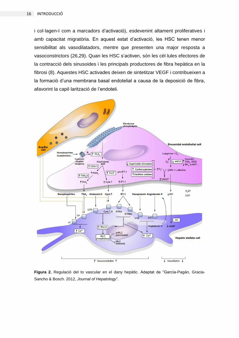
i col·lagen-I com a marcadors d’activació), esdevenint altament proliferatives i
amb capacitat migratòria. En aquest estat d’activació, les HSC tenen menor
sensibilitat als vasodilatadors, mentre que presenten una major resposta a
vasoconstrictors (26,29). Quan les HSC s’activen, són les cèl·lules efectores de
la contracció dels sinusoides i les principals productores de fibra hepàtica en la
fibrosi (8). Aquestes HSC activades deixen de sintetitzar VEGF i contribueixen a
la formació d’una membrana basal endotelial a causa de la deposició de fibra,
afavorint la capil·larització de l’endoteli.
Figura 2. Regulació del to vascular en el dany hepàtic. Adaptat de “García-Pagán, Gracia-
Sancho & Bosch. 2012, Journal of Hepatology”.

17 INTRODUCCIÓ
2.3. KLF2
Les proteïnes de la família Krüppel-like factor (KLF) són factors de transcripció
del tipus zinc finger que regulen el desenvolupament tissular i el creixement
cel·lular (30). KLF2, a més a més d’expressar-se al pulmó i als limfòcits, té una
elevada expressió endotelial (31) i té com a diana gens que confereixen un
fenotip vasoprotector a l’endoteli. Entre els més rellevants, s’hi troben gens
vasodilatadors (eNOS, pèptid natriurètic de tipus C (CNP)), anti-trombòtics
(trombomodulina) i anti-oxidants (Hemo-oxigenasa-1 (HO-1), nuclear factor
erythroid 2-related factor 2 (Nrf2)), mentre que inhibeix l’expressió de molècules
d’adhesió leucocitària (E-selectina i VCAM-1) i l’apoptosi (15,32–35). Un dels
activadors més potents de KLF2 és l’estrès per fricció, ocasionat pel fregament
continu del flux sanguini sobre l’endoteli (14), el qual n’augmenta la transcripció
a través de la via de MEK5-ERK5-MEF2 (36). En canvi, l’alteració del flux
sanguini en zones vasculars turbulentes o el cessament del flux provoquen la
ràpida davallada de la seva expressió i de la protecció endotelial que confereix
(15,35,37).
Al fetge, l’expressió de KLF2 és mediadora dels efectes vasoprotectors del flux
sanguini en el manteniment del fenotip especialitzat de l’endoteli sinusoïdal i és
responsable, per tant, de la protecció paracrina de les HSC (38) i de gran part
dels mecanismes moleculars que regulen el to vascular hepàtic (39), esmentats
anteriorment en aquesta tesi. Tot això i el fet que actualment està acceptat que
el dany endotelial és una causa inicial del desenvolupament de les malalties
hepàtiques més importants (1–3,7) fa que KLF2 sigui un actor principal en la
prevenció i el tractament d’aquestes malalties. En aquest sentit, estudis recents
del nostre grup han estat dirigits a modular farmacològicament l’expressió
d’aquest factor de transcripció emprant estatines (38,40–46).
2.3.1. Estatines
Les estatines són fàrmacs aprovats per les agències americana i europea de
medicaments, dissenyades per a reduir els nivells de colesterol a causa de la
seva activitat inhibidora de la 3-hydroxi-3-metil-glutaril-coenzim A (HMG-CoA)
reductasa, bloquejant així la síntesi de mevalonat (precursor del colesterol).

18 INTRODUCCIÓ
Tanmateix, les estatines han demostrat tenir importants efectes vasoprotectors
independents de la reducció dels nivells de colesterol, derivats de la davallada
dels nivells de geranil-geranil pirofosfat (GGPP) (47–49). S’ha demostrat que
aquests efectes són mediats principalment per la potent inducció que causen en
l’expressió de KLF2 (50,51); al fetge, el tractament amb simvastatina manté
l’expressió de KLF2 i el fenotip endotelial de manera similar a l’estímul d’estrès
per fricció i protegeix la resta de cèl·lules hepàtiques davant del dany hepàtic
agut i crònic (3,38,40–42,44–46). Tanmateix, els mecanismes moleculars que
vinculen directament la reducció del GGPP derivada del tractament amb
estatines amb l’increment de l’expressió de KLF2 encara són desconeguts, tot i
que la inhibició de la isoprenilació de les GTPases RhoA, Rac1 i Cdc42 per acció
de les estatines hi podria estar involucrada (48,52) (Figura 3).
Figura 3. Esquema de la via d’acció de les estatines independent de la síntesi de colesterol.
Adaptat de “Wang CY et al. 2008, Trends in Molecular Medicine”

19 INTRODUCCIÓ
2.4. Cirrosi
2.4.1. Fisiopatologia de la cirrosi
La cirrosi és la quarta causa de mortalitat en adults a Europa (53). Es produeix
per diferents etiologies, com el consum abusiu d’alcohol, la infecció per virus de
l’hepatitis o malalties no alcohòliques d’incidència creixent com l’obesitat.
Aquests insults crònics alteren el fenotip i la funció sinusoïdal pels mecanismes
descrits anteriorment en aquesta tesi, provocant disfunció microvascular i la
deposició excessiva de fibra (7,10,24). Les alteracions en la microvasculatura i
l’arquitectura hepàtiques causen un increment de la resistència vascular
intrahepàtica (HVR) al pas de la sang, provocant l’augment de la pressió portal,
la complicació més comuna de la cirrosi que desencadenarà les complicacions
clíniques més rellevants (2,54).
El territori esplàncnic, immediatament previ al fetge, detecta aquest augment
significatiu de pressió portosinusoïdal i incrementa la síntesi de vasodilatadors
(10) per tal de mitigar la hipertensió portal, però la baixa resposta del fetge fibròtic
als vasodilatadors acaba provocant una vasodilatació sistèmica, que sumada a
l’increment de la despesa cardíaca associada provoca l’increment del flux
sanguini, agreujant encara més la hipertensió portal (55). Per altra banda,
l’increment significatiu de pressió portosinusoïdal activa mecanismes
d’angiogènesi a nivell esplàncnic i la formació de vasos col·laterals extrahepàtics
(56) que alliberen pressió derivant part de la sang portal a la vena cava o cap a
l’aparell digestiu, de manera que en alguns casos fins a un 90% del flux portal és
desviat a través de les col·laterals (57). Aquesta “solució” fisiològica, però, limita
encara més l’oxigen que arriba al fetge i també és causa de l’aparició de varius
gastro-esofàgiques. Altres complicacions associades a la desviació porto-
sistèmica de la sang i a l’anomenat síndrome hiperdinàmic són l’encefalopatia
(produïda pels tòxics que es desvien per les col·laterals i no s’eliminen al fetge)
i l’aparició d’ascites (conseqüència de la retenció de líquids causada per la
hipovolèmia efectiva) (53,58) (Figura 4).
20 INTRODUCCIÓ
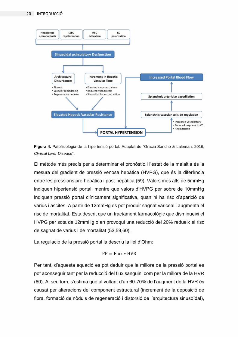
Figura 4. Patofisiologia de la hipertensió portal. Adaptat de “Gracia-Sancho & Laleman. 2016,
Clinical Liver Disease”.
El mètode més precís per a determinar el pronòstic i l’estat de la malaltia és la
mesura del gradient de pressió venosa hepàtica (HVPG), que és la diferència
entre les pressions pre-hepàtica i post-hepàtica (59). Valors més alts de 5mmHg
indiquen hipertensió portal, mentre que valors d’HVPG per sobre de 10mmHg
indiquen pressió portal clínicament significativa, quan hi ha risc d’aparició de
varius i ascites. A partir de 12mmHg es pot produir sagnat variceal i augmenta el
risc de mortalitat. Està descrit que un tractament farmacològic que disminueixi el
HVPG per sota de 12mmHg o en provoqui una reducció del 20% redueix el risc
de sagnat de varius i de mortalitat (53,59,60).
La regulació de la pressió portal la descriu la llei d’Ohm:
PP = Flux ∗ HVR
Per tant, d’aquesta equació es pot deduir que la millora de la pressió portal es
pot aconseguir tant per la reducció del flux sanguini com per la millora de la HVR
(60). Al seu torn, s’estima que al voltant d’un 60-70% de l’augment de la HVR és
causat per alteracions del component estructural (increment de la deposició de
fibra, formació de nòduls de regeneració i distorsió de l’arquitectura sinusoïdal),

21 INTRODUCCIÓ
mentre que el 30-40% és causat pel component dinàmic de la HVR (disfunció de
les cèl·lules sinusoïdals, alteració del to vascular) (10,60,61). Per tant, la millora
d’un d’aquests components (o idealment d’ambdós) reduiria la hipertensió portal
i, alhora, la seva causa.
2.4.2. Aproximacions farmacològiques
Els únics tractaments actuals per a reduir la hipertensió portal consisteixen en
l’administració de vasoconstrictors sistèmics (β-bloquejadors, somatostatina,
vasopresina) (1,62). Aquests intenten contrarestar el síndrome hiperdinàmic,
reduint el flux sanguini, ja que aquest és directament proporcional a les
variacions de la pressió portal. Per altra banda, la millora de la hipertensió portal
es podria aconseguir també reduint la HVR, que des del punt de vista etiològic
seria l’aproximació farmacològica òptima, ja que estaria dirigida a l’origen de la
malaltia. Per tant, els estudis moleculars focalitzats a reduir la resistència
vascular intrahepàtica, corregint l’arquitectura hepàtica i/o el to vascular, tenen
com objectiu la millora en el fenotip de les cèl·lules sinusoïdals per tal de reduir
la fibrosi i normalitzar la microcirculació, en contraposició als tractaments actuals,
dirigits a corregir les conseqüències de la malaltia.
Com s’ha descrit anteriorment en aquesta tesi, el to vascular es troba alterat en
la cirrosi, decantat cap a una major producció de vasoconstrictors i una menor
disponibilitat d’NO, a conseqüència d’una menor activació d’eNOS (fosforilació,
increment del cofactor tetrahidrobiopterina (BH4) i interacció amb caveolina i
dimetilarginina asimètrica (ADMA)) (63–68) i per la depleció d’NO a causa de
l’elevat estrès oxidatiu i la formació de peroxinitrit (ONOO-) (69). Alguns dels
tractaments proposats fins ara per a la correcció de la disfunció microvascular
han estat, per tant, inhibidors de la via del tromboxà A2 (terutroban) (70) i
antioxidants (resveratrol, superòxid dismutasa recombinant) (71,72), així com
l’activador de KLF2 simvastatina (46,73,74), els efectes de la qual s’han
comentat anteriorment en aquesta tesi. És important destacar que aquesta última
estratègia ha demostrat reduir la pressió portal en pacients amb cirrosi i
hipertensió portal (73) i més recentment s’ha descrit com la simvastatina reduiria
la mortalitat en pacients amb cirrosi que havien sobreviscut a un episodi
d’hemorràgia variceal en un estudi clínic aleatoritzat amb placebo com a control

22 INTRODUCCIÓ
(74). Malgrat aquests avenços en l’estudi de la cirrosi i la hipertensió portal,
encara no hi ha cap tractament aplicat a la pràctica clínica per als pacients
cirròtics dirigit a millorar la microcirculació hepàtica.
2.4.3. Liraglutida
La liraglutida és un fàrmac antidiabètic amb un 97% d’homologia a l’hormona
incretina Glucagon-Like Peptide-1 (GLP-1) (75). Aquestes hormones se
sintetitzen en resposta a la ingesta de glucosa, estimulant la secreció d’insulina
des de les cèl·lules β pancreàtiques i inhibint la secreció de glucagó de les
cèl·lules α (76). Les hormones incretines tenen una vida mitja curta (fet que els
permet regular correctament l’homeòstasi de glucosa). Tanmateix, l’estructura
de la liraglutida és gairebé idèntica a la del GLP-1 humà, però aquesta és capaç
d’associar-se a la albúmina, la qual cosa li confereix una vida mitja més llarga
que la converteix en un fàrmac d’injecció única diària per al tractament de la
diabetis del tipus II (77). A banda dels efectes directes sobre la regulació de la
glicèmia, recentment s’ha observat que la liraglutida presenta propietats
antiinflamatòries i antioxidants en malalties hepàtiques com la esteatohepatitis
no alcohòlica (NASH) o fins i tot en cèl·lules endotelials (78–80). Tenint en
compte aquestes propietats i que es tracta d’un fàrmac aprovat per a l’ús humà,
els resultats derivats de l’estudi de la liraglutida en la cirrosi podrien tenir una
ràpida aplicabilitat translacional.
2.5. Isquèmia i reperfusió
El dany per isquèmia i reperfusió (I/R) es produeix per una aturada del flux
sanguini i la seva posterior restauració. Aquests processos estan associats a
cirurgia hepàtica, com la resecció i el transplantament, que es duen a terme com
a última opció possible davant d’etapes terminals de la malaltia hepàtica crònica
(81). Se sap que el dany per I/R és en sí mateix el principal condicionant de la
disfunció i la viabilitat del fetge després de la intervenció. En cas del
transplantament hepàtic, per exemple, està descrit que fins a un 20% dels
transplantaments estan associats a complicacions clíniques greus (82). A més a
més, la baixa disponibilitat d’empelts per al transplantament obliga a estendre els

23 INTRODUCCIÓ
criteris d’acceptació d’òrgans, emprant fetges esteatòsics, de donants a cor
aturat o d’edat avançada, els quals tenen un índex de fallida encara més elevat
(83). Tanmateix, els mecanismes de dany hepàtic per I/R encara no es coneixen
del tot bé. Una bona comprensió dels mecanismes moleculars involucrats en
aquests processos és essencial per a desenvolupar nous tractaments i millorar
els sistemes de preservació actuals.
2.5.1. Isquèmia
El procés d’isquèmia (aturada del flux sanguini) difereix segons el tipus
d’intervenció. En la resecció d’una part del teixit hepàtic, fins a un 70% del fetge
pateix isquèmia calenta, anomenada així en contraposició a la isquèmia freda,
que té lloc quan l’empelt es submergeix en una solució de preservació a 4ºC fins
que l’òrgan és trasplantat. En la isquèmia calenta, la normotèrmia i la presència
de sang provoquen el ràpid deteriorament hepàtic, de manera que a partir de 90
minuts d’isquèmia la viabilitat hepatocitària es veu greument compromesa. En
canvi, la viabilitat cel·lular i la funció microvascular decauen significativament a
partir de les 6h d’isquèmia freda (41,84).
Malgrat les diferències en el tipus d’isquèmia i, en el cas de la isquèmia freda, en
la solució de preservació emprada, existeixen mecanismes comuns pels quals
es produeixen els danys. Durant el temps d’isquèmia, les cèl·lules consumeixen
els nutrients i l’oxigen, trobant-se ràpidament en un ambient hipòxic. La manca
de substrats glicolítics i d’oxigen provoquen l’aturada de la cadena respiratòria,
de manera que predomina el metabolisme anaeròbic i es produeix una depleció
d’ATP. Com a conseqüència, la bomba ATPasa de Na+/K+ perd activitat, perdent-
se també el potencial de membrana i provocant un increment de Ca2+
intracel·lular que modifica l’activitat d’enzims pro-inflamatoris com les
fosfolipases (85,86). Aquestes elevades concentracions de Ca2+ fins i tot
modifiquen l’estructura d’enzims com la xantina deshidrogenasa, convertint-la en
xantina oxidasa, productora d’anió superòxid (O2-) (87–89), o provoca també
vaquolització cel·lular i la iniciació de la mort cel·lular per la via mitocondrial (90).
L’inici del procés de mort cel·lular en conjunció amb la depleció d’ATP impedeix
la culminació de la via apoptòtica, de manera que la necrosi és un dels
mecanismes de mort cel·lular més rellevant en aquest tipus de dany (91), (Figura

24 INTRODUCCIÓ
5). D’altra banda, la pèrdua d’estrès per fricció inherent a la isquèmia fa disminuir
ràpidament els nivells de KLF2 endotelial que, com hem descrit anteriorment, és
un factor de transcripció vasoprotector que regula l’expressió de gens relacionats
amb la protecció vascular, com eNOS (vasodilatadora), trombomodulina
(antitrombòtica) i Nrf2 (antioxidant) (15,32–35). Així doncs, les LSEC esdevenen
pro-inflamatòries, pro-trombòtiques i vasoconstrictores. Tots aquests efectes de
la isquèmia condicionen la protecció de les cèl·lules hepàtiques davant la
posterior reperfusió.
Figura 5. Desregulacions del fenotip hepatocitari a causa del dany per isquèmia i reperfusió.
Adaptat de “Peralta, Jiménez-Castro & Gracia-Sancho. 2013, Journal of Hepatology”.
2.5.2. Reperfusió
En etapes inicials de la reperfusió (2h), l’entrada sobtada d’oxigen incrementa
dramàticament la producció d’espècies reactives d’oxigen (ROS) (92) a causa de
les alteracions ocorregudes durant la isquèmia (increment de l’activitat xantina
oxidasa, alteració de la cadena de transport d’electrons). L’anió O2-, que és el
principal radical lliure produït, reacciona amb l’NO formant ONOO-, que modifica
tot tipus de molècules (proteïnes, àcids nucleics i lípids) i promou la secreció de
mediadors pro-inflamatoris (interleucines, factor de necrosi tumoral (TNF)-α,
factor d’activació plaquetària (PAF)) (93,94). A més a més dels efectes directes
d’aquestes ROS sobre l’estrès oxidatiu cel·lular, els alts nivells de O2-
deplecionen gran part de l’NO disponible en la reacció de formació de ONOO-

25 INTRODUCCIÓ
(95,96), contribuint així a la disfunció microvascular, ja condicionada durant la
isquèmia per la davallada de KLF2.
A temps més avançats de reperfusió (6-24h), però, els mecanismes principals de
dany hepàtic passen a ser mediats per la inflamació. Els danys a les KC activen
la producció de citocines inflamatòries (IL-1, IL-2, TNF-α, interferó (IFN)-γ)
(97,98). Aquestes citocines amplifiquen la resposta inflamatòria de l’endoteli, el
qual expressa molècules d’adhesió cel·lular (ICAM-1, VCAM-1, E i P-selectina) i
indueixen el reclutament i l’activació de neutròfils (3,99,100). Aquests
extravasaran gràcies a la interacció de les seves integrines a les molècules
d’adhesió endotelials, agreujant el dany hepàtic.
L’endoteli hepàtic és molt susceptible al dany per I/R, sobretot quan es tracta
d’isquèmia freda, de manera que fins a un 50% de les LSEC mor durant aquest
procés (101–104). Les obertures creades a la paret del sinusoide per la mort
endotelial permeten el contacte directe dels neutròfils amb el parènquima,
exacerbant encara més l’extravasació de neutròfils i l’alliberació dels seus
enzims citotòxics (105). Per altra banda, l’edema i la mort endotelials, juntament
amb l’agregació plaquetària i la depleció/manca de síntesi d’NO i l’increment en
la síntesi d’endotelina-I i tromboxà A2 provoquen la reducció del diàmetre
sinusoïdal, alterant encara més la reperfusió del fetge (3,106) (Figura 6).
Actualment està àmpliament reconegut que el dany endotelial és el primer factor
en ocórrer en el desenvolupament de la fallida hepàtica post-cirurgia (101,107).
De fet, estudis del nostre grup focalitzats a la preservació de l’endoteli han
demostrat que el manteniment de l’expressió de KLF2 i els seus gens diana amb
simvastatina millora la microcirculació hepàtica i confereix protecció a tot l’empelt
(41,43,45). Tanmateix, els mecanismes moleculars pels quals la simvastatina
manté els nivells de KLF2 encara no es coneixen amb certesa.

26 INTRODUCCIÓ
Figura 6. Desregulacions de les cèl·lules sinusoïdals a causa del dany per isquèmia i reperfusió.
Adaptat de “Peralta, Jiménez-Castro & Gracia-Sancho. 2013, Journal of Hepatology”. ESAM:
molècula d’adhesió específica d’endoteli; ET-1: endotelina-1; MMP: metal·loproteïnasa de la
matriu; PMN: cèl·lules polimorfonuclears; ROCK: quinasa de Rho; TM: trombomodulina; vWF:
factor de von Willebrand.
2.6. Autofàgia
L’autofàgia és un procés de degradació intracel·lular amb dues finalitats
principals; l’una és eliminar substàncies nocives, com orgànuls disfuncionals o
agregats proteics altrament no degradables al proteasoma per la seva mida, i
l’altra, obtenir energia a partir dels productes de degradació. Es tracta d’un
procés constitutiu, normalment associat a la supervivència cel·lular, que es pot
sobre-activar en resposta a estrès (108,109).
Existeixen tres subtipus d’autofàgia diferents i, tot i que tots ells tenen en comú
la degradació lisosomal de substàncies intracel·lulars, difereixen en la manera
com aquestes s’hi incorporen:
Per una banda, l’autofàgia mediada per xaperones consisteix en l’entrada
dirigida de proteïnes al lisosoma mitjançant canals proteics (110), mentre que la
microautofàgia es caracteritza per la incorporació de substàncies directament al

27 INTRODUCCIÓ
lisosoma per mitjà d’invaginacions de la membrana lisosomal de manera
constitutiva (111). Per últim, el tipus més rellevant d’autofàgia és la
macroautofàgia (a partir d’aquí anomenada directament “autofàgia”). Aquesta
consta de diverses etapes que permetran l’embolcallament de les substàncies a
degradar en una estructura de doble membrana lipídica anomenada
autofagosoma, la qual s’acabarà fusionant amb el lisosoma (109). Aquest últim
tipus d’autofàgia és el més estudiat i complex, regulat per les vies metabòliques
i de senyalització cel·lulars principals, com la proliferació (112), diferents tipus
d’estrès (113,114) i la mort cel·lular (115–117). Les seves etapes es detallen a
continuació.
2.6.1. Iniciació de l’autofàgia
Com a procés de reciclatge, manteniment de l’homeòstasi cel·lular i d’obtenció
d’energia, entre d’altres, l’autofàgia es troba activada constitutivament, tot i que
és ràpidament modulable en resposta a nutrients, oxigen o estrès a través de
diverses vies moleculars. En resposta a manca de nutrients, la via de la quinasa
de fetge B1 (LKB1) – proteïna-quinasa activada per AMP (AMPK) es reactiva
(118), mentre que la manca d’insulina desactiva la via de fosfatidil-inositol-3-
quinasa (PI3K) de classe I – proteïna kinasa B (Akt), la qual cosa comporta la
desactivació d’mTORC1 (de l’anglès, mammal target of rapamycin complex 1),
inhibidor de l’autofàgia (112,119,120). Això afavoreix l’activació per fosforilació
del complex d’iniciació de l’autofàgia d’unc-51-like kinase 1 (ULK1)
(117,118,121).
Per altra banda, l’estrès per manca de nutrients promou la dissociació del
complex Beclin-1 – Bcl2 (117). Així, Beclin-1 passa a formar part del complex de
Vps34 (PI3K de classe III), que activat pel complex ULK1 afavorirà la formació
de l’omegasoma, una estructura precursora de l’autofagosoma (anomenada així
per la forma de falç que recorda a la lletra omega de l’alfabet grec) a partir del
reticle endoplasmàtic.
En resum, l’autofàgia s’inicia principalment per inhibició d’mTORC, que
desencadena la formació de l’omegasoma per mitjà del complex Vps34. És per
això que un dels activadors de l’autofàgia més emprats és la rapamicina,
inhibidor d’mTOR (122–124), mentre que l’inhibidor de la formació de

28 INTRODUCCIÓ
l’autofagosoma més comú és la 3-metiladenina, inhibidor de la PI3K de classe III
(Vps34) (114,125).
2.6.2. Elongació de l’autofagosoma
Un cop iniciada la formació de l’autofagosoma, aquest pateix un procés
d’elongació i maduració per tal de tancar-se al voltant d’allò que s’ha de degradar.
Aquest procés es caracteritza per la incorporació de la proteïna LC3 a la
membrana de l’autofagosoma. Per a què això passi, la proteïna pro-LC3 ha de
madurar primer per acció d’autophagy-related protein (Atg)4 a LC3-I, que és la
forma soluble d’aquesta proteïna (109) i, seguidament, a LC3-II per acció d’un
complex format per Atg12, Atg5 i Atg16L1 (126,127). Aquesta darrera
modificació d’LC3 consisteix en l’addició d’una cadena lipídica, de manera que
LC3-II passa a associar-se a la membrana de l’autofagosoma, on interaccionarà
directament amb complexos de degradació com els que forma la proteïna p62
per a dirigir les diferents substàncies cap a l’autofagosoma (108,128). LC3-II, per
tant, es troba associat als autofagosomes des d’etapes inicials fins el final del
procés autofàgic, essent així el marcador d’autofagosomes més consolidat
(108,128). De fet, LC3-II és essencial per al funcionament de l’autofàgia, de
manera que els models animals deficients en autofàgia més emprats són
knockouts per a les diverses Atg que permeten la seva maduració (129,130).
2.6.3. Fusió i flux autofàgic
Com s’ha comentat anteriorment, l’etapa final de l’autofàgia consisteix en la fusió
de l’autofagosoma amb el lisosoma, degradant-se així el contingut d’aquell,
eliminant així les substàncies danyades i potencialment perilloses per a la cèl·lula
i alhora descomponent-les en molècules que serviran com a matèria prima per a
la síntesi de molècules noves o per a l’obtenció d’energia, principalment
aminoàcids i lípids.
En aquest procés de fusió hi intervé la GTPasa Ras-related protein (Rab)7,
ubicada a les membranes lisosomal i endosomal, la qual s’uneix directament amb
LC3-II quan està activada, permetent així la unió entre ambdós compartiments
(lisosoma i autofagosoma) i la culminació de la degradació autofàgica (131).

29 INTRODUCCIÓ
Com que els productes resultants de l’autofàgia són substàncies ja presents al
citoplasma (aminoàcids o lípids), es fa molt difícil determinar els nivells
d’autofàgia monitoritzant els seus productes finals. És per això que la mesura
més emprada en l’estudi de l’autofàgia és l’avaluació dels nivells
d’autofagosomes. Tanmateix, el nombre d’autofagosomes pot variar per un
augment en la producció d’aquests o per una inhibició en la degradació, com
passa en algunes patologies hepàtiques, com per exemple l’hepatitis vírica
(129,132,133). Per tal de poder determinar amb més precisió el flux autofàgic
(l’activació o inhibició real de l’autofàgia en un determinat moment), una de les
aproximacions més acurades és l’estimació d’aquest flux com l’increment en la
quantitat d’autofagosomes després d’inhibir la seva fusió amb inhibidors
lisosomals, com cloroquina o bafilomicina (108,128).
Flux autofàgic = Autofagosomes(+ inhibidors) – Autofagosomes(- inhibidors)
Totes aquestes etapes de l’autofàgia estan representades a la Figura 7.
Figura 7. Regulació de l’autofàgia. Adaptat de “Gracia-Sancho, Guixé-Muntet, Hide & Bosch.
2014, Expert Opinion on Investigational Drugs”.

30 INTRODUCCIÓ
2.6.4. Autofàgia hepàtica
Com que l’autofàgia s’associa a la supervivència cel·lular en resposta a diversos
tipus d’estrès, s’ha estudiat en les principals malalties hepàtiques (114,134). Per
exemple, s’ha descrit que en condicions d’esteatosi i resistència a la insulina,
l’autofàgia es troba inhibida, impedint la degradació de gotes lipídiques i
empitjorant encara més la malaltia (114,135,136). En canvi, la degradació de
gotes lipídiques a les cèl·lules estrellades representa un mecanisme d’obtenció
de l’energia necessària per a la seva activació, jugant així un paper pro-fibrogènic
a la cèl·lula estrellada (28,27).
Tot i el recent interès en l’estudi de l’autofàgia hepàtica, la dificultat en el seu
anàlisi (sobretot in vivo) fa que sovint no hi hagi consens sobre el seu grau
d’activació i el seu rol en algunes malalties hepàtiques. N’és el cas del dany per
isquèmia i reperfusió, on diferents investigadors descriuen activació o inhibició
de l’autofàgia depenent del tipus d’isquèmia (freda o calenta) o de la solució de
preservació emprada (43,137–141). Tanmateix, la majoria d’aquests estudis s’ha
realitzat in vivo, essent molt difícil el càlcul acurat del flux autofàgic, mentre que
aquells treballs on s’han realitzat anàlisis in vitro només han estudiat l’autofàgia
a l’hepatòcit, obviant la seva activació o efectes a la microvasculatura, essencial
per a entendre el dany per isquèmia i reperfusió. En aquest sentit, hi ha estudis
que descriuen que l’autofàgia és necessària per a que l’endoteli respongui a
l’estrès per fricció (142,143), mentre que d’altres suggereixen que la simvastatina
o el resveratrol, activadors de KLF2, també ho serien de l’autofàgia (144,145),
encara que cap d’aquests estudis n’ha estudiat la relació amb KLF2 ni amb la
isquèmia i la reperfusió de l’endoteli hepàtic.

3. HIPÒTESIS I OBJECTIUS


33 HIPÒTESIS I OBJECTIUS
3. HIPÒTESIS I OBJECTIUS
El sinusoide hepàtic és un llit vascular molt especialitzat. La comunicació
autocrina i paracrina entre tots els tipus cel·lulars regula els processos
d’inflamació, trombosi i el remodelat tissular i determina la correcta modulació
del to vascular hepàtic (7).
El dany hepàtic crònic, causat principalment pel consum abusiu d’alcohol, les
hepatitis víriques o l’esteatosi altera el fenotip de les cèl·lules sinusoïdals,
esdevenint pro-trombòtiques, pro-inflamatòries, pro-contràctils i pro-fibròtiques,
incrementant la resistència vascular intrahepàtica (1,2). L’elevada resistència
provoca l’increment de la pressió portal que desencadena la resta de
complicacions clíniques de la cirrosi (54,57).
Tot i que els nous tractaments antivirals i el canvi d’estil de vida per hàbits més
saludables eliminen l’agent causant de la cirrosi, en etapes avançades de la
malaltia això no és suficient per a la seva regressió. Actualment, els únics
tractaments aplicats a la pràctica clínica (β-bloquejadors) són vasoconstrictors
sistèmics dirigits a pal·liar l’increment de flux del síndrome hiperdinàmic (62).
Tanmateix, els tractaments enfocats a la protecció de les cèl·lules sinusoïdals i
a la reducció de la resistència vascular intrahepàtica serien l’alternativa
farmacològica idònia per tal de corregir les alteracions principals ocorregudes
durant la malaltia hepàtica crònica.
D’altra banda, l’única alternativa possible en etapes terminals de la cirrosi és el
transplantament hepàtic (81). Després del procés quirúrgic, però, el fetge pateix
els danys per I/R, que condicionen greument l’estat de l’endoteli i de la resta de
cèl·lules hepàtiques, essent causa de disfunció primerenca i fallida hepàtica (82).
Estudis recents del nostre grup en models animals d’isquèmia freda i reperfusió
calenta han demostrat que la preservació del fenotip i la viabilitat de l’endoteli
amb simvastatina corregeix la disfunció microvascular i millora la viabilitat de tot
l’òrgan (41,43,45). L’autofàgia, un procés de reciclatge de components
intracel·lulars relacionada amb l’homeòstasi i la supervivència cel·lulars i induïble
amb simvastatina (145), podria tenir un rol protector durant la isquèmia i la
reperfusió.

34 HIPÒTESIS I OBJECTIUS
Amb aquests antecedents, els objectius dels estudis de la present tesi doctoral
han estat els següents:
3.1. Estudi 1
Liraglutide improves liver microvascular dysfunction in cirrhosis: Evidence from
translational studies.
Com hem exposat a la introducció d’aquesta tesi, les HSC tenen un paper dual
en l’increment de la resistència vascular intrahepàtica, ja que regulen tant el seu
component dinàmic (vasoconstricció) com l’estructural (producció de fibra)
(60,61).
La liraglutida és un anàleg de la hormona incretina GLP-1. Està aprovada per a
l’ús humà i és emprada en el tractament de la diabetis de tipus II. Estudis previs
han demostrat els efectes antiinflamatoris d’aquest fàrmac en la malaltia de fetge
gras d’origen no alcohòlic (NAFLD) (79,80,146). Com que la inflamació és un
factor implicat en l’inici i la progressió de la cirrosi hepàtica (147), la liraglutida
podria tenir efectes protectors en aquest context, essent un fàrmac ràpidament
aplicable a la pràctica clínica. Per tant, l’objectiu d’aquest estudi ha estat avaluar
els efectes de la liraglutida sobre el fenotip de les HSC, la funció microvascular i
els seus efectes derivats en models pre-clínics de malaltia hepàtica crònica.
3.2. Estudi 2
Cross-talk between autophagy and KLF2 determines endothelial cell phenotype
and microvascular function in acutely injured rat livers.
L’estudi de l’autofàgia ha cobrat interès recentment a causa del seu rol protector.
El fet de ser un mecanisme implicat en la prevenció del dany cel·lular que
comparteix regulació amb vies de proliferació (mTOR) (112) i mort cel·lular (Bcl2
– Beclin-1) (115–117) i regulable pels nivells de nutrients fa que estigui implicada
en la majoria de malalties hepàtiques (134,148). Tanmateix, hi ha controvèrsia
en el rol que desenvolupa durant la I/R, on diferents investigadors en descriuen
efectes oposats segons el tipus d’isquèmia o la solució de preservació emprada
(43,137–141). A més a més, cap d’aquests estudis avalua l’autofàgia a l’endoteli

35 HIPÒTESIS I OBJECTIUS
sinusoïdal durant aquests procés. Amb aquests antecedents i tenint en compte
que l’autofàgia és induïble amb l’activador de KLF2 simvastatina (145), els
objectius d’aquest estudi van ser 1) caracteritzar la possible interrelació entre
l’autofàgia i KLF2 a l’endoteli, 2) definir l’autofàgia endotelial en el context d’I/R
hepàtica i 3) modular l’autofàgia hepàtica in vitro i ex vivo per a corregir els
efectes negatius de la I/R.


4. CÒPIA DELS ARTICLES ORIGINALS


39 CÒPIA DELS ARTICLES ORIGINALS
4.1. Estudi 1
LIRAGLUTIDE IMPROVES LIVER MICROVASCULAR DYSFUNCTION IN
CIRRHOSIS: EVIDENCE FROM TRANSLATIONAL STUDIES.
Fernanda Cristina de Mesquita*, Sergi Guixé-Muntet*, Anabel Fernández-
Iglesias*, Raquel Maeso-Díaz, Sergi Vila, Diana Hide, Martí Ortega-Ribera, José
Luís Rosa, Juan Carlos García-Pagán, Jaime Bosch, Jarbas Rodrigues de
Oliveira#, Jordi Gracia-Sancho#
Nature Scientific Reports. 2017, DOI: 10.1038/s41598-017-02866-y
IF: 5,28
* co-primer autors


Uncorre
cted
pro
of
1Scientific RepoRts | 7:_####_ | DOI:10.1038/s41598-017-02866-y
www.nature.com/scientificreports
Liraglutide improves liver microvascular dysfunction in cirrhosis: Evidence from translational studiesFernanda Cristina de Mesquita1,3, Sergi Guixé-Muntet1,2, Anabel Fernández-Iglesias1, Raquel Maeso-Díaz1, Sergi Vila1, Diana Hide1, Martí Ortega-Ribera1, José Luís Rosa4, Juan Carlos García-Pagán1,2, Jaime Bosch1,2, Jarbas Rodrigues de Oliveira3 & Jordi Gracia-Sancho 1
Hepatic stellate cells (HSC) play a key role in the development of chronic liver disease (CLD). Liraglutide, well-established in type 2 diabetes, showed anti-inflammatory and anti-oxidant properties. We evaluated the effects of liraglutide on HSC phenotype and hepatic microvascular function using diverse pre-clinical models of CLD. Human and rat HSC were in vitro treated with liraglutide, or vehicle, and their phenotype, viability and proliferation were evaluated. In addition, liraglutide or vehicle was administered to rats with CLD. Liver microvascular function, fibrosis, HSC phenotype and sinusoidal endothelial phenotype were determined. Additionally, the effects of liraglutide on HSC phenotype were analysed in human precision-cut liver slices. Liraglutide markedly improved HSC phenotype and diminished cell proliferation. Cirrhotic rats receiving liraglutide exhibited significantly improved liver microvascular function, as evidenced by lower portal pressure, improved intrahepatic vascular resistance, and marked ameliorations in fibrosis, HSC phenotype and endothelial function. The anti-fibrotic effects of liraglutide were confirmed in human liver tissue and, although requiring further investigation, its underlying molecular mechanisms suggested a GLP1-R-independent and NF-κB-Sox9-dependent one. This study demonstrates for the first time that Liraglutide improves the liver sinusoidal milieu in pre-clinical models of cirrhosis, encouraging its clinical evaluation in the treatment of chronic liver disease.
Glucagon-like peptide-1 (GLP-1) receptor agonists (GLP-1RA) are a new class of anti-diabetic medications that mimic the effects of incretin hormones1. As an incretin hormone, which is synthesized in response to food intake, GLP-1 can stimulate insulin release by pancreatic β-cells in a glucose-dependent manner and suppress glucagon secretion from α-cells2. The favourable actions of GLP-1 on glucose homeostasis are mediated through GLP-1 receptors. However, native GLP-1 is rapidly degraded in circulation3. Liraglutide, a synthetic GLP-1RA that shares 97% homology with the structure of human GLP-1, possesses a much longer circulating half-life, thereby making it a novel anti-diabetic drug suitable for once-daily injection1. Apart from the pancreatic islets, GLP-1 recep-tors are present in many other tissues and, although its expression within the liver is not clear4, 5, recent studies demonstrated efficacy of GLP-1RA in liver diseases, such as NAFLD6, 7. In this regard, studies showed other bene-ficial properties for this type of drugs, including anti-inflammatory and anti-oxidant8, 9, which are also important for the resolution of chronic liver disease (CLD).
Cirrhosis is the end stage of CLD that starts with deregulations in the phenotype of all hepatic cells leading to parenchymal and sinusoidal dysfunction10. In CLD, both architectural alterations of the liver parenchyma and
1Liver Vascular Biology Research Group, Barcelona Hepatic Hemodynamic Lab, IDIBAPS Biomedical Research Institute - CIBEREHD, Barcelona, Spain. 2University of Barcelona Medical School, Barcelona, Spain. 3Laboratório de Biofísica Celular e Inflamação, PUCRS, Porto, Alegre-RS, Brazil. 4Departament de Ciències Fisiològiques, IDIBELL, Universitat de Barcelona, L’Hospitalet de Llobregat, Barcelona, Spain. Fernanda Cristina de Mesquita, Sergi Guixé-Muntet and Anabel Fernández-Iglesias contributed equally and share first authorship. Jarbas Rodrigues de Oliveira and Jordi Gracia-Sancho co-last authors. Correspondence and requests for materials should be addressed to J.G. (email: [email protected])
Received: 1 March 2017
Accepted: 19 April 2017
Published: xx xx xxxx
OPEN

Uncorre
cted
pro
of
www.nature.com/scientificreports/
2Scientific RepoRts | 7:_####_ | DOI:10.1038/s41598-017-02866-y
sinusoidal microvascular dysfunction contribute to the development of portal hypertension11. Architectural dis-tortion of the cirrhotic liver is mainly due to excessive synthesis and deposition of extracellular matrix performed by deregulated fibrogenic cells mainly hepatic stellate cells (HSC)12. Indeed, in response to liver injury, HSC gradually transdifferentiate to an activated α-SMA-positive phenotype with extensive proliferation, and high vasoconstrictive and pro-inflammatory properties13, 14. It is widely accepted that activation of HSC is a key factor in the pathogenesis of liver fibrosis, CLD and portal hypertension15. Moreover, an intimate crosstalk between HSC and other sinusoidal cells further contribute to the development and aggravation of CLD16.
CLD may improve in response to injury cessation, blockade of pro-fibrogenic mediators or drug-induced HSC inactivation17. Unfortunately, current treatment options for CLD and its main complication portal hypertension are limited, and importantly there is no effective therapy available to efficiently ameliorate the hepatic microcir-culation of CLD18. Therefore, novel therapeutic strategies based on EMA/FDA approved drugs with no systemic adverse effects are required to improve treatments for patients with CLD.
The primary purpose of the present study was to evaluate the effects of liraglutide on HSC phenotype, liver microvascular function and underlying mechanisms in pre-clinical models of CLD.
ResultsLiraglutide improves the phenotype of Hepatic Stellate Cells. Effects of liraglutide on HSC pheno-type were assessed in diverse pre-clinical models of CLD. After preliminary dose- and time-response experiments (Supplementary Fig. 1), we characterized liraglutide’s effects promoting the de-activation of cirrhotic primary hHSC and in the prevention of activation of control primary hHSC undergoing 7-day plastic activation. Both conditions showed a marked down-regulation in the activation markers collagen I and α-SMA at a concentration of 50 μM and after 72 h of treatment (Fig. 1A left and middle panels). The anti-fibrotic effects of liraglutide were further validated in human precision-cut liver slices (PCLS) (Fig. 1A right). In addition, the amelioration in hHSC phenotype in response to liraglutide was validated using a functional assay. As shown in Fig. 1B, liraglutide significantly prevented the contraction of primary hHSC.
The effects of liraglutide on activated HSC were further analyzed in LX-2, a widely-accepted human cell line mimicking activated HSC. These experiments indeed showed de-activation of LX-2 cells in response to liraglutide (Fig. 2A), which was associated with significant reductions in the pro-inflammatory and pro-fibrogenic markers TNF-α and TGF-βR1 (Fig. 2A).
Interestingly, LX-2 cells treated with liraglutide showed no significant changes in viability when compared to controls, as observed with the double staining with AO-PI (Fig. 2B). Contrarily, using two different analysis of cell proliferation, the trypan blue exclusion assay and the expression of the proliferative marker PDGFRβ, we herein show the anti-proliferative effects of liraglutide in HSC (Fig. 2C), which were accompanied with a marked reduction in their contraction ability (Fig. 2D). Altogether, validating the global improvement in HSC phenotype in response to liraglutide.
Similar beneficial effects of liraglutide were observed in rat primary HSC (Supplementary Fig. 2).
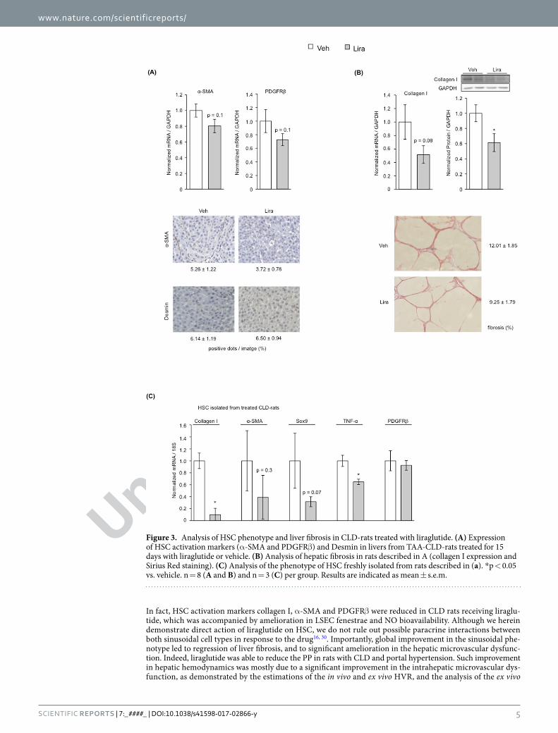
Liraglutide improves HSC phenotype and portal hypertension in CLD-rats. The potential benefi-cial effects of liraglutide as a new therapeutic strategy to improve CLD and portal hypertension were also analyzed in vivo. After 15 days of treatment, CLD-rats treated with liraglutide displayed lower expression of α-SMA and PDGFRβ (Fig. 3A), accompanied by reductions in extracellular matrix synthesis and deposition as demonstrated by diminished collagen expression and hepatic fibrosis (Fig. 3B). No significant differences in TIMPs and MMPs were observed, thus suggesting that the peak of fibrinolysis already occurred (Supplementary Fig. 3). No effects on HSC viability (desmin expression) were observed, thus supporting the results obtained in vitro. Additional analysis of HSC phenotype in cells freshly isolated from CLD-rats treated with liraglutide, or vehicle, confirmed the marked beneficial effects of the drug promoting HSC deactivation (Fig. 3C).
Possible beneficial effects of liraglutide on hepatic and systemic hemodynamic in CLD-rats were also analyzed. Table 1 shows the morphometric and hemodynamic data from these animals. As expected, CLD-rats treated with liraglutide exhibited a slight but significant reduction in body weight, which is in agreement with previous studies19. Importantly, liraglutide-treated animals showed significantly lower portal pressure than vehicle-treated animals (11.6 ± 0.8 vs. 9.3 ± 1.0 mmHg; −20%; p = 0.03) without changes in portal blood flow, thus suggesting an improvement in the hepatic vascular resistance (9.5 ± 1.8 vs. 5.7 ± 1.3 mmHg·mL·min−1·g−1; −23%; p = 0.1). No effects of liraglutide on systemic hemodynamic or biochemical tests were observed.
Intrahepatic microcirculatory amelioration in response to liraglutide was further confirmed analyzing the hepatic microvascular phenotype. Indeed, LSEC from animals receiving liraglutide showed a significant reversal in their capillarization, as suggested by marked increments in fenestrae frequency and porosity (Fig. 4A), and a trend to higher nitric oxide bioavailability (Fig. 4B). In addition, characterization of the hepatic microvascular function ex vivo confirmed the global sinusoidal improvement, as demonstrated by reduction in HVR (Table 1) and improved liver vascular response to incremental doses of acetylcholine (Fig. 4C).
Liraglutide has a complementary effect with simvastatin improving HSC. Treatment of LX-2 cells with liraglutide did not modify the expression of the simvastatin-inducible transcription factor KLF2 (Supplementary Fig. 4 left). However, LX-2 treated with liraglutide or simvastatin showed reduced lev-els of α-SMA in the same magnitude. Interestingly, when both drugs were combined further reduced α-SMA (Supplementary Fig. 4 right), altogether suggesting that liraglutide has a complementary effect to simvastatin improving the phenotype of activated HSC.
Liraglutide improves HSC phenotype and liver microcirculation probably through a GLP1-R independent mechanism. Analysis of GLP1-R expression in rat and human liver tissues and HSC showed

Uncorre
cted
pro
of
www.nature.com/scientificreports/
3Scientific RepoRts | 7:_####_ | DOI:10.1038/s41598-017-02866-y
no detectable mRNA expression (Supplementary Fig. 5A), while a band corresponding to 53 kDa (predicted GLP-1R molecular weight) was only detected in LX-2 and barely present in cirrhotic and NASH human livers, but not in control human or rat livers (either control or cirrhotic) (Supplementary Fig. 5B). Accordingly, analysis of the GLP-1R secondary messenger PKA in rHSC and LX-2 treated with liraglutide did not show differences in its phosphorylation in comparison to cells treated with vehicle (Supplementary Fig. 5C), and incubation of LX-2 with the GLP-1R antagonist Exendin 9–39 did not affect the de-activation effects of liraglutide (Supplementary Fig. 5D). Oppositely, liraglutide did repress the NF-κB molecular pathway (Supplementary Fig. 6).
DiscussionThe major findings of the current study are that liraglutide promotes a marked amelioration in the phenotype of activated HSC, which in a pre-clinical model of chronic liver disease leads to significant improvement in portal hypertension and liver fibrosis. Importantly, the de-activating effects of liraglutide are herein demonstrated in human primary HSC and human liver tissue.
Liraglutide was developed as an anti-diabetic drug predictably acting on GLP-1R in pancreatic β-cells. Interestingly, different studies have demonstrated that these receptors may not be only limited to pancreatic β-cells. Considering the beneficial anti-inflammatory effects of GLP-1R agonists on cardiac fibrosis and NASH20–22, we aimed the present study at analyzing the effects of liraglutide in chronic liver disease (CLD).
Liver cirrhosis is the end stage situation of CLD being the main triggering factor a complex multicellular response of all hepatic cells. Indeed, in front of a chronic injury both parenchymal and non-parenchymal cells undergo profound changes in their phenotype, becoming highly de-regulated and ultimately leading to fibrosis and microvascular dysfunction16. The most relevant clinical consequence of sinusoidal cells de-regulation is the
Figure 1. Amelioration of human primary HSC in response to liraglutide. (A) Expression of depicted proteins/genes after in vitro treatment with 50 µM liraglutide or vehicle in: 1-HSC isolated from cirrhotic human livers (left), 2-quiescent human HSC undergoing in vitro activation (middle), and 3-human precision-cut liver slices (PCLS) (right). (B) Effects of liraglutide, or its vehicle, on the contraction of primary human HSC. n = 3 per experimental condition. *p < 0.05 vs. vehicle.

Uncorre
cted
pro
of
www.nature.com/scientificreports/
4Scientific RepoRts | 7:_####_ | DOI:10.1038/s41598-017-02866-y
development of portal hypertension, which derives both from pathological increases in intrahepatic vascular resistance and in portal blood flow. Considering the importance of HSC in CLD progression and aggravation, many studies focused on liver-specific drugs capable of inactivating the HSC, however few studies have advanced to the clinical stage23.
Our study is the first showing that liraglutide is able to improve the phenotype of HSC. Indeed, we performed dose- and time-dependent experiments indicating that liraglutide de-activates HSC as demonstrated by reduced expression of α-SMA and collagen. Similarly, we observed prevention of HSC activation in response to lira-glutide, therefore suggesting possible beneficial effects of the drug when administered at early stages of CLD. Importantly, we planned this study as a bed to bench-side one, and not vice versa, therefore firstly evaluating the effects of liraglutide in human primary HSC, to latterly use different preclinical models of CLD to further study the molecular mechanisms of such ameliorations.
Hepatic stellate cells are activated in response to different liver injuries, or due to paracrine factors, promot-ing tissue repair. Their response includes cell mobilization, proliferation, migration towards the lesion, and pro-duction of extracellular matrix components. When continuous liver injury occurs, HSC become chronically activated, acquire high expression of pro-inflammatory, pro-fibrogenic and proliferative markers like TNF-α, TGFβ and PDGFRβ, ultimately representing the main cell-type responsible for fibrosis deposition24, 25. In the present study, we show that improvement in HSC phenotype in response to liraglutide was accompanied by marked reductions in the expression of these cytokines and proliferation markers, without affecting cell viability. Altogether suggesting that liraglutide promotes the de-activation of HSC, reduces their proliferation but does not induce cell apoptosis. Such anti-inflammatory effects, which are potentially optimal for the resolution of liver fibrosis in vivo, are quite different from previous studies showing concomitant de-activation and apoptosis/necrosis of HSC in response to certain therapeutic strategies26–28. Importantly, analysis of HSC was not limited to molecular markers, but also included the cell contraction functional assay, which further confirmed the global improvement of HSC phenotype in response to liraglutide.
Additionally, we tested the possible beneficial effects of liraglutide when administered to human liver tissue. Taking advantage of the precision cut liver slices technique, considered an excellent tool to analyze the effects of drugs within the liver29, we observed marked reductions in the expression of α-SMA and collagen in response to liraglutide therefore corroborating the anti-fibrotic effects of the drug.
Once the phenotype of HSC was characterized in vitro, we studied the effects of a physiological-relevant dose of liraglutide administered in vivo. Liraglutide treatment markedly improved both HSC and LSEC phenotypes.
Figure 2. Underlying effects of HSC deactivation due to liraglutide. After 72 h of treatment with 50 µM liraglutide, LX-2 cells were assessed for markers of HSC activation (A), cell viability by double staining with acridine orange (green dense nuclei: apoptosis, indicated by arrowheads) and propidium iodide (red cells: necrosis) (B), HSC proliferation assessed by cell counting and expression of the proliferative marker PDGFRβ (C), and cell contraction (D). n = 3 per experimental condition. *p < 0.05 vs. vehicle.
Uncorre
cted
pro
of
www.nature.com/scientificreports/
5Scientific RepoRts | 7:_####_ | DOI:10.1038/s41598-017-02866-y
In fact, HSC activation markers collagen I, α-SMA and PDGFRβ were reduced in CLD rats receiving liraglu-tide, which was accompanied by amelioration in LSEC fenestrae and NO bioavailability. Although we herein demonstrate direct action of liraglutide on HSC, we do not rule out possible paracrine interactions between both sinusoidal cell types in response to the drug16, 30. Importantly, global improvement in the sinusoidal phe-notype led to regression of liver fibrosis, and to significant amelioration in the hepatic microvascular dysfunc-tion. Indeed, liraglutide was able to reduce the PP in rats with CLD and portal hypertension. Such improvement in hepatic hemodynamics was mostly due to a significant improvement in the intrahepatic microvascular dys-function, as demonstrated by the estimations of the in vivo and ex vivo HVR, and the analysis of the ex vivo
Figure 3. Analysis of HSC phenotype and liver fibrosis in CLD-rats treated with liraglutide. (A) Expression of HSC activation markers (α-SMA and PDGFRβ) and Desmin in livers from TAA-CLD-rats treated for 15 days with liraglutide or vehicle. (B) Analysis of hepatic fibrosis in rats described in A (collagen I expression and Sirius Red staining). (C) Analysis of the phenotype of HSC freshly isolated from rats described in (a). *p < 0.05 vs. vehicle. n = 8 (A and B) and n = 3 (C) per group. Results are indicated as mean ± s.e.m.

Uncorre
cted
pro
of
www.nature.com/scientificreports/
6Scientific RepoRts | 7:_####_ | DOI:10.1038/s41598-017-02866-y
vasodilatory capacity in response to incremental doses of acetylcholine. Importantly, liraglutide did not affect systemic hemodynamics.
We next ascertained which could be the molecular pathway underlying liraglutide effects on HSC, and con-sequently on liver microcirculation and fibrosis. First, and considering that liraglutide was formulated to act on GLP-1 receptor, we analyzed the expression of this receptor both in HSC isolated from rat and humans, and also in liver tissues. Surprisingly, GLP-1R mRNA was not detected in whole liver homogenates, primary HSC
Vehicle n = 11
Liraglutide n = 11 p value
PP (mmHg) 11.6 ± 0.8 9.3 ± 1.0 0.03
MAP (mmHg) 99.5 ± 7.2 89.9 ± 7.1 0.4
PBF (mL/min) 11.9 ± 1.0 11.5 ± 2.3 0.5
HVR (mmHg·min·mL−1·g−1) 9.5 ± 1.8 5.7 ± 1.3 0.1
ex vivo HVR (mmHg·min·mL−1·g−1) 1.6 ± 0.3 0.9 ± 0.04 0.07
Body weight pre-treatment (g) 289 ± 12 288 ± 7 0.5
Body weight post-treatment (g) 310 ± 9 274 ± 9 0.03
Liver weight (g) 8.4 ± 0.7 6.7 ± 0.4 0.1
AST (U/L) 105 ± 14 126 ± 14 0.3
ALT (U/L) 61 ± 10 67 ± 4 0.5
Albumin (g/L) 15.3 ± 1.3 16.3 ± 0.5 0.5
Cholesterol (mg/dL) 54.0 ± 8.3 44.6 ± 5.8 0.4
TG (mg/dL) 31.2 ± 5.6 26.8 ± 3.4 0.5
FFA (µmol/L) 506 ± 66 477 ± 55 0.7
Table 1. Effects of Liraglutide on hepatic and systemic hemodynamic, and biochemical parameters in rats with chronic liver disease due to chronic TAA administration, represented as mean ± s.e.m. PP, portal pressure; MAP, mean arterial pressure; PBF, portal blood flow; HVR, hepatic vascular resistance; AST, a
spartate
aminotransferase; ALT, alanine aminotransferase; TG, triglycerides; FFA, free fatty acids.Q1
Figure 4. Effects of liraglutide on hepatic endothelial phenotype and microvascular function. (A) Liver sinusoidal fenestrae analysis by means of frequency (no. fenestrae/cell area) and porosity (fenestrae area/cell area) in TAA-CLD-rats treated with liraglutide or vehicle. (B) Hepatic nitric oxide (NO) bioavailability in rats described in A. (C) Hepatic microvascular function, calculated as the decrease in portal pressure in response to increasing doses of the endothelium-dependent vasodilator acetylcholine after vasoconstriction with methoxamine. *p < 0.05 vs. vehicle. n = 3 (A), n = 8 (B) and n = 5 (C) per group.

Uncorre
cted
pro
of
www.nature.com/scientificreports/
7Scientific RepoRts | 7:_####_ | DOI:10.1038/s41598-017-02866-y
or LX-2 cells using two different Taqman probes with PCR reactions going up to 60 cycles. At the protein level, western blot of hepatic samples using an antibody against GLP-1R showed signal in LX-2 lysates at the predicted GLP-1R molecular weight, but this signal was barely present in cirrhotic and NASH human tissue, while it was not detected in control human livers or rat liver tissue (either control or cirrhotic). These observations suggest that the beneficial effects of liraglutide in rat HSC and its effects in vivo would not be dependent on GLP-1R. Previous reports already showed contradictory results in terms of GLP-1R expression within the liver4, 5, and we are not totally convinced that the band detected by the antibody in human samples really corresponds to GLP-1R (as it is contradictory to the two specific mRNA TaqMan probes). In agreement, additional experiments showed lack of protein kinase A phosphorylation (marker of GLP-1R activation)31, 32 in response to liraglutide, and no differences in liraglutide-mediated HSC de-activation and proliferation when an antagonist of GLP-1R was used. Secondly, we analyzed the expression of the transcription factor Kruppel-like factor 2 (KLF2) in response to lira-glutide since the effects of liraglutide on HSC were quite similar to those previously observed using statins26, 33–35. These experiments showed no up-regulation in response to the drug, moreover a synergistic effect of liraglutide and simvastatin de-activating HSC was observed, thus suggesting that they act by different pathways and could be used in combination at the bedside. Lastly, we evaluated the NF-κB molecular pathway, which plays a major role in liver fibrosis36 and is inhibited by liraglutide in the endothelium37. Interestingly, liraglutide down-regulated the expression of NF-κB, and also of its target gene Sox9.
With these results we cannot delineate the exact molecular mechanism driving liraglutide’s beneficial effects, and represents a limitation of the study. Nevertheless, considering the main role of NF-κB and Sox9 modulating HSC phenotype38, 39, and the findings described above, we herein suggest that in the specific scenario of CLD liraglutide may improve HSC phenotype and liver microcirculation through a GLP-1R independent mechanism, being the NF-κB–Sox9 pathway a solid candidate to mediate, at least in part, these effects. Desirable future exper-iments, out of the scope of the present study, will clarify the specific receptor and the molecular mechanisms that mediate the beneficial effects of this drug in chronic liver disease.
In conclusion, the present study describes the anti-fibrotic effects of liraglutide both in rodent and, impor-tantly, human pre-clinical models of chronic liver disease, therefore encouraging its application at the bedside as new therapeutic tool to improve cirrhosis and portal hypertension. Moreover, the potential results of the LEAN trial opens the possibility to use liraglutide not only to improve mild NASH7, but promote regression of advanced chronic liver disease. Desirable future trials will clarify this promising therapeutic alternative.
Materials and MethodsAnimal models of chronic liver disease (CLD). The study was carried out in male Wistar rats (Charles River Laboratories, Barcelona, Spain). Animals were kept in environmentally controlled animal facilities. All procedures were approved by the Laboratory Animal Care and Use Committee of the University of Barcelona and were conducted in accordance with the European Community guidelines for the protection of animals used for experimental and other scientific purposes (EEC Directive 86/609). The personnel who prepared and adminis-tered treatments and those that performed the experimental studies were different. Treatment’s codes were not open for interpretation of the results until the inclusion of all animals.
Induction of CLD by thioacetamide (TAA). TAA (Sigma Chemical Co) was dissolved in 0.9% normal saline approximately one hour before injection. Treatment groups received 200 mg/kg of TAA twice per week for a total of 12 weeks while control groups received the same volume of 0.9% normal saline18.
Induction of CLD by Carbon Tetrachloride (CCl4). Rats underwent inhalation exposure to CCl4 (Sigma) and received phenobarbital (0.3 g/L) in the drinking water. When rats developed ascites, toxicants administration was stopped40.
HSC isolation, culture and treatments. Isolation and culture of HSC. HSC were isolated from human (hHSC: control or cirrhotic) and rat (rHSC: control, TAA-CLD or CCl4-CLD) livers as described26 with minor modifications. Briefly, liver tissues were perfused with collagenase A, pronase and DNase (all Roche) in Gey’s Balanced Salt Solution (GBSS; Sigma), and dispersed cells were fractionated by density gradient centrifugation using 11.5% Optiprep (Sigma). HSC were cultured in Iscove’s Modified Dulbecco’s Media (IMDM, Invitrogen, Gibco) supplemented with fetal bovine serum, glutamine, antibiotics and amphotericin B. Results using primary HSC derived from at least 3 independent isolations and 3 replicates.
Immortalized human-activated HSC LX-2 were cultured as described11. Results using LX-2 derived from at least three replicates per experimental condition.
HSC treatments. HSC were incubated with liraglutide (Novo Nordisk), or its vehicle PBS, at different con-centrations (1 μM, 10 μM, 50 μM and 100 μM) and for different periods of time (24 h, 48 h and 72 h). In vitro dosing of liraglutide was based on previous literature41. Considering that liraglutide exerted beneficial effects at 50 μM and after 72 h of treatment, all subsequent experiments were performed using these experimental condi-tions. Simvastatin was used in 10 µM dose alone and in combination with liraglutide. Exendin fragment 9–39 (Sigma-Aldrich; 10 nM–1 uM) was used as a GLP-1R antagonist41.
Cell viability & proliferation. Equal number of LX-2 were seeded and after 72 h of liraglutide or vehicle, floating and adhered cells were collected and counted using a hemocytometer with trypan blue dye exclusion (FLUKA).
Cell apoptosis. Cells were incubated with fresh medium containing 800 ng/mL Acridine Orange (AO) and 5 μg/mL Propidium Iodide (PI) for 10 min at 37 °C and then washed with PBS. Fresh medium was added and cell

Uncorre
cted
pro
of
www.nature.com/scientificreports/
8Scientific RepoRts | 7:_####_ | DOI:10.1038/s41598-017-02866-y
death was examined using a fluorescence microscope (Olympus BX51, Tokyo, Japan) equipped with a digital camera (Olympus, DP72). AO is a metachromatic dye that stains both viable and apoptotic cells by intercalating into DNA and emits green fluorescence upon excitation at 480–490 nm. Nevertheless, nuclear condensation that occurs during apoptosis glares a more intense fluorescence. PI is excluded by viable cells but can penetrate cell membranes of dying or dead cells due to necrosis, emitting red fluorescence. Positive controls (in vitro ischemia and reperfusion), and negative controls (without dye) were included26.
Cell contraction. Contraction of HSC was performed as previously described with some modifications42. Briefly, culture plates were incubated with 1% BSA-PBS and afterwards filled with a mix of collagen (2 mg/mL) and human HSC (1–2 × 10⁵ cells/mL). Once the gels were solidified, serum free IMDM with 50 µM liraglutide or vehicle was added. After 24 h, contraction was induced by adding 10% FBS for 24 h. Finally, the contraction area was digitalized and measured with ImageJ software. The results are expressed as % of contraction relative to the initial area of the gel.
Characterization of CLD-rats treated with liraglutide. Liraglutide administration. TAA-CLD-rats received by subcutaneous injections, twice a day, either liraglutide (0.5 mg/kg/day; n = 11) or vehicle (0.9% NaCl; n = 11) during 15 days. The dose was selected based on a conversion calculation starting from the dose used in humans and agreed with previous publications19, 43, 44. Administration of liraglutide to CLD-rats started one week after stopping the administration of TAA.
In vivo hemodynamics. Rats (n = 8 per group) were anesthetized with ketamine hydrochloride (100 mg/Kg; Merial Laboratories) plus midazolam (5 mg/kg; Laboratorios Reig Jofré) intraperitoneally. A tracheotomy was performed and a polyethylene tube PE-240 was inserted into the trachea to ensure a patent airway. PE-50 cathe-ters were introduced into the femoral artery to measure mean arterial pressure (MAP; mmHg) and into ileocolic vein to measure portal pressure (PP; mmHg). A perivascular ultrasonic flow probe (Transonic System) was placed around the portal vein, as close as possible to the liver to avoid portal-collateral blood flow, in order to measure portal blood flow (PBF; mL∙min−1). Hepatic vascular resistance (mmHg·min·mL−1·g−1) was calculated as: PP/PBF. Blood pressures and flows were registered on a multichannel computer based recorder (Power Lab; AD Instruments). Temperature of the animals was maintained at 37 ± 0.5 °C and hemodynamic data were collected after 20 min stabilization40, 45. Blood serum and plasma samples were stored for biochemical analysis.
Liver microvascular function. Immediately after recording in vivo hemodynamics, rat livers were isolated and perfused with Krebs buffer as previously described (n = 5 per group)45, 46. The perfused rat liver preparation was allowed to stabilize for 20 min before vasoactive substances were added. Intrahepatic microcirculation was pre-constricted by adding the α1-adrenergic agonist methoxamine (Mtx; 10−4 M; Sigma) to the reservoir, and liver microvascular function was assessed as concentration–response curves to cumulative doses of acetylcholine (Ach; 10–7–10−5 M; Sigma). Liver tissue was snap-frozen for subsequent molecular analysis.
Evaluation of hepatic fibrosis. CLD-rat livers were fixed in 10% formalin, embedded in paraffin, sectioned, and stained with 0.1% Sirius Red, photographed, and analyzed using a microscope equipped with a digital camera. The red-stained area was measured using Axiovision software26. Values are expressed as the mean of 8 fields per sample.
Sinusoidal characterization using Scanning Electron Microscope. In a sub-group of animals (n = 3 per group), after obtaining in vivo hemodynamics, livers were perfused through portal vein with a solution containing 2.5% glutaraldehyde and 2% paraformaldehyde and fixed overnight at 4 °C. Samples were washed 3 times with 0.1 M cacodylate buffer. Liver sections were fixed with 1% osmium in cacodylate buffer, dehydrated in ethanol, and dried with hexamethyldisilazane. Six randomly selected blocks from each animal were mounted onto stubs, and sputter coated with gold. 10 images per animal were acquired at a resolution of 15,000x using a Jeol 6380 Scanning Electron Microscope (JEOL Ltd, Tokyo, Japan). Liver sinusoidal fenestrations were quantified using ImageJ Software (NIH)47.
Nitric Oxide bioavailability. Levels of cGMP, a marker of NO bioavailability, were analyzed in liver homogenates using an enzyme immunoassay (Cayman Chemical Co., Ann Arbor, MI) as previously described48.
RNA isolation and quantitave PCR. RNA from cells and tissue were extracted using RNeasy mini kit (Qiagen) and Trizol (Life Technologies), respectively. RNA quantification was performed using a NanoDrop spec-trophotometer. cDNA was obtained using QuantiTect reverse transcription kit (Qiagen). Real-Time PCR were performed in an ABI PRISM 7900HT Fast Real-Time PCR System, using TaqMan predesigned probes for Col1A1 (Hs00164004_m1, Rn01463848_m1), α-SMA (Hs00426835_g1, Rn01759928_g1), PDGFRβ (Hs01019589_m1, Rn01491838_m1), GLP1-R (Hs00157705_m1, Hs01006326_m1), Sox9 (Hs00165814_m1, Rn01751070_m1) and GAPDH or 18 S as endogenous controls. Results, expressed as 2−ΔΔCt, represent the x-fold increase of gene expression compared with the corresponding control group.
Western blot analysis. Cells were rinsed twice with PBS and lysed with Triton lysis buffer. Livers were homogenized in triton-lysis buffer for whole protein extraction. Aliquots from each sample containing equal amounts of protein were run on a sodium dodecylsulphate polyacrylamide gel, and transferred to a nitrocel-lulose membrane. After the transfer, blots were blocked with Tris buffered saline containing 0.05% Tween-20 and 5% non-fat dry milk or 3% albumin and subsequently incubated overnight at 4 °C with primary antibodies

Uncorre
cted
pro
of
www.nature.com/scientificreports/
9Scientific RepoRts | 7:_####_ | DOI:10.1038/s41598-017-02866-y
against collagen I (ABT123, Millipore), α-SMA (A2547, Sigma), TNF-α (sc-1351, Santa Cruz Biotechnology), TGFβR (sc-398, Santa Cruz Biotechnology), GAPDH (sc-32233, Santa Cruz Biotechnology), p-PKA (4781, Cell Signaling), NF-κB (6956, Cell Signaling) and IκB (4812, Cell Signaling), all at 1:1000 dilution. Then membranes were incubated with the appropriate horseradish peroxidase-conjugated secondary antibody at room tempera-ture. Protein expression was determined by densitometric analysis using the LAS4000 (GE Healthcare) and Image Studio Lite software (LI-COR). Quantitative densitometric values of all proteins were normalized to GAPDH.
Precision-cut liver slices of human livers (PCLS). Fresh human liver biopsies were used to obtain 250 μm slices using a Vibratome VT1000S (Leica Microsystems, Wetzlar, Germany). Samples were washed in PBS, soaked in 4% agarose solution (Ultrapure LMP Agarose, Invitrogen, Carlsbad, California, USA) for 20 min, and then orientated, mounted and immobilized using cyanoacrylate glue. Tissue slices were placed on organotypic tissue culture plate inserts (Millicell®-CM; Millipore). Tissues were maintained at 37 °C in a 5% CO2 humidified incubator using 1.1 mL of Williams’ Medium E supplemented with 1% inactivated fetal bovine serum, 2 mM L-Glutamine, 50 U/mL penicillin and 50 μg/mL streptomycin. Tissue slices were incubated with 50 µM liraglutide for 24 h. Sections were then transferred to a 1.5 mL tube and lysed for RNA isolation and qPCR29.
Ethics information. Quiescent hHSC (for prevention of in vitro activation) and PCLS were obtained from remnants from partial hepatectomy. Livers were considered control, but exhibited fibrosis staging between F1 and F2. In all cases surgery was recommended to excise tumor metastasis from colon carcinoma. Cirrhotic hHSC (for in vitro de-activation) were isolated from remnant cirrhotic livers (all alcoholic aetiology) obtained after transplantation. The Ethics Committee of the Hospital Clinic de Barcelona approved the experimental protocol, samples manipulation and isolation procedures were carried out following good laboratory practices; in all cases patients signed the informed consent.
Statistical analysis. Statistical analysis was performed with the SPSS V.23.0 for Windows statistical package (IBM, Armonk, New York, USA). Results are expressed as mean ± s.e.m. Normality of samples was assessed using the Kolmogorov Smirnov test. For samples following a normal distribution, comparisons between groups were performed with the Student t test or analysis of variance, followed by a Bonferroni Post Hoc test when adequate. Otherwise, comparisons were assessed with the non-parametric Mann-Whitney U or Kruskal Wallis test when adequate. Differences were considered significant at a p value < 0.05.
References 1. Nauck, M. A. Incretin-based therapies for type 2 diabetes mellitus: properties, functions, and clinical implications. Am. J. Med. 124,
S3–18 (2011). 2. Kazafeos, K. Incretin effect: GLP-1, GIP, DPP4. Diabetes Res. Clin. Pract. 93(Suppl 1), S32–6 (2011). 3. Drucker, D. J. The biology of incretin hormones. Cell Death Differ. 3, 153–165 (2006). 4. Gupta, N. A. et al. Glucagon-like peptide-1 receptor is present on human hepatocytes and has a direct role in decreasing hepatic
steatosis in vitro by modulating elements of the insulin signaling pathway. Hepatology 51, 1584–1592 (2010). 5. Pyke, C. et al. GLP-1 Receptor Localization in Monkey and Human Tissue: Novel Distribution Revealed With Extensively Validated
Monoclonal Antibody. Endocrinology 155, 1280–1290 (2014). 6. Wang, X.-C., Gusdon, A. M., Liu, H. & Qu, S. Effects of glucagon-like peptide-1 receptor agonists on non-alcoholic fatty liver disease
and inflammation. World J. Gastroenterol. 20, 14821–14830 (2014). 7. Armstrong, M. J. et al. Liraglutide safety and efficacy in patients with non-alcoholic steatohepatitis (LEAN): a multicentre, double-
blind, randomised, placebo-controlled phase 2 study. Lancet (London, England) 387, 679–90 (2016). 8. McClean, P. L., Jalewa, J. & Hölscher, C. Prophylactic liraglutide treatment prevents amyloid plaque deposition, chronic
inflammation and memory impairment in APP/PS1 mice. Behav. Brain Res. 293, 96–106 (2015). 9. Shiraki, A. et al. The glucagon-like peptide 1 analog liraglutide reduces TNF-α-induced oxidative stress and inflammation in
endothelial cells. Atherosclerosis 221, 375–82 (2012). 10. Fernández-Iglesias, A. & Gracia-Sancho, J. How to face chronic liver disease: the sinusoidal perspective. Front. Med. 4, 7 (2017). 11. Marrone, G. et al. The transcription factor KLF2 mediates hepatic endothelial protection and paracrine endothelial-stellate cell
deactivation induced by statins. J. Hepatol. 58, 98–103 (2013). 12. Pinzani, M. & Gentilini, P. Biology of hepatic stellate cells and their possible relevance in the pathogenesis of portal hypertension in
cirrhosis. Semin. Liver Dis. 19, 397–410 (1999). 13. Török, N. J. Recent advances in the pathogenesis and diagnosis of liver fibrosis. J. Gastroenterol. 43, 315–21 (2008). 14. Soon, R. K. & Yee, H. F. Stellate Cell Contraction: Role, Regulation, and Potential Therapeutic Target. Clin. Liver Dis. 12, 791–803
(2008). 15. Reynaert, H. Hepatic stellate cells: role in microcirculation and pathophysiology of portal hypertension. Gut 50, 571–581 (2002). 16. Marrone, G., Shah, V. H. & Gracia-Sancho, J. Sinusoidal communication in liver fibrosis and regeneration. J. Hepatol. 65, 608–17
(2016). 17. Kisseleva, T. et al. Myofibroblasts revert to an inactive phenotype during regression of liver fibrosis. Proc. Natl. Acad. Sci. USA 109,
9448–9453 (2012). 18. Cerini, F. et al. Enoxaparin reduces hepatic vascular resistance and portal pressure in cirrhotic rats. J. Hepatol. 64, 834–42 (2016). 19. Gao, H. et al. The Glucagon-Like Peptide-1 Analogue Liraglutide Inhibits Oxidative Stress and Inflammatory Response in the Liver
of Rats with Diet-Induced Non-alcoholic Fatty Liver Disease. Biol. Pharm. Bull 694, 694–702 (2015). 20. Gaspari, T. et al. Molecular and cellular mechanisms of glucagon-like peptide-1 receptor agonist-mediated attenuation of cardiac
fibrosis. Diabetes Vasc. Dis. Res. 13, 56–68 (2016). 21. Armstrong, M. J. et al. Glucagon-like peptide 1 decreases lipotoxicity in non-alcoholic steatohepatitis. J. Hepatol. 64, 399–408
(2016). 22. Steven, S. et al. Gliptin and GLP-1 analog treatment improves survival and vascular inflammation/dysfunction in animals with
lipopolysaccharide-induced endotoxemia. Basic Res. Cardiol. 110, 6 (2015). 23. Schuppan, D. & Kim, Y. O. Evolving therapies for liver fibrosis. J. Clin. Invest. 123, 1887–1901 (2013). 24. Weiskirchen, R. & Tacke, F. Cellular and molecular functions of hepatic stellate cells in inflammatory responses and liver
immunology. Hepatobiliary Surg Nutr 3, 344–363 (2014). 25. Trautwein, C., Friedman, S. L., Schuppan, D. & Pinzani, M. Hepatic fibrosis: Concept to treatment. J. Hepatol. 62, S15–S24 (2015). 26. Marrone, G. et al. KLF2 exerts antifibrotic and vasoprotective effects in cirrhotic rat livers: behind the molecular mechanisms of
statins. Gut 64, 1434–43 (2015).

Uncorre
cted
pro
of
www.nature.com/scientificreports/
1 0Scientific RepoRts | 7:_####_ | DOI:10.1038/s41598-017-02866-y
27. Klein, S. et al. Atorvastatin inhibits proliferation and apoptosis, but induces senescence in hepatic myofibroblasts and thereby attenuates hepatic fibrosis in rats. Lab. Investig. 92, 1440–1450 (2012).
28. Wright, M. C. et al. Gliotoxin Stimulates the Apoptosis of Human and Rat Hepatic Stellate Cells and Enhances the Resolution of Liver Fibrosis in Rats. Gastroenterology 121, 685–698 (2001).
29. Olinga, P. & Schuppan, D. Precision-cut liver slices: A tool to model the liver ex vivo. J. Hepatol. 58, 1252–1253 (2013). 30. Xie, G. et al. Role of Differentiation of Liver Sinusoidal Endothelial Cells in Progression and Regression of Hepatic Fibrosis in Rats.
Gastroenterology 142, 918–927 (2012). 31. Laviola, L. et al. Glucagon-like peptide-1 counteracts oxidative stress-dependent apoptosis of human cardiac progenitor cells by
inhibiting the activation of the c-Jun N-terminal protein kinase signaling pathway. Endocrinology 153, 5770–81 (2012). 32. Wang, L. et al. GLP-1 analog liraglutide enhances proinsulin processing in pancreatic β-cells via a PKA-dependent pathway.
Endocrinology 155, 3817–28 (2014). 33. Abraldes, J. G. et al. Simvastatin treatment improves liver sinusoidal endothelial dysfunction in CCl4 cirrhotic rats. J. Hepatol. 46,
1040–6 (2007). 34. Trebicka, J. et al. Atorvastatin lowers portal pressure in cirrhotic rats by inhibition of RhoA/Rho-kinase and activation of endothelial
nitric oxide synthase. Hepatology 46, 242–253 (2007). 35. La Mura, V. et al. Effects of simvastatin administration on rodents with lipopolysaccharide-induced liver microvascular dysfunction.
Hepatology 57, 1172–1181 (2013). 36. Luedde, T. & Schwabe, R. F. NF-κB in the liver—linking injury, fibrosis and hepatocellular carcinoma. Nat. Rev. Gastroenterol.
Hepatol. 8, 108–118 (2011). 37. Gaspari, T. et al. A GLP-1 receptor agonist liraglutide inhibits endothelial cell dysfunction and vascular adhesion molecule
expression in an ApoE-/- mouse model. Diabetes Vasc. Dis. Res. 8, 117–24 (2011). 38. Pritchett, J. et al. Osteopontin is a novel downstream target of SOX9 with diagnostic implications for progression of liver fibrosis in
humans. Hepatology 56, 1108–1116 (2012). 39. Martin, K. et al. PAK proteins and YAP-1 signalling downstream of integrin beta-1 in myofibroblasts promote liver fibrosis. Nat.
Commun. 7, 12502 (2016). 40. Gracia-Sancho, J. et al. Endothelial expression of transcription factor Kruppel-like factor 2 and its vasoprotective target genes in the
normal and cirrhotic rat liver. Gut 60, 517–24 (2011). 41. Cantini, G. et al. Effect of liraglutide on proliferation and differentiation of human adipose stem cells. Mol. Cell. Endocrinol. 402,
43–50 (2015). 42. Perri, R. E. et al. Defects in cGMP-PKG pathway contribute to impaired NO-dependent responses in hepatic stellate cells upon
activation. Am. J. Physiol. Gastrointest. Liver Physiol. 290, G535–42 (2006). 43. Lu, N. et al. Glucagon-like peptide-1 receptor agonist Liraglutide has anabolic bone effects in ovariectomized rats without diabetes.
PLoS One 10, e0132744 (2015). 44. Hoang, V., Bi, J., Mohankumar, S. M. & Vyas, A. K. Liraglutide Improves Hypertension and Metabolic Perturbation in a Rat Model
of Polycystic Ovarian Syndrome. PLoS One 10, e0126119 (2015). 45. Hide, D. et al. Effects of warm ischemia and reperfusion on the liver microcirculatory phenotype of rats: underlying mechanisms
and pharmacological therapy. Sci. Rep. 6, 22107 (2016). 46. Gracia-Sancho, J. et al. Evidence Against a Role for NADPH Oxidase Modulating Hepatic Vascular Tone in Cirrhosis.
Gastroenterology 133, 959–966 (2007). 47. Hilmer, S. N. et al. Age-related changes in the hepatic sinusoidal endothelium impede lipoprotein transfer in the rat. Hepatology 42,
1349–1354 (2005). 48. Hide, D. et al. A novel form of the human manganese superoxide dismutase protects rat and human livers undergoing ischaemia and
reperfusion injury. Clin. Sci. (Lond). 127, 527–37 (2014).
AcknowledgementsAuthors are in debt with Drs Garcia-Valdecasas and Molina for providing human biopsies, Dr Bataller for providing LX-2 cells, and Bibiana Rius and Dr Clària for their expertise in PCLS. This work was funded by the Ministerio de Economía y Competitividad – Instituto de Salud Carlos III, FIS (PI14/00029 and PI13/00341), and the European Union (Fondos FEDER, “una manera de hacer Europa”). FCdM has a Fellowship from CAPES/Ciência sem Fronteiras - Brazil, and SG-M from the Fundació Catalana de Trasplantament. AF-I has a Sara Borrell contract from the Instituto de Salud Carlos III. CIBEREHD is funded by the Instituto de Salud Carlos III.
Author ContributionsF.C.dM., S.G.-M. and A.F.-I. designed the research, conceived ideas, performed experiments, and wrote the manuscript. R.M.-D., S.V., D. H. and M. O.-R. performed experiments and analyzed data. J.L.R. and J.C.G.-P. critically revised the manuscript. J.B. critically revised the manuscript and obtained funding. J.R.dO. conceived ideas and critically revised the manuscript. J.G.-S. designed the research, conceived ideas, wrote the manuscript, obtained funding and directed the study. All authors edited and reviewed the final manuscript.
Additional InformationSupplementary information accompanies this paper at doi:10.1038/s41598-017-02866-yCompeting Interests: The authors declare that they have no competing interests.Publisher's note: Springer Nature remains neutral with regard to jurisdictional claims in published maps and institutional affiliations.
Open Access This article is licensed under a Creative Commons Attribution 4.0 International License, which permits use, sharing, adaptation, distribution and reproduction in any medium or
format, as long as you give appropriate credit to the original author(s) and the source, provide a link to the Cre-ative Commons license, and indicate if changes were made. The images or other third party material in this article are included in the article’s Creative Commons license, unless indicated otherwise in a credit line to the material. If material is not included in the article’s Creative Commons license and your intended use is not per-mitted by statutory regulation or exceeds the permitted use, you will need to obtain permission directly from the copyright holder. To view a copy of this license, visit http://creativecommons.org/licenses/by/4.0/. © The Author(s)

51 CÒPIA DELS ARTICLES ORIGINALS
Supplementary Figures
Supplementary figure 1. Analysis of LX-2 de-activation in terms of -SMA
expression in response to liraglutide administered at different doses (a) and times
(b). n=3 per experimental condition. *p<0.05 vs. vehicle.

52 CÒPIA DELS ARTICLES ORIGINALS
Supplementary figure 2. Effects of 72h-liraglutide on the in vitro activation of
primary quiescent HSC isolated from healthy rats (a), and in the amelioration of
activated HSC isolated from TAA-CLD (b) and CCl4-CLD (c) rats. n=3 per group.
*p<0.05 vs. vehicle.

53 CÒPIA DELS ARTICLES ORIGINALS
Supplementary figure 3. Expression of TIMPs and MMPs in livers from TAA-
CLD-rats treated for 15 days with liraglutide, or vehicle. n=8 per group.

54 CÒPIA DELS ARTICLES ORIGINALS
Supplementary figure 4. (a) Expression of the transcription factor KLF2 in LX-2
cells treated with 50 µM liraglutide, or vehicle. (b) Expression of α-SMA in LX-2
cells treated with 50 µM liraglutide, or vehicle, in combination with 10µM
simvastatin, or its vehicle. n=3 per experimental condition. *p<0.05 vs. vehicle

55 CÒPIA DELS ARTICLES ORIGINALS
Supplementary figure 5. (a) 60-cycle amplification charts of GLP-1R mRNA
expression analyzed in human liver & cells samples using the human cell line SH-
SY5Y as positive control. (b) Western blot of different hepatic samples using a
GLP-1R antibody (Abcam ab189397). From left to right, brain tissue as positive
control, LX-2 cells, human liver tissue (control, cirrhotic, NASH) and rat liver
tissue (control, cirrhotic). (c) p-PKA protein expression in primary cirrhotic HSC
from TAA-CLD (left), CCl4-CLD (middle) and in LX-2 cells (right) in response to
50M liraglutide. (d) Effects of the GLP-1R antagonist exendin 9-39 on α-SMA
mRNA expression (left) and proliferation (right) in LX-2 cells. n=3 per
experimental condition. *p<0.05 vs. vehicle.

56 CÒPIA DELS ARTICLES ORIGINALS
Supplementary figure 6. (a) Protein expression of NF-κB and its inhibitor IκB
and (b) mRNA expression of Sox9 in livers from TAA-CLD-rats treated with
liraglutide, or vehicle. n=8 per group.
57 CÒPIA DELS ARTICLES ORIGINALS
4.2. Estudi 2
CROSS-TALK BETWEEN AUTOPHAGY AND KLF2 DETERMINES
ENDOTHELIAL CELL PHENOTYPE AND MICROVASCULAR FUNCTION IN
ACUTELY INJURED RAT LIVERS
Sergi Guixé-Muntet, Fernanda Cristina de Mesquita, Sergi Vila, Virginia
Hernández-Gea, Carmen Peralta, Juan Carlos García-Pagán, Jaime Bosch,
Jordi Gracia-Sancho
Journal of Hepatology. 2017, 66(1); 86-94
IF: 10,59


Cross-talk between autophagy and KLF2 determines endothelialcell phenotype and microvascular function in acute liver injury
Sergi Guixé-Muntet1, Fernanda Cristina de Mesquita1,2, Sergi Vila1, Virginia Hernández-Gea1,Carmen Peralta3, Juan Carlos García-Pagán1, Jaime Bosch1, Jordi Gracia-Sancho1,⇑
1Barcelona Hepatic Hemodynamic Lab., IDIBAPS – Hospital Clínic – CIBEREHD, University of Barcelona Medical School, Barcelona, Spain;2Laboratório de Biofisica Celular e Inflamação, Pontificia Universidade Católica do Rio Grande do Sul, PUCRS, Porto Alegre,
RS, Brazil; 3IDIBAPS & CIBEREHD, Spain
Background & Aims: The transcription factor Krüppel-like factor2 (KLF2), inducible by simvastatin, confers endothelial vasopro-tection. Considering recent data suggesting activation ofautophagy by statins, we aimed to: 1) characterize the relation-ship between autophagy and KLF2 in the endothelium, 2) assessthis relationship in acute liver injury (cold ischemia/reperfusion)and 3) study the effects of modulating KLF2-autophagy in vitroand in vivo.Methods: Autophagic flux, the vasoprotective KLF2 pathway, cellviability and microvascular function were assessed in endothelialcells and in various pre-clinical models of acute liver injury (coldstorage and warm reperfusion).Results: Positive feedback between autophagy and KLF2 wasobserved in the endothelium: KLF2 inducers, pharmacological(statins, resveratrol, GGTI-298), biomechanical (shear stress) orgenetic (adenovirus containing KLF2), caused endothelial KLF2overexpression through a Rac1-rab7-autophagy dependentmechanism, both in the specialized liver sinusoidal endothelialcells (LSEC) and in human umbilical vein endothelial cells. Inturn, KLF2 induction promoted further activation of autophagy.
Cold ischemia blunted autophagic flux. Upon reperfusion, LSECstored in University of Wisconsin solution did not reactivateautophagy, which resulted in autophagosome accumulationprobably due to impairment in autophagosome-lysosome fusion,ultimately leading to increased cell death and microvasculardysfunction.
Simvastatin pretreatment maintained autophagy (through theupregulation of rab7), resulting in increased KLF2, improved cellviability, and ameliorated hepatic damage and microvascularfunction.Conclusions:We herein describe for the first time the complexautophagy-KLF2 relationship, modulating the phenotype and sur-vival of the endothelium. These results help understanding themechanisms of protection conferred by KLF2-inducers, such assimvastatin, in hepatic vascular disorders.
Lay summary: Autophagy and the transcription factor KLF2 sharea common activation pathway in the endothelium, being able toregulate each other.
Statins maintain microvascular function through the inhibitionof Rac1, which consequently liberates Rab7, activates autophagyand increments the expression of KLF2.� 2016 European Association for the Study of the Liver. Publishedby Elsevier B.V. All rights reserved.
Introduction
Autophagy is a constitutive process that maintains cellularhomeostasis in a wide variety of cell types through the encapsu-lation of damaged proteins or organelles in double-membranevesicles called autophagosomes, which fusing with lysosomesallow degradation of the cargo. As a result, cells obtain amino-acids, lipids and other components that will serve as a sourceof energy and new building blocks for synthesis. Regulation ofautophagy is complex since it shares molecular signaling withcell proliferation and apoptosis [1,2]. Cells activate autophagyas a mechanism of cellular recycling and survival in response tocellular stresses like low nutrients, low ATP or hypoxia [3,4].
Autophagy plays a role in the pathophysiology of diverse liverdiseases, including non-alcoholic steatohepatitis, viral hepatitis,fibrosis, and hepatocellular carcinoma [5–7]. However, there isno consensus about the role of autophagy in ischemia and reper-fusion injury, where autophagy may be differentially regulated,and have opposite effects, depending on type of ischemia (warmor cold) or the preservation solution used [7–11]. Moreover, thepossible role of autophagy in the maintenance of liver sinusoidalendothelial cells (LSEC) phenotype is completely unknown.
The vasoprotective transcription factor Krüppel-like factor 2(KLF2) is expressed in the liver endothelium in response to bloodflow-derived shear stress and plays a key role in the pathophys-iology of hepatic ischemia and reperfusion injury [12,13]. In fact,when the liver is ischemic due to cold preservation or to surgicalprocedures, the endothelium rapidly loses its KLF2 expressionleading to the dysregulation of its specialized phenotype (capil-larization), development of hepatic microvascular dysfunction,and ultimately hepatic injury.
Journal of Hepatology 2017 vol. 66 j 86–94
Keywords: Statins; Simvastatin; Ischemia/reperfusion; LSEC; HUVEC; Rab7; Rac1.Received 21 December 2015; received in revised form 27 July 2016; accepted 30 July2016; available online 18 August 2016⇑ Corresponding author. Address: Barcelona Hepatic Hemodynamic Lab., Rosselló149, 4th floor, 08036 Barcelona, Spain. Tel.: +34 932275707x4306.E-mail address: [email protected] (J. Gracia-Sancho).
Research Article

Pharmacologically, KLF2 can be efficiently induced by statins.Although these FDA/EMA approved drugs were designed toreduce cholesterol synthesis, several reports demonstrated thatstatins are potent KLF2 activators independently of their lipidlowering effects but dependent on the geranylgeranyl-pyrophosphate (GGPP) pathway [14,15]. In the field of acute liverinjury, previous studies demonstrated that simvastatin protectshealthy and steatotic rat livers through the maintenance of LSECvasoprotective phenotype [13,16,17].
Considering this background and recent studies suggestingthat KLF2 activators such as simvastatin and resveratrol mightbe able to increase autophagic flux in the endothelium [18–20],and that autophagy is required for the endothelium to respondto shear stress [21], the present study aimed at: 1) characterizingthe possible cross-link between autophagy and KLF2 in theendothelium; 2) defining endothelial autophagy in the contextof liver ischemia/reperfusion; and 3) modulating hepaticautophagy in vitro and ex vivo to revert the negative effects ofischemia/reperfusion.
Materials and methods
Animals
Male Wistar rats (Charles River) weighting 300–350 g were kept in environmen-tally controlled facilities at the IDIBAPS. All experiments were approved by theLaboratory Animal Care and Use Committee of the University of Barcelona andwere conducted in accordance with the European Community guidelines forthe protection of animals used for experimental and other scientific purposes(European Economic Community (EEC) Directive 86/609).
Liver sinusoidal endothelial cells (LSEC) isolation
Rat LSEC were isolated as described in Supplementary methods. LSEC used in thepresent study showed average viability of 96% (by trypan blue exclusion) and 92%purity (by acetylated-low density lipoprotein (Ac-LDL) incorporation and Reca-1staining) [22,23].
In vitro cold ischemia and warm reperfusion (I/R)
Freshly isolated LSEC were washed twice with warm phosphate balanced saline(PBS) and cultured at 4 �C in cold Celsior or University of Wisconsin (UWS) solu-tions. Using an oxygen microsensor (Unisense OX-NP), O2 availability was mea-sured demonstrating that cold storage solutions have a 35% reduction in O2
saturation in comparison to standard culture conditions, therefore ensuring anappropriate hypoxic environment.
After ischemia time, cells were washed twice with cold PBS and in vitro reper-fusion was mimicked incubating LSEC in complete media during 2 h at 37 �C innormoxic humid atmosphere.
Endothelial cells treatments
Primary rat LSEC or human umbilical vein endothelial cells (HUVEC) (Lonza) weretreated with 5 lM simvastatin (Calbiochem), 5 lM mevastatin (Calbiochem),10 lM GGPP (Sigma), 5 lM GGTI-298 (Sigma), 20 lM chloroquine (CQ; Sigma),2 lM rapamycin (Santa Cruz), 1 lM resveratrol (Sigma), 50 lM NSC23766(Sigma) or 50 nM bafilomycin (Baf; Sigma) when appropriate. Drugs concentra-tions derive from previously published reports [14,19,23,24] or from preliminarystudies performed by our team.
For each experiment, culture medium was aliquoted and complemented withthe corresponding drug or vehicle and added to the corresponding wells. Resultsfrom endothelial cells derive from at least n = 3 independent experiments (differ-ent isolations for LSEC; different commercial batches for HUVEC always belowpassage 6) with n = 2–3 replicates for each experimental condition.
Adenoviral overexpression of KLF2
Cells were seeded at a 60% confluence in complete medium. At the time of infec-tion, plates were washed twice with PBS and were incubated with 10 MOI of ade-novirus containing KLF2 (AdKLF2) or adenovirus containing green fluorescentprotein (AdGFP) (kindly provided by Prof Garcia-Cardena) for 2 h in culture med-ium containing 2% FBS. After infection, the medium containing the adenoviruswas removed and cells were incubated for 24 h [25].
Endothelial shear stress
HUVEC were seeded in gelatin-coated lSlide flow chambers (IBIDI) at a conflu-ence of 100,000 cells/chamber and maintained at the incubator for 12 h. After-wards, culture media (Medium 199 with 20% FBS, 2 mM L-glutamine, 1%penicillin/streptomycin, 0.1 mg/ml heparin, 0.05 mg/ml endothelial mitogenand 2% dextran) was added and cells were cultured for 24 h in static or dynamicconditions (12 dyn/cm2) [23].
Endothelial viability
Cell death was analyzed by double staining with acridine orange and propidiumiodide (AO-PI) and by trypan blue exclusion assay, as previously described [25].Briefly, for AO-PI staining, endothelial cells were incubated with 800 ng/ml acri-dine orange and 5 lg/ml propidium iodide for 10 min. Plates were washed twicewith PBS and complete Roswell Park Memorial Institute (RPMI) without phenolred media was added. Cells were observed with a fluorescence microscope (Olym-pus BX51 with a U-LH100HG light source). Four fields were randomly selected perwell and pictures of visible light, green light (488 nm emission) and red light(555 nm emission) were taken at 200x magnification. Images were merged usingImageJ software (NIH). Cells stained in plain green were counted as viable, cellswith bright and green dotted nuclei were counted as apoptotic, and cells stainedin red were counted as necrotic.
The trypan blue exclusion assay was performed on plate. Trypan blue (Fluka)was added to the medium at a final concentration of 0.04 mg/ml. Cells were incu-bated at 37 �C for 10 min and pictures were taken for further analysis.
Two independent researchers (SG-M and FM) performed the analysis of allpictures blindly.
Immunofluorescence
Endothelial cells were seeded onto 12 mm confocal coverglasses (Electron Micro-scopy Sciences). At the end of treatments, cells were fixed with 4% paraformalde-hyde for 10 min, rinsed with PBS and permeabilized with 0.1% triton X-100(Sigma) for 5 min. Cells were blocked for 30 min with 1% BSA in PBS and subse-quently incubated with primary antibodies against LC3B (1:200, cell signaling)and Lamp2 (1:50, Santa Cruz) overnight at 4 �C. Incubation with secondary anti-bodies conjugated with Alexa Fluor 488/555 (1:300, Invitrogen) was performed atroom temperature for 1 h along with DAPI (3 ng/ml, Invitrogen). Preparationswere then mounted using Fluoromount-G (Bionovacientífica) and dried over-night. Six images per preparation and channel (visible; green, 488 nm; red,555 nm) were obtained with a spectral confocal microscope (Leica TCS-SP5).Images were then analyzed with the ImageJ software (NIH).
Ex vivo model of ischemia/reperfusion
Male Wistar rats (250–300 g; n = 8 per experimental condition) were pre-treated with the autophagy inhibitor CQ (60 mg/kg, i.p., t = �24 h and �2 h)[26], or its vehicle, followed by the KLF2 inducer simvastatin (1 mg/kg, i.v.,t = �1 h) or vehicle. Afterwards, rats were anesthetized with ketamine(80 mg/kg i.p; Merial Laboratories, Barcelona, Spain) + midazolam (5 mg/kg i.p; Laboratorios Reig Jofré, Barcelona, Spain). Livers were exsanguinated withKrebs buffer, flushed through the portal vein with 10 ml of ice-cold UWS andexplanted. Grafts were kept submerged in this solution for 16 h and reperfusedfor 2 h with warm Krebs buffer [13,16]. Then, liver microvascular function wasevaluated analyzing endothelium-dependent vasorelaxation to incrementaldoses of acetylcholine (10�7–10�5 M) after pre-constriction with methoxamine(10�4 M) [27].
Samples of perfusion buffer and liver tissue were stored for molecularanalyses.
JOURNAL OF HEPATOLOGY
Journal of Hepatology 2017 vol. 66 j 86–94 87

mRNA and protein analysis
Please see Supplementary methods for detailed information regarding RNAextraction, qPCR and Western blotting.
LC3 turnover assay
Autophagic flux was calculated as the accumulation of autophagosomes afterinhibition of autophagosomes-lysosomes fusion with CQ or Baf for 2 h. Autopha-gic flux = LC3-II (CQ or Baf)/glyceraldehyde 3-phosphate dehydrogenase(GAPDH) – LC3-II (vehicle)/GAPDH for each experimental condition.
Transmission electron microscopy
Livers were perfused through portal vein with a fixation solution containing 2.5%glutaraldehyde and 2% paraformaldehyde and fixed overnight at 4 �C. Sampleswere washed 3 times with 0.1 M cacodylate buffer. Liver sections were fixed with1% osmium in cacodylate buffer and, after dehydration in acetone gradients,embedded in Spurr resin. Ultrathin sections (50 nm) were counterstained withuranyl acetate and lead citrate [28]. Samples were analyzed using a JEOL J1010microscope and an ORIUS camera (Gatan, Inc.; Roper Technologies, Inc.).Ultrastructural analysis derived from n = 3 rats per group, with 3 different tissueareas from each liver and at least 10 pictures of each one.
Statistical analysis
Statistics were performed using the SPSS V.19.0 software for Windows (IBM,Armonk, New York, USA). All results are expressed as mean ± SEM. Comparisonsbetween groups were performed as follows: normality was assessed using theKolmogorov-Smirnov test. For experimental data following a normal distributionwe performed the one-way ANOVA test followed by least significant difference(LSD) and Bonferroni post hoc tests. Otherwise, non-parametric Kruskal-Wallistest followed by Mann-Whitney U test were performed when groups did not fol-low normality. Differences were considered significant at a p value <0.05.
Results
KLF2 upregulation induces autophagy in endothelial cells
Effects of KLF2 upregulation on the autophagy axis were analyzedusing pharmacological, adenoviral and biomechanical strategies.Treatment of primary LSEC with the KLF2 activators simvastatinand resveratrol did not modify the protein expression of theautophagosome-formation mediator Atg7, or the ubiquitin-binding autophagic adaptor p62, but significantly upregulatedautophagic flux as demonstrated by the LC3 turnover assay andautophagosome-lysosome colocalization (Fig. 1A, B and Supple-mentary Fig. 1). Rapamycin was used as positive control, showingcomparable activation of autophagy. Simvastatin effects onautophagic flux were validated using a different statin formula-tion, mevastatin (Supplementary Fig. 2).
Modulation of the KLF2 protective pathway by inhibiting ger-anylgeranylation with GGTI-298 (activator of KLF2) mimicked theeffects of simvastatin on autophagy (Fig. 1C left), while additionof the KLF2 inhibitor GGPP abrogated simvastatin effects(Fig. 1C right).
In addition, specific KLF2 overexpression using adenoviralconstructs codifying for this transcription factor (AdKLF2)(Fig. 1D) or biomechanical stimulation by shear stress (Fig. 1E)markedly increased autophagic flux, altogether demonstratingthat KLF2 per se activates autophagy in the endothelium.
Effects of I/R on endothelial viability and autophagy
LSEC cultured under cold ischemia conditions, without reperfu-sion, did not exhibit significant changes in viability when com-pared to controls. However, cells that after cold preservationwere subsequently warm reperfused displayed increased celldeath, as observed with double staining with AO-PI and by trypanblue exclusion assay, and its magnitude was dependent on timeof ischemia (Fig. 2A, B).
Analysis of autophagic flux in LSEC undergoing I/R revealedprofound inhibition of autophagy in response to cold ischemia(both at 6 h, 12 h or 24 h of cold storage) using either Celsior orUW solutions. Upon reperfusion, LSEC stored in Celsior reacti-vated autophagy in an ischemia time-dependent manner. How-ever, those cells kept in UWS failed to reactivate autophagicflux. In fact, they displayed marked accumulation of autophago-somes, suggesting that fusion of autophagosomes with lysosomeswas impaired under these conditions (Fig. 2C). In vitro culture ofLSEC did not modify autophagy as demonstrated analyzing cellscultured in standard conditions during the same periods of time.
Simvastatin stimulates the KLF2-autophagy axis and improves cellviability in endothelial cells undergoing I/R
LSEC pretreated with simvastatin 1 h before undergoing UWScold ischemia showed proper upregulation of KLF2, activationof autophagy, and consequently preservation of cell viability(Fig. 3A–C). The beneficial effects of simvastatin were notobserved when the simvastatin-KLF2 pathway was blocked withGGPP, or when autophagy was blocked with CQ, suggesting thatsimvastatin requires autophagy to induce KLF2 and confer vaso-protection. To note, endothelial cells preserved in Celsior solutiondid not show activation of autophagy in response to simvastatin(Fig. 3D), and was associated with no changes in cell viability(data not shown).
Simvastatin reactivates autophagic flux through Rab7 upregulation
Endothelial cells that underwent cold storage in UWS displayedlower levels of the autolysosome-formation mediating proteinRab7 in comparison to those cells stored in Celsior. Such decreasein Rab7 was prevented administering statins prior to cold ische-mia (Fig. 4A and Supplementary Fig. 2).
Similarly, HUVEC and LSEC cultured under standard condi-tions and treated for 24 h with simvastatin exhibited significantlyincreased protein levels of Rab7 in comparison to vehicle-treatedcells (Fig. 4B). This effect, together with activation of KLF2, was atleast in part dependent on the small GTPase Rac1. Indeed, treat-ment of LSEC and HUVEC with the Rac1 inhibitor NSC23766 sig-nificantly upregulated Rab7, the KLF2 protective pathway(measured as KLF2 and its target gene eNOS), and autophagic flux(Fig. 5 and Supplementary Fig. 3).
A more detailed analysis of Rab7 upregulation in response tosimvastatin revealed that statins do not promote Rab7 transcrip-tion, measured as mRNA expression, nor inhibit its protein degra-dation, since Rab7 protein levels still increased in the presence ofprotease inhibitors (Supplementary Fig. 4). These data suggestthat statins modulate Rab7 availability at its translational level.
Research Article
88 Journal of Hepatology 2017 vol. 66 j 86–94

Autophagy mediates the protective effects of simvastatin in liversundergoing I/R
Fig. 6 shows that livers undergoing UWS cold storage and warmreperfusion displayed marked accumulation of autophagosomes,both in the endothelium and in hepatocytes, that was accompa-nied by the development of acute microcirculatory dysfunction(Fig. 7). These detrimental effects of I/R were not observed in ratsthat received simvastatin prior to cold ischemia, which in factexhibited increased autophagosome clearance and improvedmicrocirculatory relaxation in response to acetylcholine. Impor-tantly, pharmacological inhibition of hepatic autophagy usingCQ completely abrogated the beneficial effects of simvastatin.
To denote, CQ had no effect in vehicle-treated animals, there-fore suggesting that CQ specifically affected autophagy (whichwas indeed downregulated due to cold storage and warm reper-fusion) and did not impair microcirculation independently ofsimvastatin action. Please find additional transmission electronmicroscopy (TEM) images at different magnifications in Supple-mentary Fig. 5.
Discussion
Different studies have recently reported that autophagy is induci-ble by simvastatin or resveratrol [18–20]. These drugs, in addition,are known to strongly activate KLF2 [14,29], a vasoprotectivetranscription factor determinant in the state and phenotype ofthe endothelium. Considering this background, we herein aimedto determine the possible link between KLF2 and autophagy inthe unique liver sinusoidal endothelium. Interestingly, treatmentof LSEC with pharmacological activators of KLF2 increasedautophagic flux, demonstrated via two well-accepted techniques,which are the LC3 turnover assay using two different autophagyinhibitors (CQ and Baf) and the colocalization of autophagosomeswith lysosomes using confocal microscopy. Moreover, preciseupregulation of KLF2 using an adenovirus coding for KLF2 ordue to biomechanical shear stress stimulation resulted in mark-edly increased autophagic flux not only in LSEC, but also in notso specialized endothelial cells (HUVEC), altogether demonstrat-ing for the first time that KLF2 is able to activate autophagy. Thisfinding is consistent with other results indicating that targets of
LC3-ILC3-II
GAPDH
CQBaf
- + - - + - - + - - + -- - + - - + - - + - - +
Veh Simva Rapa Resve
Vehicle
Simvastatin
Rapamycin
Resveratrol
Nor
mal
ized
LC
3-II
/GA
PD
H
*
*
*
0123456789
A
BVehicle Simvastatin RapamycinNeg. control
R = 0.27 R = 0.41 R = 0.41* *
p62Atg7
VehCQBaf
- + ++--
SimvaGGPP
- + ++--
- + ++--
- + ++--
+ +- -+ +- -+ +- -CQ
LC3-ILC3-II
GAPDH
eNOS
Simva
0.51.01.52.02.53.0
0
3.54.0
- + ++--GGPP
Nor
mal
ized
LC3-
II/G
AP
DH + CQ
- CQ*
Nor
mal
ized
eNO
S/G
AP
DH
- + ++--
SimvaGGPP
0.51.01.52.02.53.0
0
3.5 *
Veh GGTI- 298CQBaf
- - + + - - - - + + - -- - - - + + - - - - + +
LC3-ILC3-II
GAPDH
Nor
mal
ized
LC3-
II/G
AP
DH p = 0.1
0123456789
GGTI-298 Veh
VehCQBaf
1
2
3
4
5
6
0
Nor
mal
ized
auto
phag
icflu
x
GGTI-298 Veh
C
- CQ+ CQ
012345678
Nor
mal
ized
LC3-
II/ G
AP
DH
(HU
VE
C)
AdGFP
AdKLF
2
9 #
012345678
Nor
mal
ized
LC3-
II/ G
AP
DH
(LS
EC
)
AdGFP
AdKLF
2
#
eNOS
GAPDH
LC3 -I
mKLF2-CQ
AdGFP AdKLF2
LC3-II
- + + - - + +D+ +- -CQ
LC3-ILC3-II
GAPDH
Static Shear stress
Static
Shear
stres
s0
1
2
3
4
6
5
Nor
mal
ized
LC3-
II/ G
AP
DH
*
E
+ CQ- CQ
p = 0.1
Fig. 1. KLF2 upregulates autophagy in the endothelium. (A) Protein expression of autophagy markers Atg7, p62 and LC3-II in primary liver sinusoidal endothelial cells(LSEC) treated with different vasoprotective drugs. The LC3 turnover is represented in the graph below (CQ: chloroquine, Baf: bafilomycin). (B) Representative images ofautophagosomes (LC3, green) with lysosomes (Lamp2, red) colocalization in LSEC. Quantification of colocalization (R value representing autophagy) derives from n = 60–80cells per condition from n = 3 independent experiments. (C) Autophagic flux in response to KLF2 modulators. Left, GGTI-298 as KLF2 inducer. Right, GGPP as KLF2 repressor.(D) Autophagic flux in response to genetic upregulation of KLF2 (mKLF2: transfected mouse KLF2) in LSEC and HUVEC. (E) Autophagic flux in HUVEC cultured under staticconditions or biomechanical shear stress stimulation. ⁄p <0.05 vs. its corresponding control; #p <0.05 vs. AdGFP.
JOURNAL OF HEPATOLOGY
Journal of Hepatology 2017 vol. 66 j 86–94 89

KLF2 like HO-1 are associated with autophagy activation and liverprotection [26].
Considering that autophagy is a general survival process thatis involved in various types of liver injury and may confer oppo-site properties (i.e., protective or harmful) [7,30], we specificallycharacterized the viability status and the possible role ofautophagy in endothelial cells suffering acute injury by meansof cold ischemia and warm reperfusion. To properly evaluate cellviability, we performed a commonly used and well-describedtechnique as is trypan blue exclusion assay, and a more specificfluorescent staining that allows to differentiate between apopto-sis and necrosis (AO-PI) [25,31]. As expected, ischemia per se didnot affect LSEC viability. However, LSEC survival dramatically
decreased upon reperfusion depending on ischemia time, andindependent of using Celsior or Wisconsin solutions. Upon reper-fusion, cells that were stored in Celsior solution reactivatedautophagy in an ischemia time-dependent manner. However,cells stored under UWS exhibited basal accumulation ofautophagosomes and were unable to properly reactivateautoghagic flux, suggesting impairment of clearance ofautophagosomes. It is intriguing how UWS may inhibit the fusionof autophagosomes and lysosomes, especially since its composi-tion does not reveal known inhibitors of autophagy. However, itis important to denote that beyond its composition, previousstudies reported significantly better maintenance of energy sta-tus (ATP levels) in cells preserved in UWS than in Celsior [32],
0102030405060708090
Cel
l via
bilit
y (%
)
CT 6 h 12 h 24 hIsch
*
+ 2 h Rep
*
*
CT Basal CT Final
Isch Isch + rep
0102030405060708090
100
CT Basal CT Final Isch 6 h 12 h 24 h
*
**
OA)%(
sllecfoegatnecreP
-PI
+ 2 h Rep
A B
6 6 612 12 12CT 24 240
24
123456
CT Final Ischemia +2 h rep
Celsior stored LSEC*
*
0
Nor
mal
ized
au
toph
agic
flux
1
2
3
4
*
**
*
CT Final Ischemia +2 h rep
0
1
2
3
4
Nor
mal
ized
au
toph
agic
flux
CT Final Ischemia +2 h rep
100
+ CQ-CQ
*
*
0123456
UWS stored LSEC
CT Final Ischemia +2 h rep
Viable
NecroticApoptotic
C
Nor
mal
ized
LC
3-II/
LC
3-I
Nor
mal
ized
LC
3-II/
LC
3 -I
Celsior stored LSEC UWS stored LSEC
(h) 6 6 612 12 12CT 24 24 24 (h) 6 6 612 12 12CT 24 24 24 (h) 6 6 612 12 12CT 24 24 24 (h)
Fig. 2. Characterization of cell death and autophagy in in vitro acute liver injury. (A) Evaluation of cell death by double staining with acridine orange & propidium iodidein liver sinusoidal endothelial cells (LSEC) freshly isolated (control cells; CT basal), cultured in standard conditions for 24 h (CT final), cold stored for 24 h (Isch), or coldstored for 6 h, 12 h and 24 h and afterwards warm reperfused (Rep). Plain and tenuous green cells where counted as viable, cells with bright green nucleus (arrow heads)were considered apoptotic, and red cells, as necrotic. (B) Cell viability of cells described in A assessed by trypan blue exclusion assay. ⁄p <0.05 vs. CT. (C) LC3 proteinexpression in LSEC described in A using Celsior or University of Wisconsin (UWS), as cold storage solutions. Autophagosomes are represented as white bars. Autophagic flux,i.e., autophagosomes accumulation after inhibition of autophagy with chloroquine (CQ), is represented as light blue bars. ⁄p <0.05 vs. CT.
CA
- + ++--
SimvaGGPP
0.00.20.40.60.81.01.21.4
Nor
mal
ized
KLF
2 m
RN
A
*
##
#
+ CQ- CQ
B
Cel
ldea
th(%
)
10
20
30
40
50
0SimvaGGPP
- + +- - +
*
+ CQ- CQ
0.51.01.52.02.53.0
0.0
3.54.0
SimvaI/R
- +++
Nor
mal
ized
LC3-
II/
LC3
-I (U
WS
)
*
++++ ----+- +-- - + +Simva
CQLC3 -ILC3 -II
GAPDH
+ CQ- CQ
0.51.01.52.02.53.0
0.0
3.5
SimvaI/R
- +++
Nor
mal
ized
LC3-
II/
LC3-
I (C
elsi
or)
- - - -+ + + +Simva
CQ- + - +- + - +
LC3 -ILC3 -II
GAPDH
+ CQ- CQ
D
Fig. 3. Simvastatin reactivates the KLF2-autophagy vasoprotective pathway in UWS cold-stored endothelial cells. KLF2 levels (A), autophagic flux (B) and cell death(apoptotic + necrotic cells; C) in UWS cold stored/warm reperfused LSEC pretreated with simvastatin, the KLF2 inhibitor geranylgeranyl-pyrophosphate (GGPP), theautophagy inhibitor CQ, or their corresponding vehicles. (D) Autophagic flux in LSEC cold stored in Celsior solution and warm reperfused. ⁄p <0.05 vs. corresponding vehicle;#p <0.05 vs. no-CQ.
Research Article
90 Journal of Hepatology 2017 vol. 66 j 86–94

which may itself impair autophagic flux. Altogether suggests thatUWS protective capability may be further potentiated throughthe exogenous addition of autophagy activators.
In order to better understand the underlying mechanism forthis inhibited formation of autolysosomes, we analyzed theexpression of Rab7, a small GTPase responsible for fusion of cel-lular vesicles that has been reported to mediate the fusion ofautophagosomes with lysosomes [33]. These experimentsrevealed that endothelial cells stored in UWS displayed lowerprotein levels of Rab7 in comparison to Celsior stored cells.
Considering the above-described inter-relation between KLF2and autophagy, we next evaluated the possible protective effectsof simvastatin in the specific scenario of liver endothelial I/Rinjury. As expected, simvastatin significantly increased theexpression of KLF2 in endothelial cells, which prevented celldamage. It is important to note that the maintenance of cellviability achieved with simvastatin was not observed when LSECwere pretreated with the KLF2 inhibitor GGPP, therefore confirm-ing KLF2-mediated protection. However, when autophagosome-lysosome fusion was blocked using CQ, KLF2 levels and cellviability fell to a threshold level regardless of simvastatinpretreatment, suggesting that autophagy would be upstream ofKLF2 in this case. As shear stress is the natural inducer of KLF2,our findings are in agreement with a recent report demonstrating
that autophagy is required to transduce shear stress signaling tonitric oxide production [21].
In addition, regarding the underlying molecular mechanismon how KLF2 activators promote cell survival, we herein demon-strate for the first time that simvastatin activates autophagic fluxin the cold stored sinusoidal endothelium, ultimately conferringvasoprotection. It is worth noting that this KLF2 activator didnot increase autophagosomes, demonstrated by unchanged levelsof Atg7 and basal LC3-II, but promoted their fusion with lyso-somes probably through upregulation of Rab7. It is intriguing thatp62 levels remain stable in response to simvastatin or rapamycin,however our results are in line with previous data demonstratingthat p62 protein expression does not always correlate withautophagic flux, as it can be transcriptionally regulated duringautophagy and also be degraded through the proteasomal path-way [33]. Accordingly, despite protein levels of p62 being invari-ant, autophagy activators increased transcription of p62,altogether suggesting enhanced p62 turnover and thus increasedautophagic flux.
Importantly, the novel mechanism of protection of simvas-tatin was also confirmed in HUVEC and LSEC cultured under stan-dard conditions, moreover validated using mevastatin, thusproposing a new pan-endothelial mechanism by which KLF2 acti-vators may exert their vasoprotection.
0.0
0.5
1.0
1.5
2.0
Veh Simva
*
Nor
mal
ized
Rab
7/ G
AP
DH
(LS
EC
)
0.0
0.5
1.0
1.5
2.0
Nor
mal
ized
Rab
7/ G
AP
DH
(HU
VE
C)
Veh Simva
*
Rab7
GAPDH
Simva - - + + - - + +
2.5
0.0
0.5
1.0
1.5
2.0
Simva - + -CelsiorUWS
Simva - + -CelsiorUWS-+-
* *
Nor
mal
ized
Rab
7/G
AP
DH
Rab7
GAPDH
A
B
Fig. 4. Simvastatin upregulates Rab7 in endothelial cells. Rab7 proteinexpression in cold stored/warm reperfused endothelial cells (A), and endothelialcells cultured under standard conditions (B), in response to simvastatin orvehicle. ⁄p <0.05 vs. vehicle.
eNOS
LC3-ILC3-II
GAPDH
Veh NSC
Rab7
CQBaf
- - + + - - - - + + - -- - - - + + - - - - + +
*
Nor
mal
ized
eNO
S/G
AP
DH
0.5
1.0
1.5
2.0
2.5
3.0
0.0Veh NSC
*
0.2
0.40.60.81.0
1.6
0.0
1.21.4
Veh NSC
Nor
mal
ized
Rab
7/G
AP
DH
Veh NSC0
1
2
3
4
5
6
7
*
*
Nor
mal
ized
LC3-
II/G
AP
DH Veh
CQBaf
Fig. 5. Inhibition of Rac1 activates the Rab7-autophagy-KLF2 pathway. Proteinexpression, and relative quantification, of autophagic flux, eNOS (as major KLF2target gene) and Rab7 in primary LSEC treated for 24 h with the Rac1 inhibitorNSC23766, or its vehicle. ⁄p <0.05 vs. vehicle.
JOURNAL OF HEPATOLOGY
Journal of Hepatology 2017 vol. 66 j 86–94 91

To ascertain the molecular link between autophagy activationand KLF2 upregulation, we focused on the GTPase Rac1. In fact, ithas been shown that: 1) Rac1 represses autophagy [19], 2) itsactivity is modulated by geranylgeranylation [34], therefore rep-resenting a possible regulator of KLF2 expression, and 3) Rac1regulates Rab7 protein availability [35]. Considering these data,we assessed whether Rac1 inhibition could have a crucial rolein endothelial autophagy. Interestingly, we herein show thatspecific Rac1 inhibition using NSC23766 was associated with asignificant activation of the Rab7-autophagy-KLF2 axis, both inLSEC and HUVEC. Although our results are consistent, it shouldbe noted that previous studies showed opposed effects of statinson autophagy due to the inhibition of Rab11 [36], and that RabGTPases may also be post-translationally modified by geranylger-anylation [37], so further desirable studies would clarify Rab7activity and location under these circumstances. Nevertheless,our manuscript adds a significant piece of knowledge regardingthe intermediaries involved in the molecular pathway activatedby statins and ultimately leading to KLF2 expression. From thecurrent studies it is clear that Rac1 should be now consideredas the protein responsible for GGPP-mediated repression ofKLF2 expression, either direct or indirectly, and thereforedeserves further investigation.
The relevance of these results was also validated in in vivo andex vivo models: Firstly, we confirmed the abrogation of autopha-gic flux in UWS cold stored livers. Indeed, transmission electronmicroscopy images revealed that rat livers cold stored usingUWS showed marked accumulation of double-membraned struc-tures, both in the sinusoidal endothelium and the parenchyma,compared to control rat livers. An increase in the number ofautophagosomes after hepatic I/R in UWS was previouslyreported by others [8,38], but it was associated with an increasein autophagy. However, in the present study, the use of dynamictechniques to monitor autophagic flux in vitro suggests that this
increase in autophagosomes after I/R in UWS is caused by adownstream impairment of autophagy also ex vivo. This is thereason why we investigated whether the microcirculatory pro-tective effects of statins in the setting of hepatic I/R, previouslyshown by our group and others [13,16,39], may derive from thereactivation of autophagy. The results of these studies revealedthat pretreatment of animals with simvastatin shortly before coldstorage activated hepatic autophagic flux (demonstrated by ame-liorated autophagosome clearance), leading to improvement ofliver microvascular function upon reperfusion (measured asex vivo vasodilatation in response to increasing concentrationsof acetylcholine) in comparison to animals receiving vehicle.Moreover, the beneficial effects of simvastatin were dependenton autophagy, as they were not observed when autophagy waseffectively blocked in the liver by administering CQ before statin.Aside from the protective effects of autophagy activation by sim-vastatin in the hepatic microcirculation, we didn’t observe anyother endothelial protective effects in vivo, such as improvementin endothelial cell phenotype (in terms of eNOS and vWF analy-sis; data not shown), suggesting that reactivation of endothelialautophagy by statins may also have KLF2-independent effectsin the hepatic microcirculation, p.e. increased sinusoidal para-crine cross-talk due to rab7-mediated secretion of endothelialautophagic bodies [40–42]. It is also important to note that theex vivo perfusion model used to mimic reperfusion injury repro-duces only in part the detrimental effects of this process. Indeed,it lacks blood components such as polymorphonuclear neu-trophils and platelets, which may also contribute to LSEC dys-function and respond to statins.
Finally, and considering previous studies demonstratingsimvastatin-mediated KLF2 upregulation in LSEC physiologicallystimulated with shear stress [23], we herein propose that activa-tion of autophagy through a simvastatin-KLF2-mediated mecha-nism may also confer protection in ischemia-independent
Endothelium ParenchymaI/R
Sha
m
Sim
vast
atin
Veh
Veh
Fig. 6. Simvastatin prevents impairment of autophagosome clearance after I/R in rat livers. Representative TEM images of the endothelium (left panels, 12,000xmagnification) and parenchyma (right panels, 6000x magnification) of livers from sham animals, and animals pretreated with simvastatin, or vehicle, and afterwards coldstored and warm reperfused (I/R). Autophagosomes are indicated with arrowheads and amplified on the right side of each image.
Research Article
92 Journal of Hepatology 2017 vol. 66 j 86–94

situations, such as chronic liver disease or other types of acuteliver injury.
In conclusion, we herein report for the first time the intimatecross-talk between autophagy and the transcription factor KLF2in the endothelium. KLF2 per se is able to activate autophagy,but in an acute liver injury situation as is I/R, simvastatin wouldmaintain proper autophagic flux through a Rac1-Rab7 pathway,which in turn would maintain KLF2 levels, ultimately conferringendothelial, microvascular and parenchymal protection. Ourresults help understanding the molecular mechanisms of statin-mediated vasoprotection and developing new therapeutic strate-gies for the treatment of hepatic and extrahepatic vasculardiseases.
Financial support
Thisworkwas fundedby theMinisterio deEconomía yCompetitivi-dad – Instituto de Salud Carlos III, FIS (PI14/00029 and PI13/00341),and the European Union (Fondos FEDER, ‘‘una manera de hacer
Europa”). SG-M has a Fellowship from the Fundació Catalana deTrasplantament. JG-S has a Ramón y Cajal contract from the Minis-terio de Economía y Competitividad. CIBEREHD is funded by theInstituto de Salud Carlos III.
Conflict of interest
The authors who have taken part in this study declared that theydo not have anything to disclose regarding funding or conflict ofinterest with respect to this manuscript.
Authors’ contributions
S.G.-M. designed the research, conceived ideas, performed exper-iments, and wrote the manuscript. F.C.dM. and S.V. performedexperiments and analyzed data. V.H.-G., C.P. and J.C.G.-P. criti-cally revised the manuscript. J.B. critically revised the manuscriptand obtained funding. JG-S. designed the research, conceivedideas, wrote the manuscript, obtained funding and directed thestudy. All authors edited and reviewed the final manuscript.
Acknowledgments
Authors are indebted to Diana Hide for her expert help in electronmicroscopy, and Martí Ortega-Ribera and Jose Yeste for theirexpertise in O2 measurements.
Supplementary data
Supplementary data associated with this article can be found, inthe online version, at http://dx.doi.org/10.1016/j.jhep.2016.07.051.
References
Author names in bold designate shared co-first authorship
[1] Tanida I. Autophagosome formation and molecular mechanism of autop-hagy. Antioxid Redox Signal 2011;14:2201–2214.
[2] Nikoletopoulou V, Markaki M, Palikaras K, Tavernarakis N. Crosstalkbetween apoptosis, necrosis and autophagy. Biochim Biophys Acta2013;1833:3448–3459.
[3] Egan DF, Shackelford DB, Mihaylova MM, Gelino S, Kohnz RA, Mair W, et al.Phosphorylation of ULK1 (hATG1) by AMP-activated protein kinase connectsenergy sensing to mitophagy. Science 2011;331:456–461.
[4] Kim J, Kim YC, Fang C, Russell RC, Kim JH, Fan W, et al. Differential regulationof distinct Vps34 complexes by AMPK in nutrient stress and autophagy. Cell2013;152:290–303.
[5] Amir M, Czaja MJ. Autophagy in nonalcoholic steatohepatitis. Expert RevGastroenterol Hepatol 2011;5:159–166.
[6] Schneider JL, Cuervo AM. Liver autophagy: much more than just taking outthe trash. Nat Rev Gastroenterol Hepatol 2014;11:187–200.
[7] Gracia-Sancho J, Guixe-Muntet S, Hide D, Bosch J. Modulation of autophagyfor the treatment of liver diseases. Expert Opin Investig Drugs2014;23:967–977.
[8] Gotoh K, Lu Z, Morita M, Shibata M, Koike M, Waguri S, et al. Participation ofautophagy in the initiation of graft dysfunction after rat liver transplanta-tion. Autophagy 2009;5:351–360.
[9] Minor T, Stegemann J, Hirner A, Koetting M. Impaired autophagic clearanceafter cold preservation of fatty livers correlates with tissue necrosis upon
0.0
0.5
1.0
1.5
2.0
2.5
3.0
A
B
LC3 -I
LC3 -II
GAPDH
CQSimva
- - +- + +
*
Nor
mal
ized
LC
3-II
/GA
PD
H
0
-10
-20
-30
-40
-50
-60
% c
hang
e in
por
tal p
ress
ure
p <0.01
10-7 10-6 10-5Pre-Ach
Ach (M)
Veh Simva CQ + Simva CQ + Veh
Fig. 7. Autophagy mediates simvastatin-protective effects on microvascularfunction of rat livers. (A) LC3-II Western blot and quantification in livers fromrats pretreated with the autophagy inhibitor chloroquine (CQ), or vehicle,followed by simvastatin, or its vehicle, cold stored for 16 h and warm reperfused.(B) Hepatic microcirculatory function of livers described in A calculated asrelaxation in response to increasing doses of acetylcholine (Ach) after pre-constriction with methoxamine. ⁄p <0.05 vs. all.
JOURNAL OF HEPATOLOGY
Journal of Hepatology 2017 vol. 66 j 86–94 93

reperfusion and is reversed by hypothermic reconditioning. Liver Transpl2009;15:798–805.
[10] Wang JH, Ahn IS, Fischer TD, Byeon JI, Dunn Jr WA, Behrns KE, et al.Autophagy suppresses age-dependent ischemia and reperfusion injury inlivers of mice. Gastroenterology 2011;141:2188–2199.
[11] Cursio R, Colosetti P, Gugenheim J. Autophagy and liver ischemia-reperfu-sion injury. Biomed Res Int 2015;2015:417590.
[12] Gracia-Sancho J, Villarreal Jr G, Zhang Y, Yu JX, Liu Y, Tullius SG, et al. Flowcessation triggers endothelial dysfunction during organ cold storage condi-tions: strategies for pharmacologic intervention. Transplantation2010;90:142–149.
[13] Russo L, Gracia-Sancho J, Garcia-Caldero H, Marrone G, Garcia-Pagan JC,Garcia-Cardena G, et al. Addition of simvastatin to cold storage solutionprevents endothelial dysfunction in explanted rat livers. Hepatology2012;55:921–930.
[14] Parmar KM, Nambudiri V, Dai G, Larman HB, Gimbrone Jr MA, Garcia-Cardena G. Statins exert endothelial atheroprotective effects via the KLF2transcription factor. J Biol Chem 2005;280:26714–26719.
[15] Sen-Banerjee S, Mir S, Lin Z, Hamik A, Atkins GB, Das H, et al. Kruppel-likefactor 2 as a novel mediator of statin effects in endothelial cells. Circulation2005;112:720–726.
[16] Gracia-Sancho J, Garcia-Caldero H, Hide D, Marrone G, Guixe-Muntet S,Peralta C, et al. Simvastatin maintains function and viability of steatotic ratlivers procured for transplantation. J Hepatol 2013;58:1140–1146.
[17] La Mura V, Pasarin M, Meireles CZ, Miquel R, Rodriguez-Vilarupla A, Hide D,et al. Effects of simvastatin administration on rodents with lipopolysaccha-ride-induced liver microvascular dysfunction. Hepatology2013;57:1172–1181.
[18] Guo H, Chen Y, Liao L, Wu W. Resveratrol protects HUVECs from oxidized-LDL induced oxidative damage by autophagy upregulation via the AMPK/SIRT1 pathway. Cardiovasc Drugs Ther 2013;27:189–198.
[19] Wei YM, Li X, Xu M, Abais JM, Chen Y, Riebling CR, et al. Enhancement ofautophagy by simvastatin through inhibition of Rac1-mTOR signalingpathway in coronary arterial myocytes. Cell Physiol Biochem2013;31:925–937.
[20] Andres AM, Hernandez G, Lee P, Huang C, Ratliff EP, Sin J, et al. Mitophagy isrequired for acute cardioprotection by simvastatin. Antioxid Redox Signal2014;21:1960–1973.
[21] Bharath LP, Mueller R, Li Y, Ruan T, Kunz D, Goodrich R, et al. Impairment ofautophagy in endothelial cells prevents shear-stress-induced increases innitric oxide bioavailability. Can J Physiol Pharmacol 2014;92:605–612.
[22] Gracia-Sancho J, Lavina B, Rodriguez-Vilarrupla A, Garcia-Caldero H, Bosch J,Garcia-Pagan JC. Enhanced vasoconstrictor prostanoid production by sinu-soidal endothelial cells increases portal perfusion pressure in cirrhotic ratlivers. J Hepatol 2007;47:220–227.
[23] Marrone G, Russo L, Rosado E, Hide D, Garcia-Cardena G, Garcia-Pagan JC,et al. The transcription factor KLF2 mediates hepatic endothelial protectionand paracrine endothelial-stellate cell deactivation induced by statins. JHepatol 2013;58:98–103.
[24] Dutting S, Heidenreich J, Cherpokova D, Amin E, Zhang SC, Ahmadian MR,et al. Critical off-target effects of the widely used Rac1 inhibitors NSC23766and EHT1864 in mouse platelets. J Thromb Haemost 2015;13:827–838.
[25] Marrone G, Maeso-Díaz R, Garcia-Cardena G, Garcia-Pagan JC, Bosch J,Gracia-Sancho J. KLF2 exerts anti-fibrotic and vasoprotective effects in
cirrhotic rat livers: behind the molecular mechanisms of statins. Gut2015;64:1434–1443.
[26] Yun N, Cho HI, Lee SM. Impaired autophagy contributes to hepatocellulardamage during ischemia/reperfusion: heme oxygenase-1 as a possibleregulator. Free Radic Biol Med 2014;68:168–177.
[27] Gupta TK, Toruner M, Chung MK, Groszmann RJ. Endothelial dysfunction anddecreased production of nitric oxide in the intrahepatic microcirculation ofcirrhotic rats. Hepatology 1998;28:926–931.
[28] McLean AJ, Cogger VC, Chong GC, Warren A, Markus AM, Dahlstrom JE, et al.Age-related pseudocapillarization of the human liver. J Pathol2003;200:112–117.
[29] Gracia-Sancho J, Villarreal Jr G, Zhang Y, Garcia-Cardena G. Activation ofSIRT1 by resveratrol induces KLF2 expression conferring an endothelialvasoprotective phenotype. Cardiovasc Res 2010;85:514–519.
[30] Rautou PE, Mansouri A, Lebrec D, Durand F, Valla D, Moreau R. Autophagy inliver diseases. J Hepatol 2010;53:1123–1134.
[31] Foglieni C, Meoni C, Davalli AM. Fluorescent dyes for cell viability: anapplication on prefixed conditions. Histochem Cell Biol 2001;115:223–229.
[32] Janssen H, Janssen PH, Broelsch CE. UW is superior to Celsior and HTK in theprotection of human liver endothelial cells against preservation injury. LiverTranspl 2004;10:1514–1523.
[33] Mizushima N, Yoshimori T, Levine B. Methods in mammalian autophagyresearch. Cell 2010;140:313–326.
[34] Roberts PJ, Mitin N, Keller PJ, Chenette EJ, Madigan JP, Currin RO, et al. RhoFamily GTPase modification and dependence on CAAX motif-signaledposttranslational modification. J Biol Chem 2008;283:25150–25163.
[35] Carroll B, Mohd-Naim N, Maximiano F, Frasa MA, McCormack J, Finelli M,et al. The TBC/RabGAP Armus coordinates Rac1 and Rab7 functions duringautophagy. Dev Cell 2013;25:15–28.
[36] Miettinen TP, Bjorklund M. Mevalonate pathway regulates cell size home-ostasis and proteostasis through autophagy. Cell Rep 2015;13:2610–2620.
[37] Coxon FP, Ebetino FH, Mules EH, Seabra MC, McKenna CE, Rogers MJ.Phosphonocarboxylate inhibitors of Rab geranylgeranyl transferase disruptthe prenylation and membrane localization of Rab proteins in osteoclastsin vitro and in vivo. Bone 2005;37:349–358.
[38] Lu Z, Dono K, Gotoh K, Shibata M, Koike M, Marubashi S, et al. Participationof autophagy in the degeneration process of rat hepatocytes after trans-plantation following prolonged cold preservation. Arch Histol Cytol2005;68:71–80.
[39] Trocha M, Merwid-Lad A, Szuba A, Chlebda E, Piesniewska M, Sozanski T,et al. Effect of simvastatin on nitric oxide synthases (eNOS, iNOS) andarginine and its derivatives (ADMA, SDMA) in ischemia/reperfusion injury inrat liver. Pharmacol Rep 2010;62:343–351.
[40] Nilsson P, Saido TC. Dual roles for autophagy: degradation and secretion ofAlzheimer’s disease Abeta peptide. Bioessays 2014;36:570–578.
[41] Baietti MF, Zhang Z, Mortier E, Melchior A, Degeest G, Geeraerts A, et al.Syndecan-syntenin-ALIX regulates the biogenesis of exosomes. Nat Cell Biol2012;14:677–685.
[42] Jae N, McEwan DG, Manavski Y, Boon RA, Dimmeler S. Rab7a and Rab27bcontrol secretion of endothelial microRNA through extracellular vesicles.FEBS Lett 2015;589:3182–3188.
Research Article
94 Journal of Hepatology 2017 vol. 66 j 86–94


69 CÒPIA DELS ARTICLES ORIGINALS
Supplementary Materials and Methods
LSEC isolation
Rat livers were perfused through the portal vein with 200mL of Hanks buffer
(HBSS) without calcium and magnesium containing 12.5mM Hepes (pH 7.4),
0.5mM EGTA and 2% BSA, followed by perfusion of 0.02% collagenase A in
HBSS containing 12mM Hepes (pH=7.4) and 5mM CaCl2. Livers were explanted,
kindly disaggregated and further digested in vitro in this solution for 10 minutes.
Resulting cells were filtered through a 100µm nylon filter and collected in cold
Krebs buffer. Cells were centrifuged at 50g for 5 minutes in order to eliminate the
hepatocytes. The supernatant was then centrifuged at 800g for 10 minutes and
the pellet containing LSEC and Kupffer cells (KC) was resuspended in cold PBS
and centrifuged again at 800g in a two phase Percoll gradient (25%/50%). The
central fraction containing LSEC and KC was collected, washed with PBS and
resuspended in LSEC media (RPMI 10% FBS, 2mM L-glutamine, 1%
penicillin/streptomycin, 1% amphotericin B, 0.1mg/mL heparin and 0.05mg/mL
endothelial mitogen). The resulting cell suspension was seeded in a non-coated
plate for 30 minutes (37ºC, 5% CO2). KC attached to the plate were discarded
and the supernatant containing LSEC was seeded in collagen-treated culture
plates for 1h and washed afterwards. The resulting cells were incubated at 37ºC
in humid atmosphere with 5% CO2, pharmacological compounds were added to
LSEC 2h after isolation.
RNA extraction and real-time PCR
Cells were lysed and RNA was extracted using RNeasy mini kit (Qiagen). The
resulting RNA was quantified using a NanoDrop spectrophotometer. cDNA was
obtained using QuantiTect reverse transcription kit (Qiagen). Real-Time PCR
were performed in an ABI PRISM® 7900HT Fast Real-Time PCR System, using
TaqMan® predesigned probes for KLF2 and GAPDH or 18S as endogenous
controls.

70 CÒPIA DELS ARTICLES ORIGINALS
Western Blot
Cells were rinsed twice with PBS and lysed by adding 100μL of triton-lysis buffer
per well, with the aid of a scrapper. Liver samples were snap-frozen, thoroughly
minced with mortar and pestle under liquid nitrogen and added to a tube
containing triton-lysis buffer. After that, samples were centrifuged for 10 minutes
at 14000g and 4ºC and supernatants, containing the protein fraction, were
collected. Aliquots containing equal amounts of each sample (10µg) were loaded
on sodium dodecylsulfate polyacrylamide gels (12%) and transferred to
nitrocellulose membranes. Membranes were then blocked for 1h with Tris
buffered saline containing 0.05% Tween-20 and 3% BSA and subsequently
incubated overnight at 4ºC with primary antibodies (1:1000) against LC3B (Cell
Signaling), p62 (Enzo Life Science), Atg7 (Cell Signaling), KLF2 (Santa Cruz
Biotechnology), mouse KLF2 (LifeSpan BioScience), eNOS (BD Biosciences),
Rab7 (Abcam) and GAPDH (Santa Cruz Biotechnology). Secondary antibodies
conjugated with horseradish peroxidase were used at 1:10000 in 5% non-fat dried
milk for 1h. Membranes were developed in a LAS400 (GE Healthcare) and
images were analyzed using the Fujifilm Multigauge V2.1 software.

71 CÒPIA DELS ARTICLES ORIGINALS
Supplementary Figures
Supplementary Figure 1. Levels of p62 in liver sinusoidal endothelial cells.
Protein (left) and mRNA (right) expression of p62 determined in primary rat LSEC
treated with vasoprotective drugs or vehicle. * p<0.05 vs. vehicle.

72 CÒPIA DELS ARTICLES ORIGINALS
Supplementary Figure 2. Mevastatin activates the Rab7-autophagy-KLF2
protective pathway in LSEC. Treatment of LSEC with 5µM Mevastatin for 24h
increased the KLF2 axis, Rab7 and autophagic flux. * p<0.05 vs. vehicle.

73 CÒPIA DELS ARTICLES ORIGINALS
Supplementary Figure 3. Inhibition of Rac1 promotes Rab7, KLF2 and
autophagy in HUVEC.
Protein expression, and relative quantification, of Rab7 (white), KLF2 (light grey)
and LC3-II (dark grey) in endothelial cells treated for 24h with increasing
concentrations of the Rac1 inhibitor NSC23766, or its vehicle. * p<0.05 vs.
vehicle.

74 CÒPIA DELS ARTICLES ORIGINALS
Supplementary Figure 4. Characterization of induction of Rab7 in response
to simvastatin. (A) Rab7 mRNA expression in cold stored/warm reperfused
LSEC (left), and LSEC cultured under standard conditions (right), in response to
simvastatin or vehicle. (B) Levels of Rab7 protein in LSEC cultured under
standard conditions and pretreated with Protease Inhibitor Mix (PIM: 0.5µM
aprotinin, 5µM chymostatin, 5µM leupeptin, 5µM pepstatin, 3.3µg/mL trypsin
inhibitor; all from Sigma) or vehicle for 1h followed by addition of 5µM simvastatin
or vehicle for 24h. * p<0.05 vs. its vehicle.
(B) (A)

75 CÒPIA DELS ARTICLES ORIGINALS
Supplementary figure 5. Simvastatin prevents impairment of
autophagosome clearance after I/R in rat livers. Extended data from Figure 6.
TEM images (A: 4000x magnification; B: 6000x) of livers from sham rats and
animals pre-treated with simvastatin, or vehicle, and afterwards cold stored and
warm reperfused (I/R).
(A)
(B)


5. RESUM DELS RESULTATS


79 RESUM DELS RESULTATS
5. RESUM DELS RESULTATS
5.1. Estudi 1
Liraglutide improves liver microvascular dysfunction in cirrhosis: Evidence from
translational studies.
La liraglutida millora el fenotip de les HSC (primàries humanes i de rata, i
LX-2), prevenint-ne l’activació in vitro, induint-ne la desactivació
(cirròtiques) i reduint-ne la proliferació i la contracció. Aquests efectes
s’han validat també en talls hepàtics de precisió (PCLS) provinents de
teixit humà.
La liraglutida millora la hipertensió portal en rates que pateixen malaltia
hepàtica crònica, a causa d’una reducció en la HVR derivada d’una millora
en el fenotip de les HSC, LSEC i en la funció microvascular.
Els efectes de la liraglutida són independents dels de la simvastatina en
la desactivació de les HSC, suggerint efectes complementaris.
El mecanisme molecular associat seria independent del receptor de GLP-
1, ja que vam ser incapaços de veure’n l’expressió en mostres de diferents
teixits hepàtics tant per RT-PCR com per Western Blot, així com tampoc
vam observar canvis en l’activació de la seva proteïna diana PKA.
Els efectes de la liraglutida estan possiblement relacionats amb la via de
NF-κB – Sox9.
5.2. Estudi 2
Cross-talk between autophagy and KLF2 determines endothelial cell phenotype
and microvascular function in acutely injured rat livers
La inducció de KLF2, ja sigui farmacològica (simvastatina, resveratrol),
genètica (adenovirus) o fisiològica (estès per fricció), activa el flux
autofàgic, tant a les LSEC com a l’endoteli general (HUVEC).
Les LSEC que han patit I/R presenten nivells de mort cel·lular
proporcionals al temps d’isquèmia.
80 RESUM DELS RESULTATS
Les cèl·lules que han patit isquèmia en solució Celsior reactiven
l’autofàgia durant la reperfusió també de manera proporcional a la
isquèmia. En canvi, aquelles cèl·lules preservades en UWS no són
capaces de reactivar el flux autofàgic i presenten nivells d’autofagosomes
elevats, suggerint una inhibició de la fusió entre l’autofagosoma i el
lisosoma.
Les LSEC preservades en UWS presenten nivells inferiors de Rab7
(mediador de la fusió autofagosoma – lisosoma) a les que s’han preservat
en Celsior. El pretractament amb simvastatina incrementa els nivells de
Rab7 quan s’empra UWS. La simvastatina també indueix l’expressió de
Rab7 en LSEC o HUVEC en condicions basals.
La reactivació de l’autofàgia amb simvastatina confereix protecció davant
la mort cel·lular a causa del manteniment de l’expressió de KLF2.
Tanmateix, l’expressió de KLF2 és alhora depenent dels nivells
d’autofàgia, suggerint un efecte de retroalimentació positiva entre KLF2 i
autofàgia.
La inhibició de Rac1 incrementa els nivells de Rab7 i KLF2 i activa el flux
autofàgic en LSEC i HUVEC.
Els efectes beneficiosos de la simvastatina derivats de l’activació de
l’autofàgia es van validar també in vivo. La simvastatina augmenta el flux
autofàgic dels fetges preservats en UWS i en millora la funció
microvascular durant la reperfusió, però no quan l’autofàgia s’inhibeix
prèviament amb cloroquina.

6. DISCUSSIÓ


83 DISCUSSIÓ
6. DISCUSSIÓ
La cirrosi és la situació terminal de la malaltia hepàtica crònica, desencadenada
per una resposta multicel·lular de totes les cèl·lules hepàtiques (7). En resposta
al dany crònic, tant les cèl·lules sinusoïdals com les parenquimals pateixen
canvis severs en el seu fenotip, esdevenint altament desregulades i provocant
l’aparició de fibrosi i disfunció microvascular (7,10,24). La conseqüència clínica
més rellevant de la desregulació de les cèl·lules sinusoïdals és la hipertensió
portal, que deriva tant de l’increment patològic de la HVR com de l’ulterior
increment del flux portal (10,60,61). Considerant la importància de les HSC en
l’aparició i progressió de la cirrosi, s’han realitzat diversos estudis amb fàrmacs
amb especificitat hepàtica capaços d’inactivar les HSC (46,149,70,72–74).
Tanmateix, pocs d’ells han arribat a fases clíniques avançades. Quan la cirrosi
es descompensa i no hi ha alternativa terapèutica possible, l’única solució per
als pacients és el transplantament hepàtic (81). Tot i això, els danys ocasionats
pel procés d’I/R, principalment derivats de la desprotecció endotelial (per pèrdua
d’expressió de KLF2) (35,41,43,45) i per l’elevada mort cel·lular derivada d’alts
nivells d’estrès oxidatiu (101–104), són la causa principal de disfunció
microvascular i fallida hepàtica post-transplantament.
Els estudis descrits en aquesta tesi doctoral estan dirigits a caracteritzar els
mecanismes del dany hepàtic en la cirrosi i la isquèmia/reperfusió, així com a
proposar noves aproximacions farmacològiques fàcilment aplicables a la pràctica
clínica per a prevenir/revertir el dany hepàtic crònic o, en última instància, millorar
el resultat del transplantament.
L’estudi 1 de la present tesi doctoral mostra per primer cop que la liraglutida
millora el fenotip de les HSC tant desactivant-les com prevenint-ne l’activació,
suggerint beneficiosos efectes administrada tant en etapes inicials com
avançades de la malaltia hepàtica crònica. Una de les fortaleses d’aquest estudi
és que vam enfocar-lo des del punt de vista clínic cap al bàsic (no a l’inrevés),
avaluant primer els efectes de la liraglutida en HSC humanes primàries i talls de
teixit hepàtic humà i, posteriorment, emprant models pre-clínics de malaltia
hepàtica crònica per a caracteritzar-ne els efectes in vivo. És destacable, a més

84 DISCUSSIÓ
a més, que l’estudi molecular compta amb assajos de contracció que permeten
validar els efectes observats sobre la funció real de les HSC.
A diferència d’altres estudis que mostren la desactivació de les HSC associada
a la inducció de la seva mort (44,150,151), la millora del fenotip de les HSC en
resposta a liraglutida s’acompanya d’efectes antiinflamatoris sense canvis en la
viabilitat, suggerint que la liraglutida promou la desactivació de les HSC i en
redueix la proliferació sense induir-ne l’apoptosi, essent aquests efectes
potencialment òptims per a la resolució de la fibrosi in vivo.
Els efectes de la liraglutida es van validar en un model animal de malaltia
hepàtica crònica mostrant millores en el fenotip de les HSC i l’endoteli, nivells
inferiors de fibrosi i una reducció de la pressió portal associada a la reducció de
la HVR i a la millora de la funció microvascular, sense afectar a la circulació
sistèmica.
Finalment, vam voler avaluar les possibles vies involucrades en els efectes de la
liraglutida sobre les HSC. La liraglutida va ser dissenyada com a fàrmac
antidiabètic, actuant sobre el receptor de GLP-1 (GLP-1R) a les cèl·lules
pancreàtiques β (77). Considerant que hi ha estudis emprant aquest fàrmac en
models de fibrosi cardíaca (78) i fins i tot de NASH (79,80,146), esperàvem trobar
expressió de GLP-1R als diferents teixits avaluats en aquest estudi com a
mediador dels efectes de la liraglutida. Sorprenentment, no vam ser capaços de
detectar-ne l’expressió a les mostres hepàtiques, ni d’RNA (en cap dels tipus
cel·lulars) ni de proteïna (només detectable en alguns, però no tots els teixits en
els quals observàvem efecte de la liraglutida). De fet, això està en concordança
amb altres estudis que qüestionen l’expressió de GLP-1R al fetge (152). En línia
amb aquests resultats, la liraglutida no va tenir efecte en l’activació de PKA
(diana de GLP-1R) i la inhibició farmacològica de GLP-1R amb exendina9-39 no
va ser capaç d’impedir la desactivació de les HSC a causa de la liraglutida. En
conjunt, aquests resultats suggereixen que la liraglutida actuaria amb
independència de GLP-1R.
Com que els canvis obtinguts amb el tractament amb liraglutida són similars als
observats prèviament amb estatines, vam comprovar si aquells podrien ser
derivats també de l’activació de KLF2. La liraglutida no va afectar l’expressió de

85 DISCUSSIÓ
KLF2 en cèl·lules LX-2 i va tenir un efecte sinèrgic al de la simvastatina, obrint la
possibilitat d’emprar una combinació de liraglutida i simvastatina a la pràctica
clínica. És interessant com ambdós fàrmacs tenen efectes protectors sobre el
fenotip microvascular amb mecanismes d’acció independents de les vies per les
quals es van dissenyar (la simvastatina per la síntesi de colesterol i la liraglutida
per GLP-1R).
Amb aquests resultats, no podem concloure quina és la via molecular implicada
en els efectes observats de la liraglutida, però la via de NF-κB – Sox9, rellevant
en l’activació de les HSC i la cirrosi (147,153) i inhibida en resposta a liraglutida,
podria estar-hi implicada.
Al segon estudi, vam voler aprofundir en els mecanismes involucrats en la
prevenció del dany hepàtic agut per I/R mediats per estatines i KLF2.
Considerant que simvastatina i resveratrol (inductors de KLF2) (36,51) són
capaços d’activar l’autofàgia (144,145), la qual és un mecanisme de
supervivència cel·lular involucrada en les principals malalties hepàtiques (134),
primer vam voler determinar quina era la interrelació entre KLF2 i l’autofàgia a
l’endoteli.
Com hem comentat a la introducció de la present tesi doctoral, un dels reptes
més importants en l’estudi de l’autofàgia és la correcta monitorització del flux
autofàgic; com que les substàncies resultants del procés són molècules
abundants al citosol (aminoàcids, lípids) que també s’obtenen com a producte
d’altres vies metabòliques, no resulta plausible determinar el flux autofàgic
mesurant la quantitat dels seus productes de degradació. Per altra banda, la
mesura del nombre d’autofagosomes no és un indicador fiable del flux autofàgic,
ja que el seu nombre pot variar per increments en la seva síntesi, però també per
diferències en les etapes finals del procés. És per això que les tècniques òptimes
per al seu estudi consisteixen en observar la colocalització dels autofagosomes
amb els lisosomes o en provocar una aturada de l’autofàgia i mesurar el grau
d’acumulació d’autofagosomes. Per a dur a terme aquestes tècniques, la
caracterització in vitro és gairebé obligada (108,128). Per tant, vam aïllar LSEC
primàries i en vam analitzar l’autofàgia en resposta a activadors de KLF2
farmacològics (simvastatina, resveratrol), genètics (adenovirus KLF2) i fisiològics
(estrès per fricció). Els resultats d’aquests estudis suggereixen que KLF2, per se,

86 DISCUSSIÓ
és capaç d’induir l’autofàgia a l’endoteli. Aquest mecanisme no només seria propi
de l’endoteli sinusoïdal hepàtic, sinó que s’estendria a tots tipus d’endotelis
vasculars. Els nostres resultats són els primers en demostrar l’efecte de KLF2
sobre l’autofàgia i concordarien amb d’altres que mostren l’associació de l’estrès
per fricció i gens diana de KLF2 com hemooxigenasa-1 amb l’activació de
l’autofàgia endotelial (143,154).
Com que l’autofàgia és un procés generalment associat a supervivència cel·lular
però que juga diferents rols en les diverses malalties hepàtiques, vam voler
caracteritzar la viabilitat endotelial en condicions d’I/R emprant dues tècniques
diferents. Com esperàvem, la isquèmia sola no va afectar la viabilitat de les
LSEC. Tanmateix, vam observar un augment dramàtic de la mortalitat durant la
reperfusió que era dependent del temps d’isquèmia, independentment de la
solució de preservació emprada. De manera similar, vam observar que les LSEC
reactiven l’autofàgia de manera depenent del temps que havien estat
preservades en Celsior, suggerint un mecanisme compensatori en resposta a
l’increment de mortalitat. Tanmateix, la preservació en UWS altera la fusió dels
lisosomes amb els autofagosomes, provocant l’acumulació d’aquests últims i
bloquejant el flux autofàgic, probablement a causa de la pèrdua de l’expressió de
Rab7, una GTPasa petita present als lisosomes i endosomes que s’ha descrit
que permet directament la fusió amb l’autofagosoma (131). Com que la
composició d’ambdues solucions de preservació no difereix en components
moduladors de l’autofàgia, hipotetitzem que el bloqueig de l’autofàgia que ocorre
en la preservació en UWS deriva de la seva capacitat de mantenir elevats els
nivells cel·lulars d’ATP (155), el qual és un inhibidor de l’autofàgia.
El pretractament amb simvastatina activaria l’autofàgia en aquesta etapa final del
procés, ja que indueix l’expressió de Rab7 sense incrementar la síntesi
d’autofagosomes i confereix protecció envers la mort cel·lular de manera
depenent de l’autofàgia. Sorprenentment, en aquestes condicions KLF2 es troba
regulat per l’autofàgia, suggerint un cicle de retroalimentació positiva entre
autofàgia i KLF2. De fet, com que l’estrès per fricció és l’activador natural de
KLF2 (15), aquests resultats concordarien amb estudis recents que suggereix
que l’autofàgia és necessària per a la resposta a l’estrès per fricció o que aquest
activa l’autofàgia (142,143).

87 DISCUSSIÓ
Per a determinar quin és l’enllaç entre l’expressió de Rab7 i KLF2 i l’activació de
l’autofàgia en resposta a simvastatina, ens vam centrar en la GTPasa Rac1, que
s’ha descrit que és capaç d’inhibir l’autofàgia (145), s’activa per
geranilgeranilació (156) (per tant, essent un possible mediador de la inducció de
KLF2 per simvastatina) i regula la disponibilitat de Rab7 (131). De manera
interessant, la inhibició farmacològica de Rac1 tant en LSEC com en endoteli
vascular reprodueix els efectes observats derivats de la simvastatina (augment
de Rab7, KLF2 i flux autofàgic), proposant així a Rac1 com a mediador d’aquests
efectes i ampliant així el coneixement dels intermediaris de l’activació de KLF2
per simvastatina.
Finalment, la rellevància d’aquests resultats es va validar in vivo, de manera que
el pretractament amb simvastatina va reduir l’acumulació hepàtica
d’autofagosomes, tant a l’endoteli com als hepatòcits, i va millorar la funció
microvascular de manera dependent de l’autofàgia. Seria interessant, però,
estudiar els efectes de l’autofàgia hepàtica mediada per simvastatina en un
model que també reproduís els components sanguinis inflamatoris durant la
reperfusió.
Per tant, aquest estudi descriu per primera vegada el rol de l’autofàgia endotelial
en la I/R, representant una nova diana terapèutica per a la millora de la
preservació hepàtica, ampliant sobretot el potencial protector de la solució de
preservació UWS.
Els resultats derivats de la present tesi doctoral reforcen la importància del
manteniment del fenotip de les cèl·lules sinusoïdals, ja sigui durant el dany agut
o crònic. Tot i que cada un dels estudis exposats se centra en un tipus cel·lular
sinusoïdal diferent (el primer estudi se centra en la millora de les HSC per mitjà
de liraglutida mentre que el segon se centra en la protecció de les LSEC
mantenint l’autofàgia endotelial amb simvastatina), ambdós tractaments han
demostrat tenir efectes protectors in vivo als respectius models animals estudiats
a nivell de funció i fenotip microvasculars. Això suggereix que ambdós
tractaments millorarien la comunicació entre els diferents tipus cel·lulars
sinusoïdals. De fet, estudis recents del nostre grup ja han demostrat efectes
beneficiosos de la simvastatina en el cross-talk sinusoïdal per efecte de KLF2
(38,44). En aquest sentit, la bibliografia indica que l’autofàgia també intervé en el

88 DISCUSSIÓ
procés de comunicació intercel·lular induint la secreció d’exosomes, els quals
són vesícules extracel·lulars implicades en la comunicació paracrina entre
cèl·lules i la secreció dels quals depèn de les GTPases Rab i altres proteïnes
involucrades en l’autofàgia (157). Els resultats del segon estudi mostren per
primera vegada com la simvastatina indueix l’expressió de Rab7 endotelial, no
només en el context d’isquèmia i reperfusió, sinó que també en condicions
basals, de manera que part dels efectes de la simvastatina sobre la comunicació
sinusoïdal podrien ser mediats per l’autofàgia.
Per altra banda, també hi ha estudis que suggereixen que la liraglutida és capaç
d’activar l’autofàgia (158), obrint la possibilitat a un mecanisme de comunicació
també depenent de la maquinària autofàgica. Tanmateix, un efecte directe de la
liraglutida sobre el flux autofàgic sembla menys plausible, almenys a les HSC, ja
que aquestes cèl·lules utilitzarien l’autofàgia com a mecanisme d’obtenció
d’energia per a la seva activació.
En conjunt, els resultats d’ambdós estudis exposats en aquesta tesi doctoral
ajuden a ampliar el coneixement sobre la cirrosi i la I/R i proposen nous
tractaments i dianes terapèutiques fàcilment aplicables a la pràctica clínica.
Aquestes podrien prevenir o corregir el desenvolupament de la cirrosi i, en
aquells casos que requereixin transplantament, prevenir la disfunció primerenca.

7. CONCLUSIONS


91 CONCLUSIONS
7. CONCLUSIONS
7.1. Estudi 1
Liraglutide improves liver microvascular dysfunction in cirrhosis: Evidence from
translational studies.
La liraglutida prevé l’activació i promou la desactivació de les HSC
humanes.
En un model pre-clínic de malaltia hepàtica crònica, la liraglutida millora
significativament la hipertensió portal i la fibrosi hepàtica, així com el
fenotip endotelial.
Els efectes de la liraglutida serien independents de GLP-1R i
possiblement derivats de la inhibició de la via NF-κB – Sox9.
La liraglutida té efectes independents de la simvastatina, suggerint que el
co-tractament amb ambdós fàrmacs podria tenir efectes sinèrgics en la
millora de la cirrosi.
La liraglutida és un fàrmac amb un potencial prometedor per al tractament
de la cirrosi, ja que té efectes beneficiosos en mostres humanes i ja està
aprovada per a la pràctica clínica.
7.2. Estudi 2
Cross-talk between autophagy and KLF2 determines endothelial cell phenotype
and microvascular function in acutely injured rat livers.
Descrivim per primera vegada la relació entre KLF2 i l’autofàgia endotelial.
Després de la reperfusió calenta, les LSEC preservades en UWS
presenten una inhibició en la formació de l’autolisosoma.
La simvastatina millora el fenotip hepàtic i la supervivència durant la I/R,
reactivant l’autofàgia i mantenint l’expressió de KLF2.
Aquests efectes podrien ser mediats per la inhibició de Rac1 i el
conseqüent increment en la GTPasa Rab7.
Aquests resultats ajuden a la comprensió dels mecanismes moleculars de
la vasoprotecció mediada per simvastatina i a desenvolupar noves
estratègies terapèutiques per al tractament de les malalties hepàtiques i
extrahepàtiques.


8. REFERÈNCIES


95 REFERÈNCIES
8. REFERÈNCIES
1. Gracia-Sancho J, Maeso-Díaz R, Fernández-Iglesias A, Navarro-Zornoza
M, Bosch J. New cellular and molecular targets for the treatment of portal
hypertension. Hepatol Int. 2015 Apr 5;9(2):183–91.
2. Fernández-Iglesias A, Gracia-Sancho J. How to face chronic liver disease:
the sinusoidal perspective. Front Med. 2017;4:7.
3. Peralta C, Jiménez-Castro MB, Gracia-Sancho J. Hepatic ischemia and
reperfusion injury: effects on the liver sinusoidal milieu. J Hepatol. 2013
Nov;59(5):1094–106.
4. Aird WC. Phenotypic Heterogeneity of the Endothelium: II. Representative
Vascular Beds. Circ Res. 2007 Feb 2;100(2):174–90.
5. Smedsrød B, Pertoft H, Gustafson S, Laurent TC. Scavenger functions of
the liver endothelial cell. Biochem J. 1990 Mar 1;266(2):313–27.
6. Robson SC. Liver Endothelial Cells: Hemostasis, Thrombosis, and Hepatic
Vascular Diseases. In: Vascular Liver Disease. New York, NY: Springer
New York; 2011. p. 3–23.
7. Marrone G, Shah VH, Gracia-Sancho J. Sinusoidal communication in liver
fibrosis and regeneration. J Hepatol. 2016 Sep;65(3):608–17.
8. Friedman SL, Roll FJ, Boyles J, Bissell DM. Hepatic lipocytes: the principal
collagen-producing cells of normal rat liver. Proc Natl Acad Sci U S A. 1985
Dec;82(24):8681–5.
9. Tacke F. Targeting hepatic macrophages to treat liver diseases. J Hepatol.
2017 Mar 4;
10. García-Pagán J-C, Gracia-Sancho J, Bosch J. Functional aspects on the
pathophysiology of portal hypertension in cirrhosis. J Hepatol.
2012;57:458–61.
11. Wisse E, Braet F, Dianzhong Luo D, De Zanger R, Jans D, Crabbe E, et
al. Structure and Function of Sinusoidal Lining Cells in the Liver. Toxicol

96 REFERÈNCIES
Pathol. 1996 Jan 1;24(1):100–11.
12. Svistounov D, Zykova SN, Cogger VC, Warren A, Fraser R, Smedsrød B,
et al. Pseudocapillarization and the Aging Liver. In: Vascular Liver Disease.
New York, NY: Springer New York; 2011. p. 41–50.
13. Shah V, Haddad FG, Garcia-Cardena G, Frangos JA, Mennone A,
Groszmann RJ, et al. Liver sinusoidal endothelial cells are responsible for
nitric oxide modulation of resistance in the hepatic sinusoids. J Clin Invest.
1997 Dec 1;100(11):2923–30.
14. Dekker RJ, van Soest S, Fontijn RD, Salamanca S, de Groot PG, VanBavel
E, et al. Prolonged fluid shear stress induces a distinct set of endothelial
cell genes, most specifically lung Krüppel-like factor (KLF2). Blood. 2002
Sep 1;100(5):1689–98.
15. Parmar KM, Larman HB, Dai G, Zhang Y, Wang ET, Moorthy SN, et al.
Integration of flow-dependent endothelial phenotypes by Kruppel-like factor
2. J Clin Invest. 2006 Jan 8;116(1):49–58.
16. Gimbrone MA, García-Cardeña G, García-Cardeña G. Endothelial Cell
Dysfunction and the Pathobiology of Atherosclerosis. Circ Res. 2016 Feb
19;118(4):620–36.
17. DeLeve LD, Wang X, Hu L, McCuskey MK, McCuskey RS. Rat liver
sinusoidal endothelial cell phenotype is maintained by paracrine and
autocrine regulation. Am J Physiol Gastrointest Liver Physiol. 2004 Oct
1;287(4):G757-63.
18. Yamane A, Seetharam L, Yamaguchi S, Gotoh N, Takahashi T, Neufeld G,
et al. A new communication system between hepatocytes and sinusoidal
endothelial cells in liver through vascular endothelial growth factor and Flt
tyrosine kinase receptor family (Flt-1 and KDR/Flk-1). Oncogene. 1994
Sep;9(9):2683–90.
19. March S, Hui EE, Underhill GH, Khetani S, Bhatia SN. Microenvironmental
regulation of the sinusoidal endothelial cell phenotype in vitro. Hepatology.
2009 Sep;50(3):920–8.

97 REFERÈNCIES
20. Xie G, Choi SS, Syn W-K, Michelotti GA, Swiderska M, Karaca G, et al.
Hedgehog signalling regulates liver sinusoidal endothelial cell
capillarisation. Gut. 2013 Feb;62(2):299–309.
21. DeLeve LD. Liver sinusoidal endothelial cells in hepatic fibrosis.
Hepatology. 2015 May;61(5):1740–6.
22. Sørensen KK, Simon-Santamaria J, McCuskey RS, Smedsrød B. Liver
Sinusoidal Endothelial Cells. In: Comprehensive Physiology. Hoboken, NJ,
USA: John Wiley & Sons, Inc.; 2015. p. 1751–74.
23. Jarnagin WR, Rockey DC, Koteliansky VE, Wang SS, Bissell DM.
Expression of variant fibronectins in wound healing: cellular source and
biological activity of the EIIIA segment in rat hepatic fibrogenesis. J Cell
Biol. 1994 Dec;127(6 Pt 2):2037–48.
24. Friedman SL. Mechanisms of hepatic fibrogenesis. Gastroenterology. 2008
May;134(6):1655–69.
25. Rockey DC, Chung JJ. Reduced nitric oxide production by endothelial cells
in cirrhotic rat liver: endothelial dysfunction in portal hypertension.
Gastroenterology. 1998 Feb;114(2):344–51.
26. Gracia-Sancho J, Laviña B, Rodríguez-Vilarrupla A, García-Calderó H,
Bosch J, García-Pagán JC. Enhanced vasoconstrictor prostanoid
production by sinusoidal endothelial cells increases portal perfusion
pressure in cirrhotic rat livers. J Hepatol. 2007 Aug;47(2):220–7.
27. Thoen LFR, Guimarães ELM, Dollé L, Mannaerts I, Najimi M, Sokal E, et
al. A role for autophagy during hepatic stellate cell activation. J Hepatol.
2011 Dec;55(6):1353–60.
28. Hernández-Gea V, Ghiassi-Nejad Z, Rozenfeld R, Gordon R, Fiel MI, Yue
Z, et al. Autophagy releases lipid that promotes fibrogenesis by activated
hepatic stellate cells in mice and in human tissues. Gastroenterology. 2012
Apr;142(4):938–46.
29. Rockey DC, Weisiger RA. Endothelin induced contractility of stellate cells
from normal and cirrhotic rat liver: implications for regulation of portal

98 REFERÈNCIES
pressure and resistance. Hepatology. 1996 Jul;24(1):233–40.
30. Bieker JJ. Krüppel-like factors: three fingers in many pies. J Biol Chem.
2001 Sep 14;276(37):34355–8.
31. Atkins GB, Jain MK. Role of Krüppel-like transcription factors in endothelial
biology. Circ Res. 2007 Jun 22;100(12):1686–95.
32. SenBanerjee S, Lin Z, Atkins GB, Greif DM, Rao RM, Kumar A, et al. KLF2
Is a novel transcriptional regulator of endothelial proinflammatory
activation. J Exp Med. 2004 May 17;199(10):1305–15.
33. Lin Z, Kumar A, SenBanerjee S, Staniszewski K, Parmar K, Vaughan DE,
et al. Kruppel-Like Factor 2 (KLF2) Regulates Endothelial Thrombotic
Function. Circ Res. 2005 Mar 18;96(5):e48–57.
34. Dai G, Vaughn S, Zhang Y, Wang ET, Garcia-Cardena G, Gimbrone MA.
Biomechanical forces in atherosclerosis-resistant vascular regions regulate
endothelial redox balance via phosphoinositol 3-kinase/Akt-dependent
activation of Nrf2. Circ Res. 2007 Sep 28;101(7):723–33.
35. Gracia-Sancho J, Villarreal G, Zhang Y, Yu JX, Liu Y, Tullius SG, et al.
Flow cessation triggers endothelial dysfunction during organ cold storage
conditions: strategies for pharmacologic intervention. Transplantation.
2010 Jul 27;90(2):142–9.
36. Gracia-Sancho J, Villarreal G, Zhang Y, García-Cardeña G. Activation of
SIRT1 by resveratrol induces KLF2 expression conferring an endothelial
vasoprotective phenotype. Cardiovasc Res. 2010 Feb 1;85(3):514–9.
37. Dekker RJ, van Thienen J V, Rohlena J, de Jager SC, Elderkamp YW,
Seppen J, et al. Endothelial KLF2 links local arterial shear stress levels to
the expression of vascular tone-regulating genes. Am J Pathol. 2005
Aug;167(2):609–18.
38. Marrone G, Russo L, Rosado E, Hide D, García-Cardeña G, García-Pagán
JC, et al. The transcription factor KLF2 mediates hepatic endothelial
protection and paracrine endothelial-stellate cell deactivation induced by
statins. J Hepatol. 2013 Jan;58(1):98–103.

99 REFERÈNCIES
39. Gracia-Sancho J, Russo L, García-Calderó H, García-Pagán JC, García-
Cardeña G, Bosch J. Endothelial expression of transcription factor Kruppel-
like factor 2 and its vasoprotective target genes in the normal and cirrhotic
rat liver. Gut. 2011 Apr 1;60(4):517–24.
40. Abraldes JG, Rodríguez-Vilarrupla A, Graupera M, Zafra C, García-
Calderó H, García-Pagán JC, et al. Simvastatin treatment improves liver
sinusoidal endothelial dysfunction in CCl4 cirrhotic rats. J Hepatol. 2007
Jun;46(6):1040–6.
41. Russo L, Gracia-Sancho J, García-Calderó H, Marrone G, García-Pagán
JC, García-Cardeña G, et al. Addition of simvastatin to cold storage
solution prevents endothelial dysfunction in explanted rat livers.
Hepatology. 2012 Mar;55(3):921–30.
42. La Mura V, Pasarín M, Meireles CZ, Miquel R, Rodríguez-Vilarrupla A, Hide
D, et al. Effects of simvastatin administration on rodents with
lipopolysaccharide-induced liver microvascular dysfunction. Hepatology.
2013;57(3):1172–81.
43. Gracia-Sancho J, García-Calderó H, Hide D, Marrone G, Guixé-Muntet S,
Peralta C, et al. Simvastatin maintains function and viability of steatotic rat
livers procured for transplantation. J Hepatol. 2013 Jun;58(6):1140–6.
44. Marrone G, Maeso-Díaz R, García-Cardena G, Abraldes JG, García-
Pagán JC, Bosch J, et al. KLF2 exerts antifibrotic and vasoprotective
effects in cirrhotic rat livers: behind the molecular mechanisms of statins.
Gut. 2015 Sep;64(9):1434–43.
45. Hide D, Ortega-Ribera M, Garcia-Pagan J-C, Peralta C, Bosch J, Gracia-
Sancho J. Effects of warm ischemia and reperfusion on the liver
microcirculatory phenotype of rats: underlying mechanisms and
pharmacological therapy. Sci Rep. 2016;6(February):22107.
46. Meireles CZ, Pasarin M, Lozano JJ, García-Calderó H, Gracia-Sancho J,
García-Pagán JC, et al. Simvastatin Attenuates Liver Injury in Rodents with
Biliary Cirrhosis Submitted to Hemorrhage/Resuscitation. SHOCK. 2017
Mar;47(3):370–7.
100 REFERÈNCIES
47. Beckman JA, Creager MA. The nonlipid effects of statins on endothelial
function. Trends Cardiovasc Med. 2006 Jul;16(5):156–62.
48. Wang C-Y, Liu P-Y, Liao JK. Pleiotropic effects of statin therapy: molecular
mechanisms and clinical results. Trends Mol Med. 2008 Jan;14(1):37–44.
49. Gracia-Sancho J. Enhancing organ pool by statins: Is this the future? J
Gastroenterol Hepatol. 2012 Aug;27(8):1259–60.
50. Sen-Banerjee S, Mir S, Lin Z, Hamik A, Atkins GB, Das H, et al. Kruppel-
like factor 2 as a novel mediator of statin effects in endothelial cells.
Circulation. 2005 Aug 2;112(5):720–6.
51. Parmar KM, Nambudiri V, Dai G, Larman HB, Gimbrone MA, García-
Cardeña G. Statins exert endothelial atheroprotective effects via the KLF2
transcription factor. J Biol Chem. 2005 Jul 22;280(29):26714–9.
52. Trebicka J, Hennenberg M, Laleman W, Shelest N, Biecker E, Schepke M,
et al. Atorvastatin lowers portal pressure in cirrhotic rats by inhibition of
RhoA/Rho-kinase and activation of endothelial nitric oxide synthase.
Hepatology. 2007;46(1):242–53.
53. Tsochatzis EA, Bosch J, Burroughs AK. Liver cirrhosis. Lancet.
2014;383(9930):1749–61.
54. Bosch J, Groszmann RJ, Shah VH. Evolution in the understanding of the
pathophysiological basis of portal hypertension: How changes in paradigm
are leading to successful new treatments. J Hepatol. 2015 Apr;62(1):S121–
30.
55. Colombato LA, Albillos A, Groszmann RJ. The role of central blood volume
in the development of sodium retention in portal hypertensive rats.
Gastroenterology. 1996 Jan;110(1):193–8.
56. Iwakiri Y. Pathophysiology of portal hypertension. Clin Liver Dis. 2014
May;18(2):281–91.
57. Bosch J, Abraldes JG. Portal Hypertension: Extrahepatic Mechanisms. In:
Vascular Liver Disease. New York, NY: Springer New York; 2011. p. 91–
100.

101 REFERÈNCIES
58. Groszmann RJ. Hyperdynamic circulation of liver disease 40 years later:
pathophysiology and clinical consequences. Hepatology. 1994
Nov;20(5):1359–63.
59. Bosch J, Abraldes JG, Berzigotti A, García-Pagan JC. The clinical use of
HVPG measurements in chronic liver disease. Nat Rev Gastroenterol
Hepatol. 2009 Oct 1;6(10):573–82.
60. Bosch J, García-Pagán JC. Complications of cirrhosis. I. Portal
hypertension. J Hepatol. 2000;32(1 Suppl):141–56.
61. Bhathal PS, Grossman HJ. Reduction of the increased portal vascular
resistance of the isolated perfused cirrhotic rat liver by vasodilators. J
Hepatol. 1985;1(4):325–37.
62. Tsochatzis EA, Bosch J, Burroughs AK. New therapeutic paradigm for
patients with cirrhosis. Hepatology. 2012 Nov;56(5):1983–92.
63. Feron O, Dessy C, Opel DJ, Arstall MA, Kelly RA, Michel T. Modulation of
the endothelial nitric-oxide synthase-caveolin interaction in cardiac
myocytes. Implications for the autonomic regulation of heart rate. J Biol
Chem. 1998 Nov 13;273(46):30249–54.
64. Shah V, Toruner M, Haddad F, Cadelina G, Papapetropoulos A, Choo K,
et al. Impaired endothelial nitric oxide synthase activity associated with
enhanced caveolin binding in experimental cirrhosis in the rat.
Gastroenterology. 1999 Nov;117(5):1222–8.
65. Morales-Ruiz M, Cejudo-Martín P, Fernández-Varo G, Tugues S, Ros J,
Angeli P, et al. Transduction of the liver with activated Akt normalizes portal
pressure in cirrhotic rats. Gastroenterology. 2003 Aug;125(2):522–31.
66. Fleming I, Busse R. Molecular mechanisms involved in the regulation of the
endothelial nitric oxide synthase: Fig. 1. Am J Physiol - Regul Integr Comp
Physiol. 2003 Jan 1;284(1):R1–12.
67. Matei V, Rodríguez-Vilarrupla A, Deulofeu R, Colomer D, Fernández M,
Bosch J, et al. The eNOS cofactor tetrahydrobiopterin improves endothelial
dysfunction in livers of rats with CCl4 cirrhosis. Hepatology. 2006

102 REFERÈNCIES
Jul;44(1):44–52.
68. Yang Y-Y, Lee T-Y, Huang Y-T, Chan C-C, Yeh Y-C, Lee F-Y, et al.
Asymmetric dimethylarginine (ADMA) determines the improvement of
hepatic endothelial dysfunction by vitamin E in cirrhotic rats. Liver Int. 2012
Jan;32(1):48–57.
69. Gracia-Sancho J, Laviña B, Rodríguez-Vilarrupla A, García-Calderó H,
Fernández M, Bosch J, et al. Increased oxidative stress in cirrhotic rat
livers: A potential mechanism contributing to reduced nitric oxide
bioavailability. Hepatology. 2007 Dec 10;47(4):1248–56.
70. Rosado E, Rodríguez-Vilarrupla A, Gracia-Sancho J, Tripathi D, García-
Calderó H, Bosch J, et al. Terutroban, a TP-receptor antagonist, reduces
portal pressure in cirrhotic rats. Hepatology. 2013;58(4):1424–35.
71. Guillaume M, Rodriguez-Vilarrupla A, Gracia-Sancho J, Rosado E, Mancini
A, Bosch J, et al. Recombinant human manganese superoxide dismutase
reduces liver fibrosis and portal pressure in CCl4-cirrhotic rats. J Hepatol.
2013 Feb;58(2):240–6.
72. Di Pascoli M, Diví M, Rodríguez-Vilarrupla A, Rosado E, Gracia-Sancho J,
Vilaseca M, et al. Resveratrol improves intrahepatic endothelial dysfunction
and reduces hepatic fibrosis and portal pressure in cirrhotic rats. J Hepatol.
2013 May;58(5):904–10.
73. Abraldes JG, Albillos A, Bañares R, Turnes J, González R, García–Pagán
JC, et al. Simvastatin Lowers Portal Pressure in Patients With Cirrhosis and
Portal Hypertension: A Randomized Controlled Trial. Gastroenterology.
2009 May;136(5):1651–8.
74. Abraldes JG, Villanueva C, Aracil C, Turnes J, Hernandez-Guerra M,
Genesca J, et al. Addition of Simvastatin to Standard Therapy for the
Prevention of Variceal Rebleeding Does Not Reduce Rebleeding but
Increases Survival in Patients With Cirrhosis. Gastroenterology. 2016
May;150(5):1160–1170.e3.
75. Nauck MA. Incretin-based therapies for type 2 diabetes mellitus: properties,
functions, and clinical implications. Am J Med. 2011 Jan;124(1 Suppl):S3-

103 REFERÈNCIES
18.
76. Drucker DJ. The biology of incretin hormones. Cell Death Differ.
2006;3(3):153–65.
77. Jin T, Weng J. Hepatic functions of GLP-1 and its based drugs: current
disputes and perspectives. Am J Physiol Endocrinol Metab. 2016 Sep
1;311(3):E620-7.
78. Gaspari T, Liu H, Welungoda I, Hu Y, Widdop RE, Knudsen LB, et al. A
GLP-1 receptor agonist liraglutide inhibits endothelial cell dysfunction and
vascular adhesion molecule expression in an ApoE-/- mouse model.
Diabetes Vasc Dis Res. 2011 Apr;8(2):117–24.
79. Wang X-C, Gusdon AM, Liu H, Qu S. Effects of glucagon-like peptide-1
receptor agonists on non-alcoholic fatty liver disease and inflammation.
World J Gastroenterol. 2014;20(40):14821–30.
80. Armstrong MJ, Hull D, Guo K, Barton D, Hazlehurst JM, Gathercole LL, et
al. Glucagon-like peptide 1 decreases lipotoxicity in non-alcoholic
steatohepatitis. J Hepatol. 2016;64(2):399–408.
81. Prieto M, Clemente G, Casafont F, Cuende N, Cuervas-Mons V, Figueras
J, et al. [Consensus document on indications for liver transplantation.
2002]. Gastroenterol Hepatol. 26(6):355–75.
82. Pirenne J, Van Gelder F, Aerts R, Monbaliu D, Van Hees D, de Roey J, et
al. Abdominal Transplant Surgery and Transplant Coordination University
Hospitals Leuven 1997-2007: an overview. Acta Chir Belg. 108(1):15–21.
83. Busuttil R, Tanaka K. The utility of marginal donors in liver transplantation.
Liver Transplant. 2003 Jul;9(7):651–63.
84. Gracia-Sancho J, Casillas-Ramírez A, Peralta C. Molecular pathways in
protecting the liver from ischaemia/reperfusion injury: a 2015 update. Clin
Sci. 2015 Aug 1;129(4):345–62.
85. Tani M, Neely JR. Role of intracellular Na+ in Ca2+ overload and
depressed recovery of ventricular function of reperfused ischemic rat
hearts. Possible involvement of H+-Na+ and Na+-Ca2+ exchange. Circ

104 REFERÈNCIES
Res. 1989 Oct;65(4):1045–56.
86. Gasbarrini A, Borle AB, Farghali H, Bender C, Francavilla A, Van Thiel D.
Effect of anoxia on intracellular ATP, Na+i, Ca2+i, Mg2+i, and cytotoxicity
in rat hepatocytes. J Biol Chem. 1992 Apr 5;267(10):6654–63.
87. Chambers DE, Parks DA, Patterson G, Roy R, McCord JM, Yoshida S, et
al. Xanthine oxidase as a source of free radical damage in myocardial
ischemia. J Mol Cell Cardiol. 1985 Feb;17(2):145–52.
88. Fernández L, Heredia N, Grande L, Gómez G, Rimola A, Marco A, et al.
Preconditioning protects liver and lung damage in rat liver transplantation:
Role of xanthine/xanthine oxidase. Hepatology. 2002 Sep;36(3):562–72.
89. Jaeschke H, Mitchell JR. Mitochondria and xanthine oxidase both generate
reactive oxygen species in isolated perfused rat liver after hypoxic injury.
Biochem Biophys Res Commun. 1989 Apr 14;160(1):140–7.
90. Fujii Y, Johnson ME, Gores GJ. Mitochondrial dysfunction during
anoxia/reoxygenation injury of liver sinusoidal endothelial cells.
Hepatology. 1994 Jul;20(1 Pt 1):177–85.
91. Massip-Salcedo M, Roselló-Catafau J, Prieto J, Avíla MA, Peralta C. The
response of the hepatocyte to ischemia. Liver Int. 2007 Feb;27(1):6–16.
92. Novo E, Parola M. Redox mechanisms in hepatic chronic wound healing
and fibrogenesis. Fibrogenesis Tissue Repair. 2008 Oct 13;1(1):5.
93. Zhou W, McCollum MO, Levine BA, Olson MS. Inflammation and platelet-
activating factor production during hepatic ischemia/reperfusion.
Hepatology. 1992 Nov;16(5):1236–40.
94. Teoh NC, Farrell GC. Hepatic ischemia reperfusion injury: Pathogenic
mechanisms and basis for hepatoprotection. J Gastroenterol Hepatol. 2003
Aug;18(8):891–902.
95. Abdennebi H Ben, Zaoualí MA, Alfany-Fernandez I, Tabka D, Roselló-
Catafau J. How to protect liver graft with nitric oxide. World J Gastroenterol.
2011 Jun 28;17(24):2879.

105 REFERÈNCIES
96. Elias-Miró M, Jiménez-Castro MB, Rodés J, Peralta C. Current knowledge
on oxidative stress in hepatic ischemia/reperfusion. Free Radic Res. 2013
Aug 26;47(8):555–68.
97. Wanner GA, Ertel W, Müller P, Höfer Y, Leiderer R, Menger MD, et al. Liver
ischemia and reperfusion induces a systemic inflammatory response
through Kupffer cell activation. Shock. 1996 Jan;5(1):34–40.
98. Lentsch AB, Kato A, Yoshidome H, McMasters KM, Edwards MJ.
Inflammatory mechanisms and therapeutic strategies for warm hepatic
ischemia/reperfusion injury. Hepatology. 2000 Aug;32(2):169–73.
99. Jaeschke H, Bautista AP, Spolarics Z, Spitzer JJ. Superoxide generation
by neutrophils and Kupffer cells during in vivo reperfusion after hepatic
ischemia in rats. J Leukoc Biol. 1992 Oct;52(4):377–82.
100. Takada M, Nadeau KC, Shaw GD, Marquette KA, Tilney NL. The cytokine-
adhesion molecule cascade in ischemia/reperfusion injury of the rat kidney.
Inhibition by a soluble P-selectin ligand. J Clin Invest. 1997 Jun
1;99(11):2682–90.
101. Clavien P-A. Sinusoidal endothelial cell injury during hepatic preservation
and reperfusion. Hepatology. 1998 Aug;28(2):281–5.
102. Caldwell-Kenkel JC, Currin RT, Tanaka Y, Thurman RG, Lemasters JJ.
Reperfusion injury to endothelial cells following cold ischemic storage of rat
livers. Hepatology. 1989 Sep;10(3):292–9.
103. Marzi I, Zhong Z, Lemasters JJ, Thurman RG. Evidence that graft survival
is not related to parenchymal cell viability in rat liver transplantation. The
importance of nonparenchymal cells. Transplantation. 1989
Sep;48(3):463–8.
104. Miyagawa Y, Imamura H, Soeda J, Matsunaga K, Mochida S, Fujiwara K,
et al. Fate of hepatocyte and sinusoidal lining cell function and kinetics after
extended cold preservation and transplantation of the rat liver. Liver
Transplant. 2002 Apr;8(4):370–81.
105. Jaeschke H, Smith CW. Mechanisms of neutrophil-induced parenchymal

106 REFERÈNCIES
cell injury. J Leukoc Biol. 1997 Jun;61(6):647–53.
106. Vollmar B, Glasz J, Leiderer R, Post S, Menger MD. Hepatic
microcirculatory perfusion failure is a determinant of liver dysfunction in
warm ischemia-reperfusion. Am J Pathol. 1994 Dec;145(6):1421–31.
107. Klar E, Angelescu M, Zapletal C, Kraus T, Herfarth C. Impairment of hepatic
microcirculation as an early manifestation of acute rejection after clinical
liver transplantation. Transplant Proc. 31(1–2):385–7.
108. Mizushima N, Yoshimori T, Levine B. Methods in mammalian autophagy
research. Cell. 2010 Feb 5;140(3):313–26.
109. Tanida I. Autophagosome formation and molecular mechanism of
autophagy. Antioxid Redox Signal. 2011;14(11):2201–14.
110. Cuervo AM, Wong E. Chaperone-mediated autophagy: roles in disease
and aging. Cell Res. 2014 Jan 26;24(1):92–104.
111. Santambrogio L, Cuervo AM. Chasing the elusive mammalian
microautophagy. Autophagy. 2011 Jun;7(6):652–4.
112. Sarbassov DD, Ali SM, Sabatini DM. Growing roles for the mTOR pathway.
Curr Opin Cell Biol. 2005 Dec;17(6):596–603.
113. Ogata M, Hino S, Saito A, Morikawa K, Kondo S, Kanemoto S, et al.
Autophagy is activated for cell survival after endoplasmic reticulum stress.
Mol Cell Biol. 2006 Dec 15;26(24):9220–31.
114. Singh R, Kaushik S, Wang Y, Xiang Y, Novak I, Komatsu M, et al.
Autophagy regulates lipid metabolism. Nature. 2009 Apr
30;458(7242):1131–5.
115. Domart M-C, Esposti DD, Sebagh M, Olaya N, Harper F, Pierron G, et al.
Concurrent induction of necrosis, apoptosis, and autophagy in ischemic
preconditioned human livers formerly treated by chemotherapy. J Hepatol.
2009 Nov;51(5):881–9.
116. Wang Y, Han C, Lu L, Magliato S, Wu T. Hedgehog signaling pathway
regulates autophagy in human hepatocellular carcinoma cells. Hepatology.
107 REFERÈNCIES
2013 Sep;58(3):995–1010.
117. Kim J, Kim YC, Fang C, Russell RC, Kim JH, Fan W, et al. Differential
regulation of distinct Vps34 complexes by AMPK in nutrient stress and
autophagy. Cell. 2013;152(1–2):290–303.
118. Egan DF, Shackelford DB, Mihaylova MM, Gelino S, Kohnz RA, Mair W, et
al. Phosphorylation of ULK1 (hATG1) by AMP-Activated Protein Kinase
Connects Energy Sensing to Mitophagy. Science (80- ). 2010 Jan
28;331(2011):456–61.
119. Scott PH, Brunn GJ, Kohn AD, Roth RA, Lawrence JC. Evidence of insulin-
stimulated phosphorylation and activation of the mammalian target of
rapamycin mediated by a protein kinase B signaling pathway. Proc Natl
Acad Sci U S A. 1998 Jun 23;95(13):7772–7.
120. Neufeld TP. TOR-dependent control of autophagy: biting the hand that
feeds. Curr Opin Cell Biol. 2010 Apr;22(2):157–68.
121. Avruch J, Long X, Ortiz-Vega S, Rapley J, Papageorgiou A, Dai N. Amino
acid regulation of TOR complex 1. Am J Physiol Endocrinol Metab.
2009;1(98):592–602.
122. Ding W-X, Li M, Chen X, Ni H-M, Lin C-W, Gao W, et al. Autophagy reduces
acute ethanol-induced hepatotoxicity and steatosis in mice.
Gastroenterology. 2010 Nov;139(5):1740–52.
123. Wu D, Wang X, Zhou R, Yang L, Cederbaum AI. Alcohol steatosis and
cytotoxicity: The role of cytochrome P4502E1 and autophagy. Free Radic
Biol Med. 2012 Sep 15;53(6):1346–57.
124. Lin C-W, Zhang H, Li M, Xiong X, Chen X, Chen X, et al. Pharmacological
promotion of autophagy alleviates steatosis and injury in alcoholic and non-
alcoholic fatty liver conditions in mice. J Hepatol. 2013 May;58(5):993–9.
125. Shinohara Y, Imajo K, Yoneda M, Tomeno W, Ogawa Y, Kirikoshi H, et al.
Unfolded protein response pathways regulate Hepatitis C virus replication
via modulation of autophagy. Biochem Biophys Res Commun. 2013 Mar
8;432(2):326–32.

108 REFERÈNCIES
126. Mizushima N, Noda T, Ohsumi Y. Apg16p is required for the function of the
Apg12p-Apg5p conjugate in the yeast autophagy pathway. EMBO J. 1999
Jul 15;18(14):3888–96.
127. Klionsky DJ, Baehrecke EH, Brumell JH, Chu CT, Codogno P, Cuervo AM,
et al. A comprehensive glossary of autophagy-related molecules and
processes (2 nd edition). Autophagy. 2011 Nov 27;7(11):1273–94.
128. Klionsky DJ, Abdelmohsen K, Abe A, Abedin MJ, Abeliovich H, Acevedo
Arozena A, Adachi H, Adams CM, Adams PD, Adeli K, Adhihetty PJ, Adler
SG, Agam G, Agarwal R, Aghi MK, Agnello M, Agostinis P, Aguilar PV,
Aguirre-Ghiso J, Airoldi EM, Ait-Si-Ali S, Akemat ZS. Guidelines for use and
interpretation of assays for monitoring autophagy (3rd edition). Autophagy.
2016;12(1):1–222.
129. Sir D, Chen W, Choi J, Wakita T, Yen TSB, Ou JJ. Induction of incomplete
autophagic response by hepatitis C virus via the unfolded protein response.
Hepatology. 2008 Oct;48(4):1054–61.
130. Dreux M, Gastaminza P, Wieland SF, Chisari F V. The autophagy
machinery is required to initiate hepatitis C virus replication. Proc Natl Acad
Sci. 2009 Aug 18;106(33):14046–51.
131. Carroll B, Mohd-Naim N, Maximiano F, Frasa MA, McCormack J, Finelli M,
et al. The TBC/RabGAP Armus coordinates Rac1 and Rab7 functions
during autophagy. Dev Cell. 2013 Apr 15;25(1):15–28.
132. Mohl B-P, Tedbury PR, Griffin S, Harris M. Hepatitis C virus-induced
autophagy is independent of the unfolded protein response. J Virol. 2012
Oct 1;86(19):10724–32.
133. Sir D, Tian Y, Chen W -l., Ann DK, Yen T-SB, Ou J -h. J. The early
autophagic pathway is activated by hepatitis B virus and required for viral
DNA replication. Proc Natl Acad Sci. 2010 Mar 2;107(9):4383–8.
134. Gracia-Sancho J, Guixé-Muntet S, Hide D, Bosch J. Modulation of
autophagy for the treatment of liver diseases. Expert Opin Investig Drugs.
2014;3784(December):1–13.

109 REFERÈNCIES
135. Koga H, Kaushik S, Cuervo AM. Altered lipid content inhibits autophagic
vesicular fusion. FASEB J. 2010 Aug 1;24(8):3052–65.
136. Papáčková Z, Daňková H, Páleníčková E, Kazdová L, Cahová M. Effect of
short- and long-term high-fat feeding on autophagy flux and lysosomal
activity in rat liver. Physiol Res. 2012;61 Suppl 2:S67-76.
137. Minor T, Stegemann J, Hirner A, Koetting M. Impaired autophagic
clearance after cold preservation of fatty livers correlates with tissue
necrosis upon reperfusion and is reversed by hypothermic reconditioning.
Liver Transpl. 2009 Jul;15(7):798–805.
138. Gotoh K, Lu Z, Morita M, Shibata M, Koike M, Waguri S, et al. Participation
of autophagy in the initiation of graft dysfunction after rat liver
transplantation. Autophagy. 2009 Apr;5(3):351–60.
139. Kim J-S, Nitta T, Mohuczy D, O’Malley KA, Moldawer LL, Dunn WA, et al.
Impaired autophagy: A mechanism of mitochondrial dysfunction in anoxic
rat hepatocytes. Hepatology. 2008 May;47(5):1725–36.
140. Wang J-H, Ahn I-S, Fischer TD, Byeon J-I, Dunn WA, Behrns KE, et al.
Autophagy suppresses age-dependent ischemia and reperfusion injury in
livers of mice. Gastroenterology. 2011 Dec;141(6):2188–2199.e6.
141. Sun K, Xie X, Liu Y, Han Z, Zhao X, Cai N, et al. Autophagy lessens
ischemic liver injury by reducing oxidative damage. Cell Biosci. 2013 Jun
10;3(1):26.
142. Bharath LP, Mueller R, Li Y, Ruan T, Kunz D, Goodrich R, et al. Impairment
of autophagy in endothelial cells prevents shear-stress-induced increases
in nitric oxide bioavailability. Can J Physiol Pharmacol. 2014 Jul;92(7):605–
12.
143. Liu J, Bi X, Chen T, Zhang Q, Wang S-X, Chiu J-J, et al. Shear stress
regulates endothelial cell autophagy via redox regulation and Sirt1
expression. Cell Death Dis. 2015 Jul 16;6(7):e1827.
144. Guo H, Chen Y, Liao L, Wu W. Resveratrol Protects HUVECs from
Oxidized-LDL Induced Oxidative Damage by Autophagy Upregulation via

110 REFERÈNCIES
the AMPK/SIRT1 Pathway. Cardiovasc Drugs Ther. 2013 Jun
29;27(3):189–98.
145. Wei Y-M, Li X, Xu M, Abais JM, Chen Y, Riebling CR, et al. Enhancement
of autophagy by simvastatin through inhibition of Rac1-mTOR signaling
pathway in coronary arterial myocytes. Cell Physiol Biochem.
2013;31(6):925–37.
146. Armstrong MJ, Gaunt P, Aithal GP, Barton D, Hull D, Parker R, et al.
Liraglutide safety and efficacy in patients with non-alcoholic steatohepatitis
(LEAN): a multicentre, double-blind, randomised, placebo-controlled phase
2 study. Lancet (London, England). 2016 Feb 13;387(10019):679–90.
147. Luedde T, Schwabe RF. NF-κB in the liver—linking injury, fibrosis and
hepatocellular carcinoma. Nat Rev Gastroenterol Hepatol. 2011;8(2):108–
18.
148. Schneider JL, Cuervo AM. Liver autophagy: much more than just taking out
the trash. Nat Rev Gastroenterol Hepatol. 2013 Nov 5;11(3):187–200.
149. Lavina B, Gracia-Sancho J, Rodriguez-Vilarrupla A, Chu Y, Heistad DD,
Bosch J, et al. Superoxide dismutase gene transfer reduces portal pressure
in CCl4 cirrhotic rats with portal hypertension. Gut. 2009 Jan 1;58(1):118–
25.
150. Wright MC, Issa R, Smart DE, Trim N, Murray GI, Primrose JN, et al.
Gliotoxin Stimulates the Apoptosis of Human and Rat Hepatic Stellate Cells
and Enhances the Resolution of Liver Fibrosis in Rats. Gastroenterology.
2001;121(3):685–98.
151. Klein S, Klö sel J, Schierwagen R, Kö rner C, Granzow M, Huss S, et al.
Atorvastatin inhibits proliferation and apoptosis, but induces senescence in
hepatic myofibroblasts and thereby attenuates hepatic fibrosis in rats. Lab
Investig. 2012;92:1440–50.
152. Pyke C, Heller RS, Kirk RK, Ørskov C, Reedtz-Runge S, Kaastrup P, et al.
GLP-1 Receptor Localization in Monkey and Human Tissue: Novel
Distribution Revealed With Extensively Validated Monoclonal Antibody.
Endocrinology. 2014;155(4):1280–90.

111 REFERÈNCIES
153. Pritchett J, Harvey E, Athwal V, Berry A, Rowe C, Oakley F, et al.
Osteopontin is a novel downstream target of SOX9 with diagnostic
implications for progression of liver fibrosis in humans. Hepatology.
2012;56(3):1108–16.
154. Yun N, Cho H-I, Lee S-M. Impaired autophagy contributes to hepatocellular
damage during ischemia/reperfusion: heme oxygenase-1 as a possible
regulator. Free Radic Biol Med. 2014 Mar;68:168–77.
155. Janssen H, Janssen PHE, Broelsch CE. UW is superior to Celsior and HTK
in the protection of human liver endothelial cells against preservation injury.
Liver Transpl. 2004 Dec;10(12):1514–23.
156. Roberts PJ, Mitin N, Keller PJ, Chenette EJ, Madigan JP, Currin RO, et al.
Rho Family GTPase modification and dependence on CAAX motif-signaled
posttranslational modification. J Biol Chem. 2008 Sep 12;283(37):25150–
63.
157. Papandreou M-E, Tavernarakis N. Autophagy and the endo/exosomal
pathways in health and disease. Biotechnol J. 2017 Jan;12(1):1600175.
158. He Q, Sha S, Sun L, Zhang J, Dong M. GLP-1 analogue improves hepatic
lipid accumulation by inducing autophagy via AMPK/mTOR pathway.
Biochem Biophys Res Commun. 2016 Aug 5;476(4):196–203.


9. ALTRES PUBLICACIONS


115 ALTRES PUBLICACIONS
9. ALTRES PUBLICACIONS
A continuació es detallen altres publicacions en les quals he participat durant el
desenvolupament de la present tesi doctoral:
Article 1 - MODULATION OF AUTOPHAGY FOR THE TREATMENT OF LIVER
DISEASES
Gracia-Sancho J, Guixé-Muntet S, Hide D, Bosch J
Expert Opinion on Investigational Drugs, 2014; 23; 7; 965-977.
Revisió bibliogràfica. IF: 4,46
Article 2 - RESISTIN AND VISFATIN IN STEATOTIC AND NONSTEATOTIC
LIVERS IN THE SETTING OF PARTIAL HEPATECTOMY UNDER ISCHEMIA-
REPERFUSION
Elias-Miró M, Mendes-Braz M, Cereijo R, Villarroya F, Jiménez-Castro MB,
Gracia-Sancho J, Guixé-Muntet S, Massip-Salcedo M, Domingo JC, Bermudo
R, Rodés J, Peralta C;
Journal of Hepatology, 2014; 60; 1; 87-95.
Article científic. IF: 10,59.

116 ALTRES PUBLICACIONS
Article 3 - SIMVASTATIN MAINTAINS FUNCTION AND VIABILITY OF
STEATOTIC RAT LIVERS PROCURED FOR TRANSPLANTATION
Gracia-Sancho J, García-Calderó H, Hide D, Marrone G, Guixé-Muntet S,
Peralta C, García-Pagán JC, Abraldes JG, Bosch J
Journal of Hepatology, 2013; 58; 6; 1140-1146.
Article científic. IF: 10,59.

10. AGRAÏMENTS


119 AGRAÏMENTS
10. AGRAÏMENTS
Primerament, m’agradaria expressar la meva gratitud als meus directors de Tesi.
El 2011 vaig arribar al laboratori sense entrevista prèvia, per assignació unilateral
de la Universitat, així que el becari que us va tocar vaig ser jo. Tot i així, em vàreu
donar l’opció de seguir al laboratori i us estic molt agraït.
Jordi, gràcies per tenir sempre la porta del despatx oberta per qualsevol
consulta. Quan vaig començar, em meravellava com en un determinat moment
trucava a la porta del despatx, capcot, amb uns resultats desastrosos,
contradictoris i inexplicables i al cap de cinc minuts en sortia amb els mateixos
números, però pensant que eren els més biúrifuls i promisings de tots els
possibles. Gràcies per donar consells sempre en positiu i guiar-nos, però deixant-
nos marge per a pensar per nosaltres mateixos. També per portar-me de
congressos i tenir l’oportunitat d’aprendre a exposar i defensar els resultats
davant d’altres experts. Gràcies pel teu coaching, les sortides de grup, els
esmorzars i dinars o els crosanets/xuxes de les reunions. Gràcies també per
motivar-nos amb frases com “Tú mismo, es tu tesis; ¿Tú te quieres doctorar?;
Heu de llegir més; Quan jo era predoc, venia a les 4 de la matinada a recollir les
biòpsies” que ens donen ànims per treballar més dur cada dia (també ens has
ensenyat a ser sarcàstics jajaja). Broma a part, estic molt content d’haver
treballat 6 anys amb tu i poder seguir fent-ho uns quants més.
Jaume, és un honor haver pogut fer la Tesi sota la teva direcció. He après molt
de tu, tant a journal clubs, reunions, congressos o per les correccions dels
articles, i tindré la sort de seguir-ne aprenent. També vaig descobrir que ets de
les persones més divertides de l’Hemodinàmica. No sé si el grup de Suïssa té
himne, però si el tenen, no crec que el cantin amb tant de sentiment com es canta
el de l’Hemodinàmica.
També voldria fer extensiu el meu agraïment a la resta d’IPs del grup. Joan
Carles, quan vaig arribar al laboratori encara fèiem reunions de grup conjuntes
amb el grup JGS i vaig tenir l’oportunitat d’aprendre molt de la teva visió crítica
de l’anàlisi de dades. Virginia, gràcies per la teva amabilitat i per donar-me
consell quan he tingut dubtes sobre autofàgia. Gràcies també per rescatar-me
amb el cotxe a Lleida! Mercedes, sempre has tingut paraules amables per a mi

120 AGRAÏMENTS
i, encara que gairebé només hem coincidit als journal clubs i en algun congrés,
he tingut l’oportunitat d’aprendre molt de tu. Gràcies.
A continuació, voldria donar les gràcies als companys de treball que heu
compartit espai, feina i vida durant aquesta tesi.
Anabel, mai hagués pensat que riuria tant muntant un gràfic o discutint sobre un
WB de GLP-1R amb anti-chicken (faig un incís per agrair a en Jordi la paciència
que ha tingut amb nosaltres...). Treballar amb tu és molt fàcil i fa que fins i tot els
torns de dia + nit es facin més passables (sobretot quan un s’acostuma a
decantar tenint-te al costat) i m’ha permès descobrir que al darrere de la xunga
dels “Oye, ¿es una broma o qué?” i els “Eh, en serio, que no nos jodan...” hi ha
una gran persona i una amiga amb qui espero no perdre el contacte mai.
Fernanda, cuando llegaste al laboratorio, tuve la suerte de ser yo quien iba a
trabajar contigo. Al principio, sólo teníamos que plantar unas LX-2, pero al final
nos animamos a hacer experimentos y mira… ¡Un paper entero! Muchas gracias
por tu amabilidad y tu ayuda constante. Espero que puedas volver pronto al
laboratorio y que nos veamos en breve. Raquel, amb tu vaig començar a
aprendre a fer aïllaments. És una manera, si més no, original de conèixer algú.
Agraeixo el teu bon humor i les teves rialles constants (que de vegades se senten
des de l’office). Martí, m’alegro molt d’haver vist l’evolució del “Ai, vols dir?” al
“Tot ben net, eh...”. Qui ho havia de dir quan vas arribar! Gràcies per la teva
disposició a ajudar i pels teus compliments tan espontanis. Gràcies a tu, a
l’Anabel i a la secció de “coses generals” de les reunions JGS per posar-me una
mica més al dia del què passa al món. Nico, ha sido muy fácil trabajar contigo y
creo que siempre nos hemos entendido muy bien. Gracias por enseñarme
italiano en cultivos (al menos, las expresiones más usadas; porco Dio, Dio cane,
entre otras…) mientras nos regalimava el hígado por las manos. Júlia, no sé si
t’ho haurem dit alguna vegada, però és una pena que ens deixis. Ets molt llesta
i treballadora, però sobretot ets molt bona persona. Segur que facis el que facis
en un futur, t’anirà molt bé. Gabriela, nos conocemos desde hace poco, pero
siempre has sido muy amable conmigo. Espero que no te hayamos asustado
mucho con nuestras peculiares conversaciones a la hora de comer o en el office.
Sergi V, gràcies per estar sempre disposat a ajudar en qualsevol cosa i per fer-
nos el dia a dia tan fàcil, ja sigui per tema de laboratori, per tema informàtic o en

121 AGRAÏMENTS
qualsevol altre aspecte personal. Trobo a faltar els debats sobre independència
i que ens posis al dia del que es cou al Twitter. Giusi, durante mucho tiempo has
sido la becaria senior del grupo y un ejemplo para mí y me alegro de que vuelvas
al lab, aunque vayamos a coincidir poco tiempo. De ti aprendí mucho de ciencia
e intenté absorber algo de tus ganas de trabajar incansablemente (aunque aún
no he llegado a tu nivel de hacer cinco experimentos a la vez). Diana, també com
a becària sènior he après molt de tu des del primer dia que vaig arribar al
laboratori. Ara que marxo a viure sol, m’hagués anat bé que m’ensenyessis
també alguna recepta d’aquests dinars tan bons que sempre portaves. M’ho he
passat molt bé amb tu, tant al CEK com a les sortides de grup o al congrés als
famosos i típics Viñedos de San Francisco. Héctor, el primer dia de pràctiques,
em vas ensenyar a polvoritzar mostra, i recordo que em vas dir “estate atento y
toma apuntes porque yo no explico las cosas dos veces”. Com pots veure, vaig
estar tan atent que encara recordo cada paraula. Allò em va impressionar una
mica, però de seguida vaig veure que ets una gran persona. Sempre has estat
disposat a ajudar-me en tot el possible, ja sigui per punxar rates, fer estudis,
cGMP, analitzar dades... Fins i tot se’t pot demanar un tornavís, un connector o
ajuda per desembussar una bomba d’aigua, i la llista seguiria. A banda d’això,
ets un home molt savi de qui he après molt. Gràcies. Montse, al cap de poc
d’arribar al CEK vaig pensar que hi hauria d’haver miralls a les cantonades jajaja.
Tu em vas ensenyar a treballar a cultius, que és on més estona hi he passat
durant la tesi, i vas fer que els nervis del principi s’esvaïssin i m’hi acabés trobant
còmode i tot. Gràcies per tenir sempre un moment per ajudar-me en qualsevol
cosa, sempre amb un somriure. Marina, els primers anys de tesi, érem
companys de poyata i compartíem l’ordinador més atrotinat del laboratori i crec
que encara ens organitzàvem prou bé! Com que hem estat becaris
contemporanis, sempre he pogut consultar-te dubtes sobre beques, congressos
i informes del doctorat, que sempre m’has sabut resoldre. Gràcies per les
converses sobre OnePiece i d’altres frikades i per fer-me descobrir d’altres títols
que han fet que alguns dies arribés a la feina havent dormit massa poc. Dinesh,
do you remember how we met? I was told by Jordi that an Indian researcher was
coming back to the lab and that I was meant to meet with you at the police station.
Since that day, you have always had nice words and a big smile for me. Thanks
for all the interesting scientific chats we have had the moment before leaving the

122 AGRAÏMENTS
lab, so that a simple five minutes question usually became a half an hour
discussion. I want to thank Rupal too for always being so nice and kind to me.
Erica, màx razies. Quan marxi, trobaré a faltar sentir constantment els teus
laments amb veu súper-aguda, però sobretot (i encara que no t’ho sembli) la
felicitat i l’alegria que desprens. Gràcies també per abaixar el volum del reguetón
quan entrava a la sala d’animals. Laia, gràcies per les discussions sobre
autofàgia que m’han fet reflexionar sobre experiments i tècniques i per estar
sempre disposada a adaptar els calendaris aquelles setmanes amb dos o tres
aïllaments dobles diaris. Maria, estic molt content d’haver trobat algú amb qui
poder parlar de temes importants (Final Fantasy, Skyrim i The Witcher entre
d’altres). Gràcies per compartir la teva indignació amb els experiments que et
vacil·len, que acaba conduint a converses sobre westerns i aïllaments bastant
interessants. Genís, el primer dia que vaig parlar amb tu vas fer que tingués la
sensació de conèixer-te des de fa molt de temps. Gràcies per les converses
sobre autofàgia o PCRs però també sobre viatges o música (et dec una llista de
Spotify que espero haver-te enviat abans de llegir aquesta tesi). Ester, la més
matinera del lab, gràcies per haver-me suportat durant molts matins preparant
buffers al darrere teu amb el soroll de la mosca. Gràcies també pel teu tracte tan
alegre i per fer que em senti una mica més comprès quan dic que no entenc el
“jovent” del lab. Marc i Javi, us he de fer un agraïment conjunt per treure sempre
els temes importants a l’office com els cognoms dels colors, la roba d’entretemps
o la pinya a la pizza. Encara que tu, Marc, siguis un expert en cervesa artesana
y que tú, Javi, tengas el argumento en contra de beber agua, gràcies per
acceptar-me com sóc i els meus gustos estranys per la beguda (al menys no
prenc infusions de color vermell). Núria P, aviat passaré de sentir-te des de l’altra
punta del laboratori a anar a viure a un país de gent silenciosa. Em sembla que
ho trobaré a faltar. Una mica. Marta, gràcies per posar una mica de seny dins de
tota aquesta colla i per compartir alguna activitat friki amb mi. Núria S, Salva i
Carlota, us desitjo que tingueu molt d’èxit en els vostres projectes, tant actuals
com futurs.
També vull donar les gràcies a tothom amb qui vaig coincidir als meus inicis al
lab (Lucia, Marcos, Eugenio, Aina, Laura, Annalisa, Cintia, Federica, Marco
i Vincenzo), que vau acollir-me molt càlidament al laboratori i em vàreu ensenyar

123 AGRAÏMENTS
molt sobre ciència, i desitjar el millor a tots aquells que també han compartit amb
mi una temporada de les seves vides al laboratori: Cristina, Anna, Gustavo,
Maria N, Estívaliz i Alba.
Vull donar les gràcies a tota la part clínica de l’Hemodinàmica Hepàtica que,
encara que pràcticament només hem coincidit a seminaris, congressos i sopars
de Nadal, sempre heu estat molt amables amb mi i m’heu fet sentir molt còmode.
També vull donar les gràcies a tots els companys de la quarta planta del CEK,
amb qui he compartit espai de treball durant els anys de la tesi (i més) i heu fet
que estar tancat a la sala de cultius o la de microscòpia es fes molt més amè.
Vull donar les gràcies als professors de La Llauna i en especial a la Roser i
l’Elena per despertar-me la curiositat científica i el gust per la bioquímica amb
aquelles classes sobre fibrosi quística o la isomeria dels compostos orgànics.
Recordo les tardes de pràctiques de biologia o química com de les classes més
divertides de l’institut.
Gràcies a en Pol i l’Oriol per aquests sopars de pans d’olives i pizza d’unes
quantes estacions i els passejos per tirar avall que serveixen per posar-nos al
dia dels nostres projectes i arreglar el món o, si més no, passar una bona estona
de tant en tant (de vegades massa de tant en tant).
Gràcies als bioquímics, Genís, Alicia, Verònica i Laura, que encara que
estiguem una mica escampats pel món, aprofitem les ocasions per retrobar-nos
i explicar les novetats, tot i que sempre acabem tornant enrere parlant de les
classes sobre fòssils, ratolins entrepà o ginom brausers, que no deixen mai de
perdre la gràcia.
Oscar, gracias por nuestras conversaciones profundas sobre cómo funciona el
mundo. Ya sabes que aunque no suelo hablar mucho, me gusta mucho escuchar
y aprender de ti. Eso es así, como todo. Gracias también a Dana por haber
aguantado nuestra tontería desde hace tantos años. Parece que fue ayer.
Gràcies als Tokyo’s 35ers, per haver-me “fitxat” a l’equip en base a les
impressions d’un partit de comiat de solter i per haver-me despertat un altre cop
les ganes de bàsquet.

124 AGRAÏMENTS
Ja acabant, una abraçada ben forta als tiets Guixé i Muntet per interessar-vos
freqüentment per com va la tesi i donar-me tants ànims sempre, i tots els cosins
(noves incorporacions incloses) pels sopars de cosins, les tardes de cine o les
partides de play.
I per acabar, gràcies Papa i Mama per recolzar-me sempre i per haver-me donat
l’educació i els valors que m’han fet com sóc. Roger, gràcies per ser el millor
germà i amic del món. Us estimo molt.