Revisión Bibliográfica - Revista Neurocirugía / … Assessment.. Nunes Revista Latinoamericana de...
15
Neurocirurgical Assessment.. Nunes Revista Latinoamericana de Neurocirugía/Neurocirurgia Vol. 26 Nº 2 - 2017 Revisión Bibliográfica Neurocirurgical Assessment of Coma - Review Coma en Neurocirugía - Revisión Nunes N. 1 , Nunes N. 2 , Pereira C. 3 , Moura C. 1 , Alves G. 1 , França F. 1 , Alves G. 1 , Rocha D. 4 . Delourence G. 5 , Araujo L. 2 RESUMEN: El coma metabólico, es el más frecuente y su principal representante son las intoxicación exógenas. 1-2% de las consultas de pronto atendimiento son de paciente con cambio del nivel de consciencia. Describir la anatomía y fisiopatología del paciente en coma, evolución clínica del paciente comatoso y direccionar por una tabla y flujograma, los diagnósticos diferenciales posibles y tratamiento etiológico. Realizado una revisión de los bancos de dados: LILACS, Medline, PubMed, con las palabras claves: Escala de coma de Glasgow, coma post traumático da cabeza, coma, muerte cerebral. Fueron encontrados 39 trabajos para esta revisión, en 1981 hasta 2017. Tres estructuras básicas son las responsables por esa manutención de la vigilia y conocimiento: entre ellas: Sistema Reticular Activador Ascendiente (SRAA), los hemisferios cerebrales y lo sistema límbico. En el examen clínico evaluamos: 1) Conocimiento que es la validación más elemental de las funciones mentales superiores, también examinar cuanto el nivel y contenido del conocimiento. 2) El estándar respiratorio que reflete comprometimento neurológico son el Cheyne-Stokes y biot, este son un indicador indirecto de la validación. 3) Validación pupilar es por su forma, tamaño y reactividad pupilar y la 4) validación motora no se consiste en un bueno parámetro de validación de lo nivel y contenido del conocimiento. La importancia de la anamnesis y examen físico bien hechos, como la correcta definición de la etiología, lo adecuado tratamiento y inversión de lo coma en tiempo experto, sin dejar secuelas neurológicas. Palabras claves: Trastorno de la conciencia, rehabilitación, coma, tratamiento del coma, neurocirugía. 1. Medical Student - Faculdade Atenas de Medicina; Paracatu, Minas Gerais, Brazil 2. Department of Neurosurgery - Hospital Santa Casa; Ribeirão Preto, São Paulo, Brazil 3. Department of Neurosurgery of FBHC and Neurosurgery Service, Aracaju, Sergipe, Brazil 4. Department of Cardiology and Internal Medicine - Faculdade Atenas; Paracatu, Minas Gerias, Brazil 5. Medical Student - FEMA - Fundação Educacional do Município de Assis; Assis, São Paulo
Transcript of Revisión Bibliográfica - Revista Neurocirugía / … Assessment.. Nunes Revista Latinoamericana de...
Neurocirurgical Assessment.. Nunes
Revista Latinoamericana de Neurocirugía/Neurocirurgia Vol. 26 Nº 2 - 2017
Revisión Bibliográfica
Neurocirurgical Assessment of Coma - Review
Coma en Neurocirugía - Revisión
Nunes N. 1, Nunes N. 2, Pereira C. 3, Moura C. 1, Alves G. 1, França F. 1, Alves G. 1, Rocha D. 4.
Delourence G. 5, Araujo L. 2
RESUMEN:
El coma metabólico, es el más frecuente y su principal representante son las
intoxicación exógenas. 1-2% de las consultas de pronto atendimiento son de paciente
con cambio del nivel de consciencia. Describir la anatomía y fisiopatología del paciente
en coma, evolución clínica del paciente comatoso y direccionar por una tabla y
flujograma, los diagnósticos diferenciales posibles y tratamiento etiológico. Realizado
una revisión de los bancos de dados: LILACS, Medline, PubMed, con las palabras claves:
Escala de coma de Glasgow, coma post traumático da cabeza, coma, muerte cerebral.
Fueron encontrados 39 trabajos para esta revisión, en 1981 hasta 2017. Tres estructuras
básicas son las responsables por esa manutención de la vigilia y conocimiento: entre
ellas: Sistema Reticular Activador Ascendiente (SRAA), los hemisferios cerebrales y lo
sistema límbico. En el examen clínico evaluamos: 1) Conocimiento que es la validación
más elemental de las funciones mentales superiores, también examinar cuanto el nivel
y contenido del conocimiento. 2) El estándar respiratorio que reflete comprometimento
neurológico son el Cheyne-Stokes y biot, este son un indicador indirecto de la
validación. 3) Validación pupilar es por su forma, tamaño y reactividad pupilar y la 4)
validación motora no se consiste en un bueno parámetro de validación de lo nivel y
contenido del conocimiento. La importancia de la anamnesis y examen físico bien
hechos, como la correcta definición de la etiología, lo adecuado tratamiento y inversión
de lo coma en tiempo experto, sin dejar secuelas neurológicas.
Palabras claves: Trastorno de la conciencia, rehabilitación, coma, tratamiento del
coma, neurocirugía.
1. Medical Student - Faculdade Atenas de Medicina; Paracatu, Minas Gerais, Brazil
2. Department of Neurosurgery - Hospital Santa Casa; Ribeirão Preto, São Paulo, Brazil
3. Department of Neurosurgery of FBHC and Neurosurgery Service, Aracaju, Sergipe, Brazil
4. Department of Cardiology and Internal Medicine - Faculdade Atenas; Paracatu, Minas Gerias, Brazil
5. Medical Student - FEMA - Fundação Educacional do Município de Assis; Assis, São Paulo
Neurocirurgical Assessment.. Nunes
Revista Latinoamericana de Neurocirugía/Neurocirurgia Vol. 26 Nº 2 - 2017
ABSTRACT:
The metabolic coma is the most prevalent and the exogenous toxic represent 1-
2% in emergencies are by patients with level of consciousness dismissed. Describe the
anatomic and pathophysiology of comatose patient, make the clinical exam and propose
a flowchart and table directing the different possible diagnoses, and its etiologic
treatment. A review of the data base LILACS, Medline and PubMed, with the keywords:
Glasgow coma scale, coma post-head injury, coma and brain death. 39 reviews were
selected, for this article, period of 1981-2017. Three basic structures are responsible for
maintaining awake and consciousness: the ascending reticular activating system (ARAS),
the cerebral hemisphere and the limbic system. In clinical exam it is important to
evaluate: 1) The consciousness, that is the most elementary mental superior function,
examining it by the level and content of consciousness. 2) The respiratory pattern that
reflects indirectly on the neurologic damage. Two are important: Biot and Cheyne-
Stokes. 3) Pupil exam by its form, size and reactivity. 4) The motor evaluation is not a
good parameter to examine the level and content of consciousness. The importance of
a well done physical exam and anamnesis, and the correct definition of the etiology of
coma are the main concerns to the treatment and the potential reversible coma, on
time, without leaving any neurologic damage.
Key Words: Disorder of consciousness, rehabilitation, coma, coma management,
neurosurgery
INTRODUCCION
Coma is a state of
unconsciousness characterized by a lack
of arousal and awareness. The defining
clinical feature is the complete loss of
spontaneous or stimulus induced
arousal. There is no eye opening, and
EEG (electroencephalogram) testing
reveals the absence of sleep-wake
cycles. Structural lesions usually involve
diffuse cortical or white matter damage,
or a brainstem lesion. Those who
survive this stage will begin to awaken
and transition to a Vegetative State (VS),
unresponsive wakefulness state (UWS)
or Minimally Conscious State (MCS)
within 2 to 4 weeks.1
Comatose patient management
is divided into two major priorities. First,
the cause should be elucidated, so that
attempts can be made to reverse it.
Second, these extremely vulnerable
patients should be protected in every
possible way. This includes appropriate
airway management, adequate
Neurocirurgical Assessment.. Nunes
Revista Latinoamericana de Neurocirugía/Neurocirurgia Vol. 26 Nº 2 - 2017
oxygenation, and possibly mechanical
ventilation. Other abnormal vital signs
should also be corrected, such as blood
pressure and temperature. Each of
these simple tasks is essential in the
initial hours of care. In patients brought
to the emergency department, coma
can be due to traumatic brain injury,
cerebral or cerebellar hemorrhage,
acute basilar artery embolus, anoxic-
ischemic brain injury after
cardiopulmonary resuscitation, and
drug overdose. Once the cause of coma
is established, management should
proceed quickly. This is most pertinent
in patients with an expanding mass
lesion causing shift of the thalamus or
brainstem, acute obstructive
hydrocephalus, and central nervous
system (CNS) infection he capacity of
attention and vigil are the basics of our
consciousness, of the environment and
of ourselves. 2
The capacity of attention and
vigil are the basics of our consciousness,
of the environment and of ourselves.
Clinical Changes in consciousness and
the coma state are clinical specters of
the change in attention and vigil. Coma
is defined, as a clinical syndrome
characterized by a reduction of
consciousness with many possible
pathology causes. 2
Some situations in which the
level of attention is not committed, just
the content, may confuse the diagnosis.
These situations are: vegetative state,
Acute confusional state, minimally
aware states, akinetic mutism, abulia,
captivity syndrome and psychogenic
withdrawal. 1,2
Coma is studied as part of a
disease, a clinical syndrome, and not as
a clinical sign. Because of that, its real
prevalence is a challenge. The metabolic
coma is the most prevalent and its mail
representative is the exogenous
intoxication. Yet, the structural one is
represented mainly by Intracranial
hemorrhages and subdural hematomas.
In interned patients, 22% have delirium
and 1-2% of queries in Emergency are by
patients with changes in the level of
consciousness. 1,2
The objective of this paper is to
describe the anatomy and the
physiopathology of the comatose
patient, from clinical evaluation of the
comatose patient as well as to show a
table and an algorithm with many
differential diagnosis and their
respective treatment.
METHOD Review in data base was carried out:
LILACS, Medline, PubMed, by the
keywords: Glasgow Coma Scale, Head
post traumatic coma, coma, brain
death. 39 reviews were found between
1981 and 2017. Reviews about this
subject were included and analytic
articles were excluded.
DISCUSSION Three basic structures are responsible
for the maintenance of vigil and
consciousness: (1) Ascending reticular
activating system (ARAS), (2) the
cerebral hemispheres and the (3) limbic
system. The ARAS is responsible for the
cortical activation and regulation of
sleep cycle and vigil. The cortex, when
Neurocirurgical Assessment.. Nunes
Revista Latinoamericana de Neurocirugía/Neurocirurgia Vol. 26 Nº 2 - 2017
activated by ARAS, sends impulses to
the cortex, and initiates the psychic
phenomena command. This is the
principal vigil formation mechanism, but
when it is changed, it is showed by a
coma, or by the changes in the level of
consciousness, as evaluated by Glasgow
Coma Scale (GCS). The GCS was
originally developed to standardize the
evaluation in cranioencerebral traumas,
however it can be extrapolated by other
clinical conditions to quantify the level
of consciousness. 3,4,5
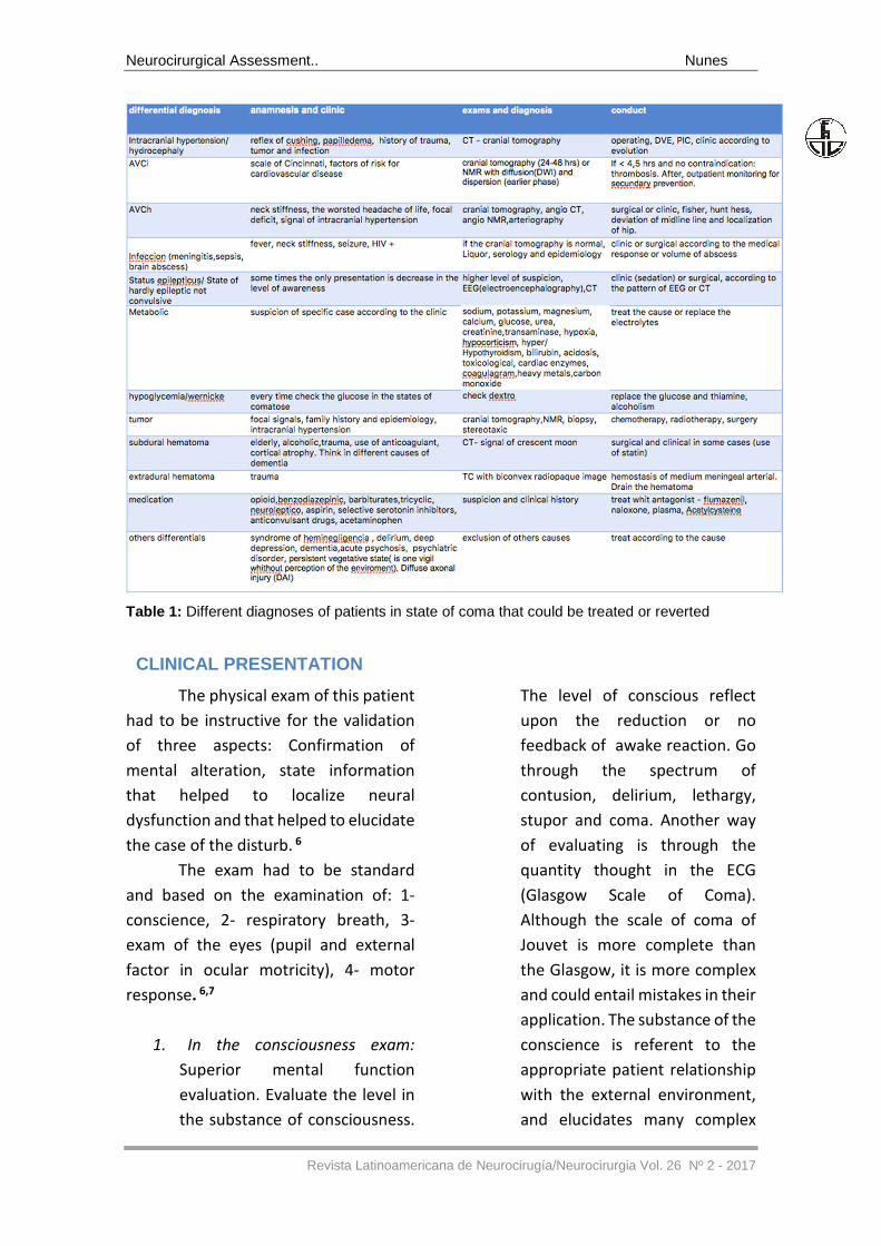
ETIOLOGY
The physiopathology of the structural
coma, which is less common, is by
compression of structures, among
which is the prototype of the
consciousness state, the ARAS. There is
the supratentorial ones, among which
are: tumor, abscess, traumatic brain
injury, ischemic stroke, hemorrhagic
stroke, hydrocephalus and the
infratentorial, represented by tumors,
pontine mielynosis, occlusion the
basilar artery. The diagnosis method is
carried out mostly by a cranial
tomography (CT) of the skull, and when
this is not sufficient, angio CT, angio MR
or arteriography are necessary.4,5
The most prevalent endocrine
metabolite is a diffuse cerebral damage,
represented by hypoglycemia,
hyperglycemia, hyponatremia,
hypernatremia, hypercalcemia, uremia,
Hepatic encephalopathy, narcosis,
hypothyroidism, Addison disease and
hypopanhipotuitarism, after drugs,
hypothermia, Inhalation of toxic gases
(carbon monoxide) and Psychiatric
causes (Table 1). 5
Neurocirurgical Assessment.. Nunes
Revista Latinoamericana de Neurocirugía/Neurocirurgia Vol. 26 Nº 2 - 2017
Table 1: Different diagnoses of patients in state of coma that could be treated or reverted
CLINICAL PRESENTATION
The physical exam of this patient
had to be instructive for the validation
of three aspects: Confirmation of
mental alteration, state information
that helped to localize neural
dysfunction and that helped to elucidate
the case of the disturb. 6
The exam had to be standard
and based on the examination of: 1-
conscience, 2- respiratory breath, 3-
exam of the eyes (pupil and external
factor in ocular motricity), 4- motor
response. 6,7
1. In the consciousness exam:
Superior mental function
evaluation. Evaluate the level in
the substance of consciousness.
The level of conscious reflect
upon the reduction or no
feedback of awake reaction. Go
through the spectrum of
contusion, delirium, lethargy,
stupor and coma. Another way
of evaluating is through the
quantity thought in the ECG
(Glasgow Scale of Coma).
Although the scale of coma of
Jouvet is more complete than
the Glasgow, it is more complex
and could entail mistakes in their
application. The substance of the
conscience is referent to the
appropriate patient relationship
with the external environment,
and elucidates many complex
Neurocirurgical Assessment.. Nunes
Revista Latinoamericana de Neurocirugía/Neurocirurgia Vol. 26 Nº 2 - 2017
mental functions like attention,
memory, language and others.
The vegetative state is one
example of lack of
consciousness, while awake. And
because of that, there had to be
attention in the evaluation of
disturbs in conscience, because
of the aphasia and the syndrome
of the confinement, that
characterizes motor disturbs
with damage in the quality of
consciousness, but not in the
level. Pay attention to this type
of evaluation in order to not
entail in wrong diagnoses on the
level of conscious. 7,8,9
2. The breath: Is one integrated
act for nerve influence of almost
all the encephalon. Thus,
breathing is one indirect
evaluation of their functions, in
spite of the low specifies. It could
be influenced by the neural gene
and metabolic. The epileptics
breath inhibition, in the pos ictal
period, in which the patient had
moments of auto-limited apnea.
The central neurogenic breath in
which PaO2 is necessarily normal
or higher, occurs in
mesencephalic lesions,
cerebellar hemispheres and
bulb. The apnea breath had long
inspired spasm followed with
total apnea in the brain bridge
lesions, hypoglycemia, lack of
cerebral oxygen, meningitis. The
Cheyne-Stokes breaths, where
periods of hyperpnea are altered
with hypopnea, occur with
diffuse involvement of the brain,
increased intracranial pressure
or cardiopulmonary impairment.
These breaths increase in depth
and volume to a peak and
decline until there is a period of
apnea. Associated respiratory
rate changes are respiratory
ataxia in which the respiratory
pattern is irregular, with
superficial and deep erratic
movements. This respiration
exists through dysfunction of the
medullary respiratory centers
and may indicate impending
agonizing breaths and apnea.
The principal cases are:
metabolic disturb, bilateral
cerebral infarction, hypertensive
encephalopathy, imminent
transtentorial herniation,
cardiac insufficiency and shock.
Central neurogenic
hyperventilation is characterized
by prolonged, rapid and regular
hyperpnea. It is associated with
diseases that affect the
paramedian reticular formation
in the mesencefalo inferior and
in the superior bridge. Apnea, a
rare form of respiratory rhythm
dysfunction, develops with a
prolonged respiratory phase,
occurs in lesions on the bridge
rostrally to the trigeminal nerve
motor nucleus or in the
cervicomedullary compression.
Systemic diseases also cause
changes in the respiratory
pattern, such as diabetic
Neurocirurgical Assessment.. Nunes
Revista Latinoamericana de Neurocirugía/Neurocirurgia Vol. 26 Nº 2 - 2017
ketoacidosis or severe
myxedema 8,9
3. The pupils were controlled by
the nucleus of brainstem
(sympathetic and
parasympathetic), when they
are intact and realize the act of
dilatation and constriction
respectively. Because of these
alterations, it is frequent in
comatose patients. The form of
evaluation is through form, size
and reactive pupil. It is important
to observe that the evaluation
observes the ocular motor that
reflects the integrity of this area
that involves the consciousness.
The unilateral alterations, in
most cases, are from structural
cases and symmetric from
metabolic cases. Some of these
reflexes could be searched
thought the evaluate of the
integrity of brainstem like the
reflex of eyelid, corneal reflex,
oculo-cephalic reflex, oculo-
vestibular reflex. These are the
most used in the protocols of
encephalic dead. The alteration
that had to be evaluated in the
urgent care is the anisocoric with
the photomotor reflex typic of
aneurism of posterior
communicant artery, for the
relationship with the pair cranial
III. 9 The main coma pupil
changes are: (1) Oculomotor
paralysis: Intracranial
compression of one of the
nerves of the third cranial nerve,
contusion of the ocular glob. (2)
Mydriasis: Anxiety, delirium,
pain, seizure, botulism, atropine,
hypermagnesemia,
sympathomimetic drugs.
Horner's syndrome: Traumatic
carotid dissection, brachial
plexus injury, internal jugular
vein catheterization. (3)
Myositis: Opiates, metabolic
encephalopathies, pontine
lesions, hypercapnia.
(4)Medium-fixed pupils typical
of encephalic death.10
4. The motor answer is separate
from the areas that regulate the
mental state, therefore, the
evaluation does not even have
relation with the depth of the
coma, in other words, they are
disproportionate. Not based in
have a good baseline of evaluate
in the level and conscience
substance. The posture of
decerebration and decortication
refers to a lesion in the
mainstream and could become
one form of evaluation oriented
for to coma. The movements like
asterix suggest uremic
encephalopathy, hypoglycemia.
The evaluation of the signal in
Babinski is of the extension of 1
year old before the reflection, if
it is not normal, reflect the
lesions of the pyramidal
structure. 11
Cerebral herniations
Inside the skull, which is a rigid
sphere, are present the brain, blood and
Neurocirurgical Assessment.. Nunes
Revista Latinoamericana de Neurocirugía/Neurocirurgia Vol. 26 Nº 2 - 2017
cerebrospinal fluid (CSF). The volume
formed by them is constant. When there
is a mass or lesion that forms an increase
volume within the cranial cavity, the
outflow of liquor initially occurs in order
to maintain this volume constant. It is
worth remembering that in this case a
pressure gradient occurs between the
two hemispheres and between the
posterior fossa and the portion above
the cerebellum tent. As the lesion
expands, more cerebrospinal fluid
outflow and in some cases decreased
blood volume. When there are
alterations of the intercompartmental
volume inside the skull there is a
decoupling of encephalic tissue to the
lower pressure compartment causing
the hernias. 12
The most important hernias are
central transtentorial hernia and lateral
hernia. The central hernia is seen in
cases where the diencephalon is pushed
down the cerebellum tent. Initially the
patient evolved with sleepiness and
Cheyne-Stokes breathing. From then on
bilateral myosis occurs, but
photoreagents, sometimes paratonia
and also motor response in flexion
(decortication). The next step of
herniation is called mesencephalic. Here
the pupils are medium fixed, with
abduction deficit of the eyes and
extension posture (decerebration).
When the bridge compromises the eye
movements are abolished, the
respiratory rhythm becomes irregular.
When the bulb is compressed the pupils
are medium-fixed, with the patient in
apnea and absence of motor reaction.
The diencephalic phase is important,
because at this time it is still possible to
reverse the process.13
Lateral herniation occurs when
the medial portion of the temporal lobe,
that is, the uncus hints between the free
edge of the tentorium of the cerebellum
and the midbrain. In this case the lesions
are in the temporal lobe or are extra-
axial. Initially, there is compression of
the opposite cerebral peduncle to the
side injured side. From this point, the
evolution is the same as that of the
central.14
Minimally Conscious State (MCS)
The MCS is characterized by a
severe impairment of consciousness,
with evidence of wakefulness and
partial preservation of awareness.
Unlike the VS, there are discernible,
purposeful behaviors that can be
differentiated from reflexive behavior.
The hallmark of MCS is inconsistent but
reproducible, command following. The
preservation of cortico thalamic
connections might explain why patients
in MCS retain the capacity for cognitive
processing. The patient may exhibit
visual pursuit, emotional responses, and
gestures to appropriate environmental
stimuli, but are unable to functionally
communicate their thoughts or
feelings.15
Acute Confusional State
Once emerged from the MCS,
patients continue to experience a
transient period of disorientation and
agitation. The full array of symptoms
associated with the acute confusional
state can also include irritability,
distractibility, anterograde amnesia,
Neurocirurgical Assessment.. Nunes
Revista Latinoamericana de Neurocirugía/Neurocirurgia Vol. 26 Nº 2 - 2017
restlessness, emotional lability,
impaired perception, attentional
abnormalities, and a disrupted sleep-
wake cycle. A key pattern to this state is
the day-to-day fluctuation of behavioral
responses. The return of behavioral
consistency despite situational stresses
may indicate a resolution of this period. 16,17
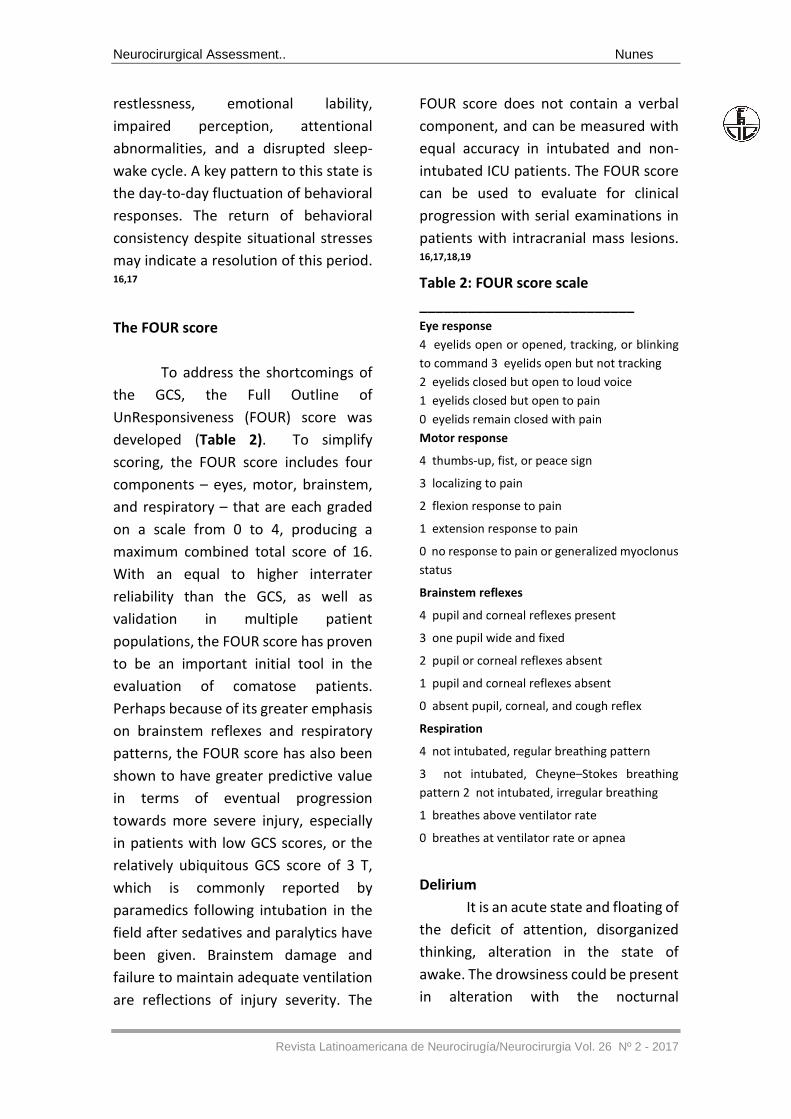
The FOUR score
To address the shortcomings of
the GCS, the Full Outline of
UnResponsiveness (FOUR) score was
developed (Table 2). To simplify
scoring, the FOUR score includes four
components – eyes, motor, brainstem,
and respiratory – that are each graded
on a scale from 0 to 4, producing a
maximum combined total score of 16.
With an equal to higher interrater
reliability than the GCS, as well as
validation in multiple patient
populations, the FOUR score has proven
to be an important initial tool in the
evaluation of comatose patients.
Perhaps because of its greater emphasis
on brainstem reflexes and respiratory
patterns, the FOUR score has also been
shown to have greater predictive value
in terms of eventual progression
towards more severe injury, especially
in patients with low GCS scores, or the
relatively ubiquitous GCS score of 3 T,
which is commonly reported by
paramedics following intubation in the
field after sedatives and paralytics have
been given. Brainstem damage and
failure to maintain adequate ventilation
are reflections of injury severity. The
FOUR score does not contain a verbal
component, and can be measured with
equal accuracy in intubated and non-
intubated ICU patients. The FOUR score
can be used to evaluate for clinical
progression with serial examinations in
patients with intracranial mass lesions. 16,17,18,19
Table 2: FOUR score scale
___________________________
Eye response
4 eyelids open or opened, tracking, or blinking
to command 3 eyelids open but not tracking
2 eyelids closed but open to loud voice
1 eyelids closed but open to pain
0 eyelids remain closed with pain
Motor response
4 thumbs-up, fist, or peace sign
3 localizing to pain
2 flexion response to pain
1 extension response to pain
0 no response to pain or generalized myoclonus
status
Brainstem reflexes
4 pupil and corneal reflexes present
3 one pupil wide and fixed
2 pupil or corneal reflexes absent
1 pupil and corneal reflexes absent
0 absent pupil, corneal, and cough reflex
Respiration
4 not intubated, regular breathing pattern
3 not intubated, Cheyne–Stokes breathing
pattern 2 not intubated, irregular breathing
1 breathes above ventilator rate
0 breathes at ventilator rate or apnea
Delirium
It is an acute state and floating of
the deficit of attention, disorganized
thinking, alteration in the state of
awake. The drowsiness could be present
in alteration with the nocturnal
Neurocirurgical Assessment.. Nunes
Revista Latinoamericana de Neurocirugía/Neurocirurgia Vol. 26 Nº 2 - 2017
agitation. The fast identification of
delirium is fundamental, because it
could pronounce the neural dysfunction
more seriously, that could be early
treated and reverted. It occurs in 10 up
50% of hospitalized patients. Some
factors of risk are advanced age,
sensorial dysfunction, drugs, dangerous
basic condition, dehydration,
immobility, dementia. The CAM-ICU
(confusion assessment method in an
intensive care until) is used for
diagnoses in critical patients. 20,21,22,23,24
Vegetative State(VS)/Unresponsive
Wakefulness State
The VS is thought of as an
unconscious, dissociative state of
wakefulness without awareness. The
patient’s eyes open spontaneously, and
EEG testing reveals the presence of
sleep-wake cycles. Patients may arouse
by provocation or external estimulation,
but they show no signs of conscious
perception or deliberate action.
Interestingly, these patients may
perform stereotyped gestural
movements such as yawning, chewing,
crying, smiling, or moaning, but these
are unrelated to context. The presence
of wakefulness suggests preserved
brainstem functioning, but the lack of
awareness suggests an underlying
cortical dysfunction. Likewise,
functional neuroimaging has shown
sensory estimulation will activate
primary cortical areas, but not the
higher order cortical areas thought
necessary for awareness. With proper
medical care, a patient in a VS can
survive for many years. 25,26,27,28
Management
Adequate management of eye,
mouth, and skin at compression sites
requires frequent change of linens.
Every day, infections may present, skin
may break down, and fluid shifts may
cause rapid imbalance of homeostasis.
Drugs (particularly antibiotics) have
potentia adverse effects. 27,28
Adequate management of eye, mouth,
and skin at compression sites requires
frequent change of linens, patches, and
protective pads. Splinting of extremities
may be needed to avoid contractures.
Inability to close eyelids completely
after trauma and, in particular,
nocturnal lagophthalmos are risk factors
for conjunctivitis and corneal erosion.
Polyethylene moisture chambers are
required to prevent early epithelial
breakdown. Filamentary keratopathy is
a common dry-eye syndrome in patients
in prolonged coma. Prolonged eyelid
contact with the cornea and reduced
blinking impair lacrimal fluid turnover
and may be contributing factors.29,30
Tracheostomy reduces
pulmonary complications and provides
easier access for pulmonary secretion.
Tracheostomy will reduce length of stay,
but it should generally be postponed
until approximately 2 weeks in patients
the most common healthcare-related
infections are pneumonia, urinary tract
infections, or infections of indwelling
venous catheters. Potentially difficult to
eradicate microorganisms include
Enterococcus faecalis or faecium,
Staphylococcus aureus, Klebsiella
pneumoniae, Acinetobacter baumannii,
Neurocirurgical Assessment.. Nunes
Revista Latinoamericana de Neurocirugía/Neurocirurgia Vol. 26 Nº 2 - 2017
Pseudomonas aeruginosa, and
Enterobacter cloacae . Clostridium
difficile infections are also on the rise,
particularly in patients with long
hospital stays. The dilemma faced by
treating physicians is that delayed
initiation of antibiotics increases
mortality, yet combination therapies to
broaden the spectrum may lead to
antibiotic resistance. Antibiotic therapy
is changing as a result of infectious
disease consultation. 31
Fever in comatose patients is
mostly caused by infections. Lingering
infections have to be excluded before
attributing fever to the brain injury.
However, Paroxysmal sympathetic
hyperactivity (PSH) syndrome is a
commonly seen in “ unexplained” fever
of comatose patients. PSH or
dysautonomic storming all too
frequently remains unrecognized and
untreated. These spells are most
common in young patients with diffuse
axonal traumatic brain injury, but can
occur with any major brain injury.
Episodes of PSH can begin during the
acute phase, often in comatose
patients, and continue into the
rehabilitation phase. Patients become
tachycardic, hypertensive (with
widened pulse pressure), tachypneic,
febrile, diaphoretic, and often develop
markedly increased tone, which may
result in dystonic posturing. 32
Pupillary dilatation, piloerection,
and skin flushing can also be seen. The
manifestations of PSH respond best to
bolus doses of morphine sulfate (2– 8
mg intravenously). This favorable
response is not related to the analgesic
effect of opiates, but rather to
modulation of central pathways that are
responsible for the autonomic
dysfunction. The response to morphine
is rapid and quite reliable in aborting
spells of PSH. Other effective
medications for the treatment of PSH
include non cardioselective beta-
blockers (such as propranolol),
clonidine, and dexmedetomidine
(central alpha 2-receptor agonists),
bromocriptine (a dopamine D2-receptor
agonist), baclofen (GABA B receptor
agonist), benzodiazepines (GABA A
receptor agonist), and gabapentin
(which binds GABA receptors and
voltage-gated calcium channels in the
dorsal horn of the spinal cord). In our
experience, beta-blockers and clonidine
are useful in controlling the tachycardia
and hypertension, but less so for the
dystonia. Baclofen and benzodiazepines
(especially diazepam) do cause muscle
relaxation, but may not improve the
other hypersympathetic features, who
can potentially be liberated from the
ventilator if they show early signs of
substantial neurologic improvement. 33,34
Gradually, after the patient is
weaned off the ventilator, the
tracheostomy can be closed, including in
patients with prolonged
unconsciousness. Pulmonary care
involves frequent culturing of sputum
when secretions change in color and
texture and immediate antibiotic
coverage to treat pneumonia and
sepsis. Pleural effusions are frequent as
a manifestation of anasarca and large
pleural collections may need to be
Neurocirurgical Assessment.. Nunes
Revista Latinoamericana de Neurocirugía/Neurocirurgia Vol. 26 Nº 2 - 2017
drained. Gastrointestinal problems vary
from gastroparesis to paralytic ileus,
resulting in distension of the colon and
increased risk of perforation. Daily
bowel care may include motility agents. 35
Continuous volume replacement
is needed for longtime care. The
adequate intravascular status is
determined by satisfactory organ
perfusion (urinary output, capillary
refill, cold or warm extremities, blood
lactate, and mixed venous oxygen
saturation). Tissue edema may form
over time, possibly as a result of
overzealous, percutaneous gastrostomy
(PEG) and Fluid administration (e.g.,
failure to adjust intravenous fluid rate
while advancing enteral nutrition,
failure to concentrate medications).
Volume depletion is less common in the
longterm but may occur, especially
when extravascular compartment is
expanded by sepsis. Hypotonic
crystalloids, such as lactated Ringer’ s or
half normal saline, should be avoided in
traumatic brain injury. Albumin (5%) is a
good volume expander, and may have a
role in sepsis resuscitation, but the
safety in acute brain injury is unclear
and may be deleterious in traumatic
brain injury. In patients who have
developed oliguria and a rise in BUN
(BUN/creatinine ratio > 20),
dehydration is very likely and should
result in discontinuation of all diuretics
and administration of normal saline.
Nutrition is eventually provided through
a PEG , which is very safe. Complications
include wound infection, leakage,
peritonitis, self-extubation or
hemorrhage in the first weeks of
placement, only in 2%. The risk of
gastrointestinal hemorrhage may be
increased. Compared with nasogastric
tubes, gastroesophageal reflux is lower
in patients with a PEG. 36,37
A bowel care regimen should be
initiated. Bowel incontinence is often
present, and the task is to keep the skin
clean and dry. Diarrhea may have many
causes, but can be attributed to certain
nutritional formulas and resolve with
reducing fiber content. Antibiotics, as
well as Escherichia coli or Clostridium
difficile infections, are other possible
causes of diarrhea. Failure to pass stool,
or marble-like stools, should be treated
with rectal enema or manual removal.
Glycerol suppository can be helpful, but
senna (10 mL) and lactulose (20 mL) are
common maintenance therapies.
Comatose patients are at risk of
adynamic ileus. Metoclopramide (10 mg
IV) or erythromycin (500 mg orally) can
be very effective to resolve the bowel
distension. Nosocomial urinary tract
infections will likely occur in comatose
patients with long-term indwelling
catheters. 38
Daily Check List in care of the comatose
patient 39
• Lungs : Mechanical ventilation
settings, Weaning option,
Tracheostomy care, Chest X-ray for
infiltrates
• Heart: Cardiac arrhythmias,
Electrocardiogram changes(i.e., QT
prolongation),
Inotropes/vasopressors/beta
blockade
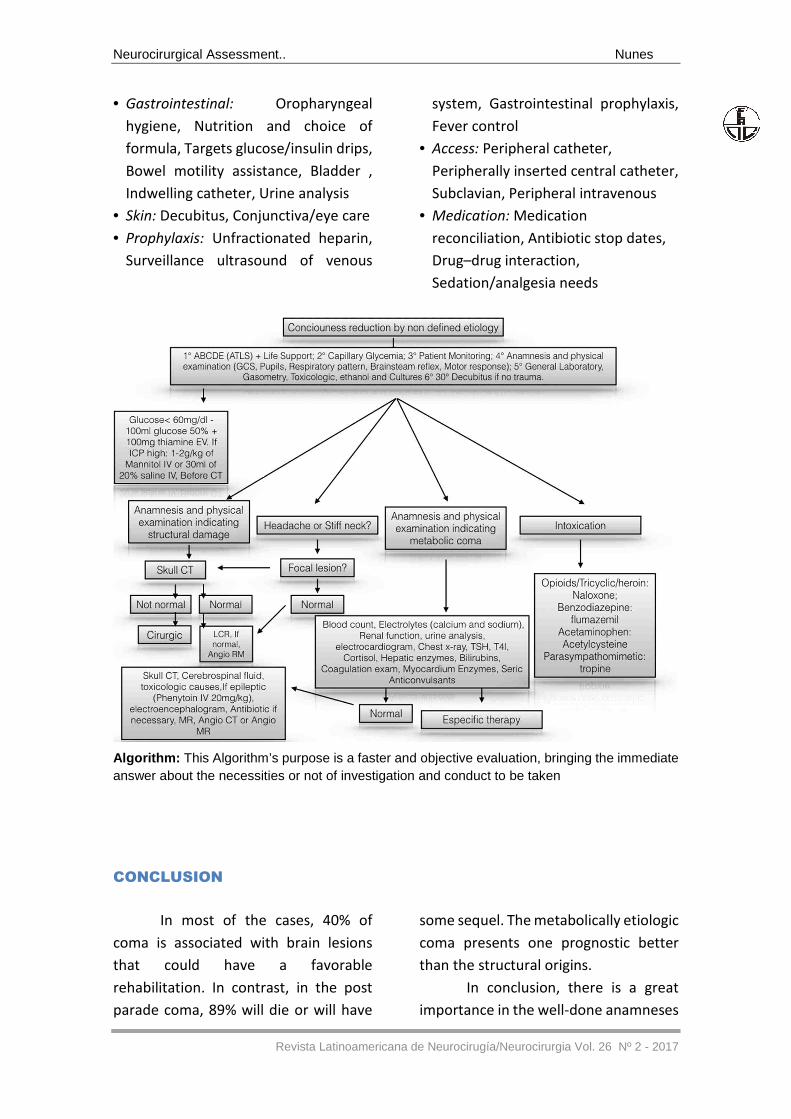
Neurocirurgical Assessment.. Nunes
Revista Latinoamericana de Neurocirugía/Neurocirurgia Vol. 26 Nº 2 - 2017
• Gastrointestinal: Oropharyngeal
hygiene, Nutrition and choice of
formula, Targets glucose/insulin drips,
Bowel motility assistance, Bladder ,
Indwelling catheter, Urine analysis
• Skin: Decubitus, Conjunctiva/eye care
• Prophylaxis: Unfractionated heparin,
Surveillance ultrasound of venous
system, Gastrointestinal prophylaxis,
Fever control
• Access: Peripheral catheter,
Peripherally inserted central catheter,
Subclavian, Peripheral intravenous
• Medication: Medication
reconciliation, Antibiotic stop dates,
Drug–drug interaction,
Sedation/analgesia needs
Algorithm: This Algorithm’s purpose is a faster and objective evaluation, bringing the immediate answer about the necessities or not of investigation and conduct to be taken
CONCLUSION
In most of the cases, 40% of
coma is associated with brain lesions
that could have a favorable
rehabilitation. In contrast, in the post
parade coma, 89% will die or will have
some sequel. The metabolically etiologic
coma presents one prognostic better
than the structural origins.
In conclusion, there is a great
importance in the well-done anamneses
Neurocirurgical Assessment.. Nunes
Revista Latinoamericana de Neurocirugía/Neurocirurgia Vol. 26 Nº 2 - 2017
and physical exam, as well as the correct
definition of etiology, for the
appropriate treatment or reversion of
the coma in an adequate time, without
neurological sequel. This chapter
suggests a flowchart that could diagnose
and treat the more prevailing causes, in
a way as to minimize the neurological
loss of patients that present a decrease
in awake and in consciousness.
REFERENCIAS:
1. Feske SK. Coma and confusional states: emergency diagnosis and management. Neurol Clin 16:237-256,1998.
2. Zeman A: Consciousness. Brain 2001; 124:v1263–1289
3. Steriade M. Arousal: revisiting the reticular activating system. Science. 1996;272:225.
4. Young GB, Pigott SE: Neurobiological basis of consciousness. Arch Neurol 1999; 56: 153–157
5. Benseñor IM, Atta JA, Matins MA. Semiologia clínica. Caramelli P. Distúrbios da consciência. 1° ed. São Paulo: Sarvier, 2002, pg-446-450.
6. Sturmann K. The neurologic examination. Emerg Med Clin North Am. 1997;15:491-506
7. Radvany J. Ferraz AC. Estados Confusionais no paciente Grave. In: Knobel E. - Condutas no Paciente Grave. 2° ed. São Paulo, Editora Atheneu, 1998. p.617-629
8. North JB, Jennet B. Abnormal breathing patterns associated with acute brain injury. Arch Neurol. 1974;32:338
9. Dandan IS. Altered Consciousness. Top Emerg Med. 2004; 11:242-253
10. Masdeu JC. The Localization of Lesions Causing Coma. In: Brazis PW, Masdeu JC, Biller J. Localization in Clinical Neurology. 2° edition. Little, Brown and Company, Boston. 1990.p.457-485.
11. Bruno M-A, Vanhaudenhuyse A, Thibaut A, et al. From unresponsive wakefulness to minimally conscious PLUS and functional locked-in syndromes: recent advances in our understanding of disorders of consciousness. J Neurol 2011; 258(7):1373–84.
12. Sherer M, Nakase-Thompson R, Yablon SA, et al. Multidimensional assessment of acute
confusion after traumatic brain injury. Arch Phys Med Rehabil 2005; 86(5): 896–904.
13. Teasdale G, Jennett B: Assessment of coma and impaired consciousness. A practical scale. Lancet 1974; 2:81–84
14. Ely EW, Inouye SK, Bernard GR et al. Delirium in mechanically ventilated patients: validity and reliability of the confusion assessment method for the intensive care unit (CAM-ICU). Jama, 2001; 286: 2703-2710.
15. Robert D, Stevens, MD. Anish Bhardwaj, MD, FCCM Approach to the comatose patient Crit Care Med 2006 Vol. 34, No. 1
16. Levy DE, Bates D, Caronna JJ, Carlidge NE, et. al. Prognosis in nontraumatic coma. Ann Intern Med. 1981, 94 293-301
17. Young GB, Wang JT, Connolly JF: Prognostic determination in anoxic-ischemic and traumatic encephalopathies. J Clin Neurophysiol 2004; 21:379–390
18. Wijdicks EFM, Varelas PN, Gronseth GS et al. (2010). American Academy of N. Evidence-based guideline update: determining brain death in adults: report of the Quality Standards Subcommittee of the American Academy of Neurology. Neurology 74: 1911–1918.
19. Wijdicks EFM (2010). The bare essentials: coma. Pract Neurol 10: 51–60.
20. Moore SA, Wijdicks EFM (2013). The acutely comatose patient: clinical approach and diagnosis. Semin Neurol 33: 110–120.
21. Stavale M. Bases da Terapia Intensiva Neurológica: Fisiopatologia e Princípios terapêuticos, 2edição, Editora Santos
22. Greer DM,Young J, Scripko PD. Clinical Examination for Outcome Prediction in Nontraumatic Coma. Crit Care Med, 40:1150-1156, 2012
Neurocirurgical Assessment.. Nunes
Revista Latinoamericana de Neurocirugía/Neurocirurgia Vol. 26 Nº 2 - 2017
23. Forsberg S, Hojer J. Metabolic vs Structural Coma In The ED: An Observational Study. American Journal of Emergency Medicine, 30:1986-1990, 2012
24. Cavanna AE, Shah S, Eddy CM, et al. Consciousness: a neurological perspective. Behav Neurol 2011;24(1):107–16.
25. Giacino JT, Fins JJ, Laureys S, et al. Disorders of consciousness after acquired brain injury: the state of the science. Nat Rev Neurol 2014;10(2):99–114.
26. Edlow JA, Rabinstein A, Traub SJ et al. (2014). Diagnosis of reversible causes of coma. Lancet 384: 2064–2076.
27. Eapen BC, Georgekutty J, Subbarao B. Disorders of Consciousness.Phys Med Rehabil Clin N Am - (2017). http://dx.doi.org/10.1016/j.pmr.2016.12.003
28. Wijdicks EFM (2001). The diagnosis of brain death. N Engl J Med 344: 1215–1221.
29. Wijdicks EFM. The bare essentials: coma. Pract Neurol 2010; 10: 51–60.
30. Wijdicks EFM, Varelas PN, Gronseth GS et al. American Academy of N. Evidence-based guideline update: determining brain death in adults: report of the Quality Standards Subcommittee of the American Academy of Neurology. Neurology 2010; 74: 1911–1918.
31. Laureys S, Owen AM, Schiff ND. Brain function in coma, vegetative state, and related disorders. Lancet Neurol 2004;3(9):537–46.
32. Wijdicks EFM (2016). Why you may need a neurologist to see a comatose patient in the ICU Crit Care 2016; 20: 193.
33. Wijdicks EFM (2017). Brain Death 3ed Oxford University Press.
34. Lavrijsen J, vaqn Rens G, van den Bosch H. Filamentary keratopathy as a chronic problem in the long-term care of patients in a vegetative state. Cornea 2005;24: 620–622.
35. Giacino JT, Ashwal S, Childs N, et al. The minimally conscious state: definition and diagnostic criteria. Neurology 2002;58(3):349–53..
36. Mijalski C, Lovett A, Mahajan R et al. Cerebral fat embolism: a case of rapid-onset coma. Stroke 2015; 46: 251–253.
37. Moore SA, Wijdicks EFM. The acutely comatose patient: clinical approach and diagnosis. Semin Neurol 2013; 33: 110–120.
38. Salottolo K, Carrick M, Levy AS et al. Aggressive operative neurosurgical management in patients with extra-axial mass lesion and Glasgow Coma Scale of 3 is associated with survival benefit: a propensity matched analysis. Injury 2016; 47: 70–76.
39. Stevens RD, Cadena RS, Pineda J. Emergency neurological life support: approach to the patient with coma. Neurocrit Care 2015; 23: 69–75.
Correspondencia:
Neiffer Nunes Rabelo
Email: [email protected]
Recibido : 13/06/17
Aprobado : 09/07/17
Conflicto de intereses : Los autores
declaran no presentar conflicto de
intereses