Dermatosis Del Embarazo 2008
8
4 Acta Dermatoven AP A Vol 17, 2008, No 1 pruritus, pregnancy dermatoses, pemphigoid gestationis, polymorphic eruption of pregnancy, intrahepatic cholestasis of pregnancy, atopic eruption of pregnancy K E Y W O R D S D e r m a t o s e s o f p re g n ancy M.-M. Pãunescu, V. Feier, M. Pãunescu, F. Dorneanu, A. Sisak, and C. M. Ambros-Rudolph The dermatoses of pregnancy represent a heterogeneous group of pruritic inflammatory skin diseases related to pregnancy and/or the postpartum period. Whereas some dermatoses are distressing only to the mother because of severe pruritus, others are associated with fetal risks including fetal distress, prematurity, and stillbirth. Early diagnosis and prompt treatment are essential for improving maternal and fetal prognosis. This review discusses the various pregnancy dermatoses in detail and offers an algorithmic approach to their diagnosis and management. In tr od uc ti on Pregnancy is associated with complex endocrino- logical, immunological, metabolic, and vascular changes that may influence the skin in various ways. In addition to physiological skin chang es and changes in the course of preexisting skin diseases, specific dermatoses of preg- nancy may develop. These represent a heterogeneous group of inflammatory skin diseases related to preg- nancy and/or the postpartum period. The leading symp- tom is severe pruritus. Over decades, attempts have been made to establish a reasonable classification (1, 2), but the rarity of these diseases, their variable clinical morphology, and the lack of unequivocal diagnostic tests has led to confusing terminologies. Based on the results of a retrospective two-center study on more than 500 pregnant patients, a new classification of these spe- cific dermatoses of pregnancy has recently been pro- posed (3) that includes the following diseases: pem- phigoid gestationis, polymorphic eruption of pregnancy, intrahepatic cholestasis of pregnancy, and atopic erup- tion of pregnancy (Table 1). Atopic eruption of preg- nancy encompasses the entities formerly described as prurigo of pregnancy and pruritic folliculitis of preg- nancy. Although the severe pruritus associated with polymorphic and atopic eruption of pregnancy is dis- tressing only for the mother, pemphigoid gestationis may be associated with small-for-date babies, and in- trahepatic cholestasis of pregnancy poses an increased risk for fetal distress, prematurity, and stillbirth (4, 5). Because specific diagnostic tests are only available for some diseases and a pregnant patient with pruritus needs information on her condition as well as the pos- sibly associated fetal risks preferentially at the first visit, before test results are available, exact knowledge of the clinical presentation of these diseases is essential. This review discusses the various pregnancy dermatoses in detail with special emphasis on epidemiology, clinical features, diagnostic tests, maternal and fetal prognosis, therapy, and management. S U M M A R Y Pregnancy dermatoses R e v i e w
Transcript of Dermatosis Del Embarazo 2008
7/23/2019 Dermatosis Del Embarazo 2008
http://slidepdf.com/reader/full/dermatosis-del-embarazo-2008 1/8
4 Acta Dermatoven APA Vol 17, 2008, No 1
pruritus,
pregnancydermatoses,
pemphigoid
gestationis,
polymorphic
eruption of
pregnancy,
intrahepatic
cholestasis of
pregnancy,
atopic eruption
of pregnancy
K E Y
W O R D S
Dermatoses of pregnancy M.-M. Pãunescu, V. Feier, M. Pãunescu, F. Dorneanu, A. Sisak, and C. M. Ambros-Rudolph
The dermatoses of pregnancy represent a heterogeneous group of pruritic inflammatory skin diseases
related to pregnancy and/or the postpartum period. Whereas some dermatoses are distressing only to
the mother because of severe pruritus, others are associated with fetal risks including fetal distress,
prematurity, and stillbirth. Early diagnosis and prompt treatment are essential for improving maternal
and fetal prognosis. This review discusses the various pregnancy dermatoses in detail and offers analgorithmic approach to their diagnosis and management.
Introduction
Pregnancy is associated with complex endocrino-logical, immunological, metabolic, and vascular changesthat may influence the skin in various ways. In additionto physiological skin changes and changes in the courseof preexisting skin diseases, specific dermatoses of preg-nancy may develop. These represent a heterogeneousgroup of inflammatory skin diseases related to preg-nancy and/or the postpartum period. The leading symp-tom is severe pruritus. Over decades, attempts havebeen made to establish a reasonable classification (1,2), but the rarity of these diseases, their variable clinicalmorphology, and the lack of unequivocal diagnostictests has led to confusing terminologies. Based on theresults of a retrospective two-center study on more than500 pregnant patients, a new classification of these spe-cific dermatoses of pregnancy has recently been pro-posed (3) that includes the following diseases: pem-phigoid gestationis, polymorphic eruption of pregnancy,intrahepatic cholestasis of pregnancy, and atopic erup-
tion of pregnancy (Table 1). Atopic eruption of preg-nancy encompasses the entities formerly described asprurigo of pregnancy and pruritic folliculitis of preg-nancy. Although the severe pruritus associated withpolymorphic and atopic eruption of pregnancy is dis-tressing only for the mother, pemphigoid gestationismay be associated with small-for-date babies, and in-trahepatic cholestasis of pregnancy poses an increasedrisk for fetal distress, prematurity, and stillbirth (4, 5).Because specific diagnostic tests are only available forsome diseases and a pregnant patient with pruritusneeds information on her condition as well as the pos-sibly associated fetal risks preferentially at the first visit,before test results are available, exact knowledge of theclinical presentation of these diseases is essential. Thisreview discusses the various pregnancy dermatoses indetail with special emphasis on epidemiology, clinicalfeatures, diagnostic tests, maternal and fetal prognosis,therapy, and management.
S U M M A R Y
Pregnancy dermatoses R e v i e w
7/23/2019 Dermatosis Del Embarazo 2008
http://slidepdf.com/reader/full/dermatosis-del-embarazo-2008 2/8
Acta Dermatoven APA Vol 17, 2008, No 1 5
Pemphigoid gestationis
Pemphigoid gestationis (PG, also known as herpesgestationis) is a rare autoimmune bullous disorder that
presents mainly in late pregnancy or the immediatepostpartum period. Its incidence is estimated to be 1 in50,000 to 60,000 pregnancies and the disease shows acorrelation with the haplotypes HLA-DR3 and DR4 (6).There is also an increased risk of developing other au-toimmune diseases, in particular Grave’s disease. PGtends to recur in subsequent pregnancies, with earlieronset and increasing severity. Very rarely (5%) a preg-nancy may be passed over (“skip pregnancies”).
Pathogenetically, circulating complement-fixing IgGantibodies of the subclass IgG1 (formerly known as“herpes gestationis factor”) bind to a 180-kDa protein,BP-180 or bullous pemphigoid antigen 2, in the
hemidesmosomes of the basal membrane zone (BMZ),leading to tissue damage and blister formation (7). Of interest, the primary site of autoimmunity seems not tobe the skin, but the placenta, because antibodies bindnot only to the BMZ of the epidermis, but also to that of chorionic and amniotic epithelia, both equally of ecto-dermal origin. Aberrant expression of MHC class IImolecules on the chorionic villi suggests an allogenicimmune reaction to a placental matrix antigen, thoughtto be of paternal origin.
Clinical features
PG presents with intense pruritus that may occa-sionally precede the appearance of skin lesions. Ini-tially, erythematous urticarial papules and plaques de- velop typically on the abdomen, but may spread to theentire skin surface. The umbilical region is almost al- ways involved. In this “pre-bullous” stage, differentia-tion between PG and polymorphic eruption of preg-nancy is almost impossible, both clinically and histo-pathologically. Diagnosis becomes clear when lesionsprogress to tense blisters that resemble those in bullouspemphigoid. Facial and mucous membranes are usu-ally spared (6).
Diagnostic tests
Histopathological findings from lesional skin de-pend on the stage and severity of the disease. Whereasthe pre-bullous stage is characterized by edema of theupper and middle dermis accompanied by a predomi-nantly perivascular inflammatory infiltrate, composedof lymphocytes, histiocytes, and a variable number of eosinophils, the bullous stage reveals subepidermal blis-tering that, ultrastructurally, may be located to the laminalucida of the BMZ (6).
Direct immunofluorescence of perilesional skin, the goldstandard in the diagnosis of PG, shows linear C3 deposition
along the BMZ in 100% of cases and additional IgG deposi-
tion in 30%. Depending on the technique used, circulatingIgG antibodies in the patient’s serum may be detected by indirect immunofluorescence in 30 to 100% of cases, bindingto the roof of the artificial cleft on salt-split skin. Antibody levels may also be monitored using modern ELISA and
immunoblot techniques, and show a good correlation withdisease activity (6, 7).
Prognosis
The natural course of PG is characterized by exacer-bations and remissions during pregnancy, with frequentimprovement in late pregnancy followed by a flare-up atthe time of delivery (75% of patients). After delivery, thelesions usually resolve within weeks to months but may recur with menstruation and hormonal contraception.Rarely, severe courses with persistence of skin lesionsover several years may occur. Fetal prognosis is gener-
ally good; there is an increase in small-for-date babiesbut not in prematurity or stillbirths (4). Due to a passivetransfer of antibodies from the mother to the fetus, 10%of newborns develop mild skin lesions that resolve spon-taneously within days to weeks (6).
Therapy and management
Treatment depends on the stage and severity of thedisease and aims to control pruritus and to prevent blis-ter formation. Only in cases of mild pre-blistering, topi-cal corticosteroids with or without oral antihistaminesmay be sufficient (6). All other cases require systemic
corticosteroids (prednisolone, usually started at a doseof 0.5–1 mg/kg/day) that are considered safe in preg-nancy (8, 9). When the disease improves, the dose canusually be reduced, but should be increased in time toprevent the flare-up common at delivery. Cases unre-sponsive to systemic corticosteroid treatment may ben-efit from immunoapheresis (10). After delivery, if nec-essary, the full range of immunosuppressive treatmentmay be administered.
Polymorphic eruption of
pregnancy Polymorphic eruption of pregnancy (PEP), also
known as pruritic urticarial papules and plaques of preg-nancy (PUPPP), toxic erythema of pregnancy, toxemicrash of pregnancy, late-onset prurigo of pregnancy) is abenign, self-limited pruritic inflammatory disorder thatusually affects primigravidae in the last weeks of preg-nancy or immediately postpartum (15%; 11). Its inci-dence is about 1:160 pregnancies and the condition isassociated with excessive maternal weight gain andmultiple pregnancies (11, 12).
The pathogenesis of PEP remains unclear. The main
theories proposed to date focus on abdominal distension,
R e v i e w Pregnancy dermatoses
7/23/2019 Dermatosis Del Embarazo 2008
http://slidepdf.com/reader/full/dermatosis-del-embarazo-2008 3/8
6 Acta Dermatoven APA Vol 17, 2008, No 1
hormonal, and immunological factors (13). The fact thatPEP starts within striae distensae at the time of greatestabdominal distension favors connective tissue damagedue to overstretching as playing a central role. It has beensuggested that previously inert structures develop anantigenic character, thus triggering the inflammatory pro-cess. Hormonal and immunological changes have not de-finitively been shown; nor has an association with increasedbirth weight or male sex of the newborn been confirmed(4, 11). (Figure 1.)
Clinical features
PEP typically starts on the abdomen, within striaedistensae, with severely pruritic urticarial papules thatcoalesce into plaques, spreading to the buttocks andproximal thighs. Often the eruption remains located tothese sites but can quickly generalize in severe cases.Unlike in PG, a characteristic finding is the sparing of the umbilical region. Later on, the clinical picture be-comes polymorphous, as vesicles (but never bullae), widespread non-urticated erythema, and targetoid and
eczematous lesions appear in more than half of patients.The rash usually resolves within 4 to 6 weeks (11).
Diagnostic tests
Histopathology is non-specific and varies with thestage of disease. Next to a superficial to mid-dermalperivascular lymphohistiocytic infiltrate intermingled with eosinophils, early biopsies show a prominent der-mal edema, and later biopsies reveal frequent epider-mal changes including spongiosis, and hyper- and par-akeratosis. Direct and indirect immunofluorescence in- vestigations are essentially negative in PEP.
Prognosis
Maternal and fetal prognosis is excellent and thereis no cutaneous involvement of the newborn (12). Le-sions are self-limited and PEP tends not to recur; theexception to this is multiple pregnancies, when bothearlier presentation and manifestation in a subsequentpregnancy may occur.
Therapy and management
Symptomatic treatment with topical corticosteroids with or without antihistamines is usually sufficient to con-trol pruritus and skin lesions (12). If systemic antihista-mines are needed during pregnancy, older sedating sub-stances such as dimethindene, clemastine, andpheniramine are preferable due to greater experience with their use. This especially applies to the first trimes-ter. If a non-sedating antihistamine is required, loratadineand cetirizine can be administered safely in the secondand third trimesters (8, 9). If topical corticosteroids areineffective to relieve pruritus, and in severe generalized
cases, a short course of systemic corticosteroids (pred-nisolone, 40–60 mg/day, for a few days) may be neces-sary and is usually very effective (12).
Intrahepatic cholestasis of pregnancy
Intrahepatic cholestasis of pregnancy (ICP), alsoknown as obstetric cholestasis, cholestasis of pregnancy,jaundice of pregnancy, pruritus/prurigo gravidarum) isa reversible form of hormonally triggered cholestasisthat typically develops in genetically predisposed indi-
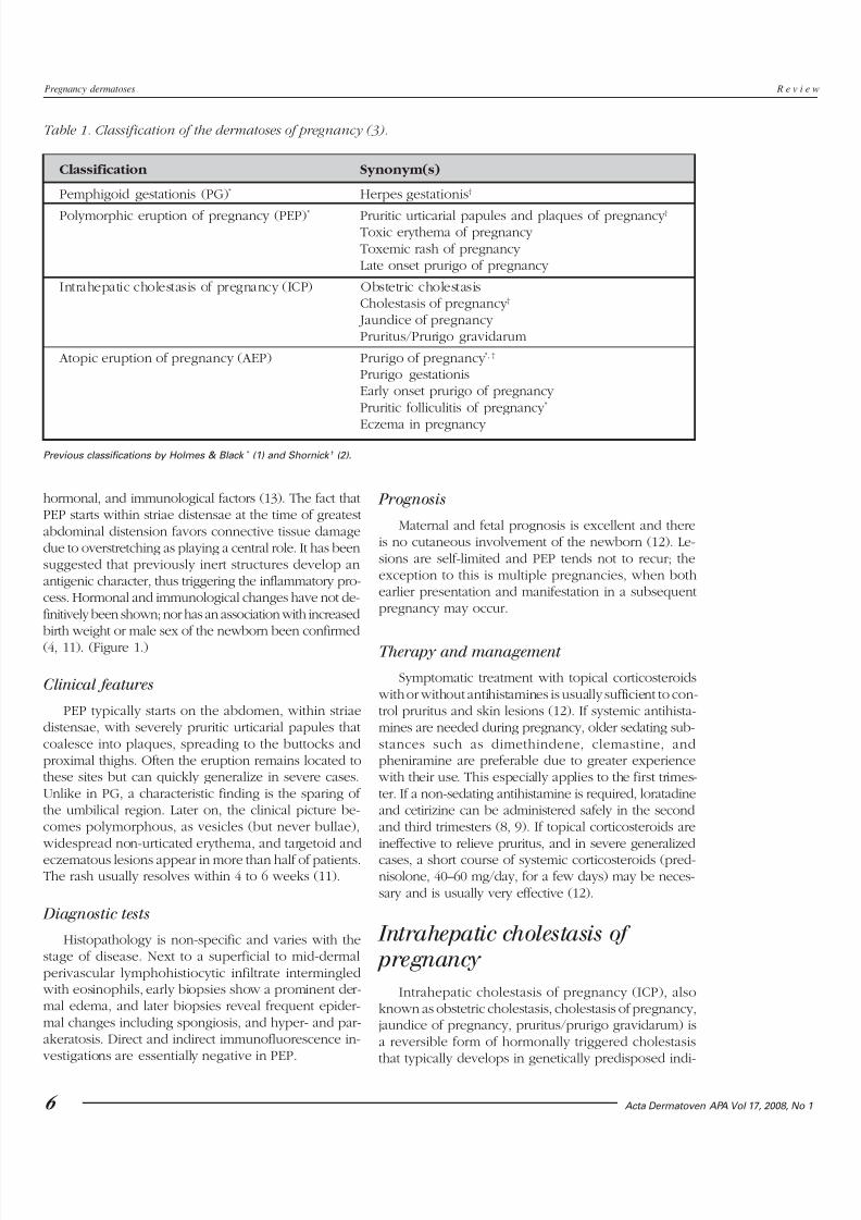
Table 1. Classification of the dermatoses of pregnancy (3).
Classification Synonym(s)
Pemphigoid gestationis (PG)* Herpes gestationis†
Polymorphic eruption of pregnancy (PEP)* Pruritic urticarial papules and plaques of pregnancy †Toxic erythema of pregnancy Toxemic rash of pregnancy Late onset prurigo of pregnancy
Intrahepatic cholestasis of pregnancy (ICP) Obstetric cholestasisCholestasis of pregnancy †
Jaundice of pregnancy Pruritus/Prurigo gravidarum
Atopic eruption of pregnancy (AEP) Prurigo of pregnancy *, †
Prurigo gestationisEarly onset prurigo of pregnancy Pruritic folliculitis of pregnancy *
Eczema in pregnancy
Previous classifications by Holmes & Black * (1) and Shornick † (2).
Pregnancy dermatoses R e v i e w
7/23/2019 Dermatosis Del Embarazo 2008
http://slidepdf.com/reader/full/dermatosis-del-embarazo-2008 4/8
Acta Dermatoven APA Vol 17, 2008, No 1 7
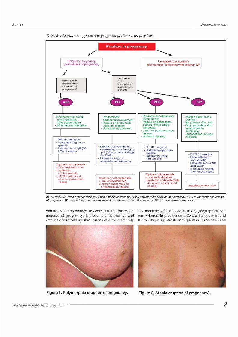
AEP = atopic eruption of pregnancy, PG = pemphigoid gestationis, PEP = polymorphic eruption of pregnancy, ICP = intrahepatic cholestasis of pregnancy, DIF = direct immunofluorescence, IIF = indirect immunofluorescence, BMZ = basal membrane zone.
Table 2. Algorithmic approach to pregnant patients with pruritus.
R e v i e w Pregnancy dermatoses
viduals in late pregnancy. In contrast to the other der-matoses of pregnancy, it presents with pruritus andexclusively secondary skin lesions due to scratching.
The incidence of ICP shows a striking geographical pat-tern; whereas its prevalence in Central Europe is around0.2 to 2.4%, it is particularly frequent in Scandinavia and
Figure 2. Atopic eruption of pregnancy).Figure 1. Polymorphic eruption of pregnancy.
7/23/2019 Dermatosis Del Embarazo 2008
http://slidepdf.com/reader/full/dermatosis-del-embarazo-2008 5/8
8 Acta Dermatoven APA Vol 17, 2008, No 1
South America, with the highest rates in Chile (15–28%)(5). ICP runs in families and tends to recur in subse-quent pregnancies (45–70%) (5, 14).
Pathogenetically, there is a defect in the excretionof bile salts resulting in elevated serum bile acids in the
serum. This leads to severe pruritus in the mother, and,because toxic bile acids can pass into fetal circulation,may have deleterious effects on the fetus due to acuteplacental anoxia and cardiac depression. The reason forthis defect seems to be multifactorial, with genetic, hor-monal, and exogenous factors being involved (5). En-demic clustering and familial occurrence has pointedtowards a genetic background; recently, mutations of certain genes encoding for transport proteins neces-sary for bile excretion (e.g., the ABCB4 [MDR 3] gene)have been identified in some ICP patients (15). Withnormal hormone levels, this defect has no clinical impli-cations; it only becomes evident with the highest con-
centrations as in late pregnancy and/or with hormonalcontraception. Furthermore, it has been demonstratedthat estrogen and progesterone metabolites arecholestatic themselves (16). Some authors also discussadditional environmental and dietary factors influenc-ing the manifestation of ICP, such as decreased serumselenium levels and others (17).
Clinical features
ICP typically presents with sudden onset of severepruritus that may start on the palms and soles but quickly becomes generalized. It persists throughout pregnancy
and may be tormenting. Of importance, ICP is not as-sociated with primary skin lesions. Clinical featurescorrelate with disease duration (18). At the beginningof pruritus, the skin may be unaffected; later on, sec-ondary skin lesions develop due to scratching that rangefrom subtle excoriations to severe prurigo nodules aspruritus persists. Skin lesions usually involve the shinsand lower arms, but may also be present on other sitessuch as on the buttocks and abdomen. Jaundice, dueto concomitant extrahepatic cholestasis, occurs in only 10% of patients, usually after 2 to 4 weeks, complicat-ing the most severe and prolonged episodes (19). Thesepatients are at risk of developing steatorrhea with mal-
absorption of fat-soluble vitamins, including vitamin K,and potential bleeding complications, as well as chole-lithiasis (5).
Diagnostic tests
Histopathology is non-specific; direct and indirectimmunofluorescence are negative. The most sensitiveindicator for the diagnosis of ICP is a rise of serum bileacid levels, whereas routine liver function tests (includ-ing transaminases) may be normal in up to 30% (5, 18).In healthy pregnancies, total serum bile acid levels areslightly higher than in non-pregnant women, and levels
up to 11.0 µmol/l are accepted as normal in late gesta-
tion (20, 21). Hyperbilirubinemia is noted in only 10 to20%; it should always lead to close surveillance of pro-thrombin time and an ultrasound examination of theliver may be necessary in such cases.
PrognosisThe prognosis for the mother is generally good. Af-
ter delivery, pruritus disappears spontaneously withindays to weeks but may recur with subsequent preg-nancies and oral contraceptive use (5). In cases of jaun-dice and vitamin K deficiency, there is an increased riskfor intra- and postpartum hemorrhage in both motherand child (5). However, the key consideration in thisdisease is not maternal pruritus but the significantly impaired fetal prognosis. ICP is associated with an in-creased risk of prematurity (19–60%), intrapartal fetaldistress (22–33%), and stillbirths (1–2%), which corre-late with higher bile acid levels, in particular if in ex-cess of 40 µmol/l (5, 22). Therefore, prompt diagnosis,specific therapy, and close obstetric monitoring as wellas maternal counseling, in particular on the expectedrecurrence in subsequent pregnancies, are essential.
Therapy and management
The aim of treatment is the reduction of serum bileacid levels in order to prolong pregnancy and reduceboth fetal risks and maternal symptoms. Ursodeo-xycholic acid (UDCA) is the only treatment that hasbeen shown not only to reduce maternal pruritus butto also improve fetal prognosis (5, 18, 23–26). It is anaturally occurring, hydrophilic, non-toxic bile acid thathas been successfully employed in Chinese medicinefor over 5,000 years in treating various liver diseasesand nowadays plays a key role in treating hepatobiliary disorders. In ICP, a dose of 15 mg/kg/day or, indepen-dent of body weight, 1 g/day is administered either assingle dose or divided into two to three doses untildelivery, when it usually can be stopped. With the ex-ception of occasional mild diarrhea, no adverse effectsoccur. However, UDCA is not licensed for use in preg-nancy; thus, it requires special patient information (“off-label use”). Other drugs, including antihistamines, S-adenosyl-L-methionine, dexamethasone, and cholesty-ramine, do not improve fetal prognosis (5). Of note,cholestyramine and other bile acid exchange resinsmay contribute to malabsorption of vitamin K withpossible consecutive bleeding complications andshould therefore be avoided (27). In addition to UDCAtreatment, close obstetric surveillance is indicated andincludes weekly fetal cardiotocographic (CTG) regis-tration at least from 34 weeks’ gestation on; early de-livery as soon as lung maturity is achieved (36–37 weeks) is recommended by some authors (28). Inter-disciplinary management of ICP by dermatologists,hepatologists, gynecologists, and pediatricians is abso-
lutely mandatory.
Pregnancy dermatoses R e v i e w
7/23/2019 Dermatosis Del Embarazo 2008
http://slidepdf.com/reader/full/dermatosis-del-embarazo-2008 6/8
Acta Dermatoven APA Vol 17, 2008, No 1 9
Atopic eruption of pregnancy
Atopic eruption of pregnancy (AEP), also knownas prurigo of pregnancy, prurigo gestationis, early-on-set prurigo of pregnancy, pruritic folliculitis of preg-nancy, eczema in pregnancy) is a benign pruritic dis-order of pregnancy which includes eczematous and/or papular lesions in patients with a personal and/orfamily history of atopy and/or elevated IgE levels afterexclusion of the other dermatoses of pregnancy. Thisterm has been introduced based on the results of alarge two-center study, in which significant overlap hasbeen observed clinically and histopathologically be-tween patients formerly diagnosed as suffering fromeczema in pregnancy, prurigo of pregnancy, and pru-ritic folliculitis of pregnancy (3). It is the most com-mon dermatosis in pregnancy, accounting for 50% of
patients, usually starts early – in 75% before the thirdtrimester – and, due to its atopic background, tends torecur in subsequent pregnancies (3).
The pathogenesis of AEP is thought to be triggeredby pregnancy-specific immunological changes; reducedcellular immunity and reduced production of Th1 cyto-kines (IL-2, interferon gamma, IL-12) stands in contrastto the dominant humoral immunity and increased se-cretion of Th2 cytokines (IL-4, IL-10) (29). Thus, theexacerbation of preexisting atopic dermatitis as well asthe first manifestation of atopic skin changes can beexplained by a predominant Th2 immune response thatis typical for pregnancy. (Figure 2.)
Clinical features
20% of patients will suffer from an exacerbation of pre-existing atopic dermatitis with a typical clinical pic-ture. The remaining 80% will experience atopic skinchanges for the first time ever or after a long remission(e.g., since childhood). Of these, two-thirds present with widespread eczematous changes (so-called E-type AEP)often affecting typical atopic sites such as face, neck,upper chest, and the flexural surfaces of the extremi-ties, whereas one-third have papular lesions (P-type
AEP) (3). These lesions include small erythematouspapules disseminated on trunk and limbs, as well astypical prurigo nodules, mostly located on the shins andarms. A key finding is the often extreme dryness of theskin and frequent atopic “minor features” according toHanifin and Rajka (30).
Diagnostic tests
Histopathology is non-specific and varies with theclinical type and stage of the disease. Direct and indi-rect immunofluorescence are negative. Laboratory testsmay reveal elevated serum IgE levels in 20 to 70% (3).
Prognosis
Maternal prognosis is good even in severe casesbecause skin lesions usually respond quickly to therapy;recurrence in subsequent pregnancies is common. Fe-tal prognosis is unaffected, but there the infant faces ahigher risk of developing atopic skin changes later on.
Therapy and management
Basic treatment is essential and consists in regularapplication of emollients, often with urea (3–10%) or an-
tipruritic additives such as menthol or polidocanol. To-gether with topical corticosteroids for several days, this will usually lead to quick improvement of skin lesions.Severe cases may require a short course of systemic cortico-steroids and antihistamines; phototherapy (UVB) is a help-ful additional measure and considered safe in pregnancy.
Algorithmic approach to pregnant patients
with pruritus
Pruritus in pregnancy should never be neglected andshould always lead to a precise work-up of the patient. Itmay be the leading symptom of the dermatoses of preg-nancy, but it can also be associated with other derma-toses coinciding by chance with pregnancy. These in-clude scabies, pityriasis rosea, drug rashes, and cutaneousinfections that, as a first step, should be excluded. In thesecond step, the four specific dermatoses of pregnancy must be differentiated. Here, the onset and localization of skin lesions as well as the history and clinical characteris-tics may offer helpful clues towards the right diagnosis(Table 2). In pemphigoid gestationis and intrahepaticcholestasis of pregnancy, specific diagnostic tests such asimmunofluorescence and laboratory investigations willfurther confirm the diagnosis. Although polymorphic andatopic eruption of pregnancy do not impair maternal orfetal prognosis, pemphigoid gestationis and intrahepaticcholestasis of pregnancy may be associated with fetalrisks. Corticosteroids and antihistamines are used to treatpemphigoid gestationis and polymorphic and atopic erup-tion of pregnancy, whereas intrahepatic cholestasis of pregnancy should be treated specifically with urso-deoxycholic acid.
1. Holmes RC, Black MM. The specific dermatoses of pregnancy. J Am Acad Dermatol. 1983;8:405–12.
2. Shornick JK. Dermatoses of pregnancy. Semin Cutan Med Surg. 1998;17:172–81.
R E F E R E N C E S
R e v i e w Pregnancy dermatoses
7/23/2019 Dermatosis Del Embarazo 2008
http://slidepdf.com/reader/full/dermatosis-del-embarazo-2008 7/8
10 Acta Dermatoven APA Vol 17, 2008, No 1
3. Ambros-Rudolph CM, Müllegger RR, Vaughan Jones SA, et al. The specific dermatoses of pregnancy revisited and reclassified: results of a retrospective two-centre study on 505 pregnant patients. J Am Acad Dermatol. 2006;54:395–404.
4. Vaughan Jones S, Hern S, Nelson-Piercy C, et al. A prospective study of 200 women with dermatosesof pregnancy correlating clinical findings with hormonal and immunopathological profiles. Br J
Dermatol. 1999;141:71–81.
5. Lammert F, Marschall HU, Glantz A, et al. Intrahepatic cholestasis of pregnancy: molecular patho-genesis, diagnosis and management. J Hepatol. 2000;33:1012–21.
6. Black MM. Pemphigoid gestationis. In: Black MM, et al., editors. Obstetric and Gynecologic Dermatol-ogy. 2nd ed. London: Mosby; 2002. p. 32–8.
7. Zilikens D. Pemphigoid gestationis: recent advances. J Eur Acad Dermatol Venereol. 2003;17(Suppl.3);7.
8. Schaefer C, Spielmann H. Arzneiverordnung in Schwangerschaft und Stillzeit, 7th ed. Munich: Urban & Fischer; 2006. 781 p.
9. Briggs GG, Freeman RK, Yaffe, SJ. Drugs in pregnancy and lactation, 7th ed, Philadelphia: Lippincot Williams & Wilkins; 2005. 1858 p.
10. Wöhrl S, Geusau A, Karlhofer F, et al. Pemphigoid gestationis: treatment with immunoapheresis. JDtsch Dermatol Ges. 2006;1:126–30.
11. Rudolph CM, Al-Fares S, Vaughan-Jones SA, et al. Polymorphic eruption of pregnancy: clinicopathology and potential trigger factors in 181 patients. Br J Dermatol. 2006;154:54–60.
12. Black MM. Polymorphic eruption of pregnancy. In: Black MM, et al., editors. Obstetric and Gyneco-logic Dermatology. 2nd ed. London: Mosby; 2002. p. 39–44.
13. Ahmadi S, Powell F. Pruritic urticarial papules and plaques of pregnancy: Current status. Australas JDermatol. 2005;46:53–60.
14. Reyes H. The spectrum of liver and gastrointestinal disease seen in cholestasis of pregnancy.Gastroenterol Clin North Am. 1992;21:905–21.
15. Ropponen A, Sund R, Riikionen S, et al. Intrahepatic cholestasis of pregnancy as an indicator of liverand biliary diseases: a population-based study. Hepatology. 2006;43:723–8.
16. Reyes H, Sjövall J. Bile acids and progesterone metabolites in intrahepatic cholestasis of pregnancy. Ann Med. 2000;32:94–106.
17. Reyes H, Báez ME, González MC, et al. Selenium, zinc and copper plasma levels in intrahepaticcholestasis of pregnancy, in normal pregnancies and in healthy individuals in Chile. J Hepatol.2000;32:542–9.
18. Ambros-Rudolph CM, Glatz M, Trauner M, et al. The importance of serum bile acid level analysis andtreatment with ursodeoxycholic acid in intrahepatic cholestasis of pregnancy. A case series from CentralEurope. Arch Dermatol. 2007;143:757–62.
19. Riosecco AJ, Ivankovic MB, Manzur A, et al. Intrahepatic cholestasis of pregnancy: a retrospectivecase-control study of perinatal outcome. Am J Obstet Gynecol. 1994;170:890–5.
20. Brites D, Rodrigues CM, Oliveira N, et al. Correction of maternal serum bile acid profile duringursodeoxycholic acid therapy in cholestasis of pregnancy. J Hepatol. 1998;28:91–8.
21. Carter J. Serum bile acids in normal pregnancy. Br J Obstet Gynaecol. 1991;98:540–3.
22. Glantz A, Marschall HU, Mattsson LA. Intrahepatic cholestasis of pregnancy: relationships betweenbile acid levels and fetal complication rates. Hepatology. 2004;40:467–74.
23. Zapata R, Sandoval L, Palma J, et al. Ursodeoxycholic acid in the treatment of intrahepatic cholestasisof pregnancy. A 12-year experience. Liver Int. 2005;25:548–54.
24. Williamson C, Hems LM, Goulis DG, et al. Clinical outcome in a series of cases of obstetric cholestasisidentified via a patient support group. Br J Obstet Gynaecol. 2004;111:676–81.
25. Palma J, Reyes H, Ribalta J, et al. Ursodeoxycholic acid in the treatment of cholestasis of pregnancy:a randomized, double-blind study controlled with placebo. J Hepatol. 1997;27:1022–8.
Pregnancy dermatoses R e v i e w
7/23/2019 Dermatosis Del Embarazo 2008
http://slidepdf.com/reader/full/dermatosis-del-embarazo-2008 8/8
Acta Dermatoven APA Vol 17, 2008, No 1 11
26. Kondrackiene J, Beuers U, Kupcinskas L. Efficacy and safety of ursodeoxycholic acid versuscholestyramine in intrahepatic cholestasis of pregnancy. Gastroenterology. 2005;29:895–901.
27. Sadler LC, Lane M, North R. Severe fetal intracranial haemorrhage during treatment withcholestyramine for intrahepatic cholestasis of pregnancy. Br J Obstet Gynaecol. 1995;102:169–70.
28. Roncaglia N, Arreghini A, Locatelli A, et al. Obstetric cholestasis: outcome with active management.Eur J Obstet Gynecol Reprod Biol. 2002;100:167–70.
29. Garcia-Gonzalez E, Ahued-Ahued R, Arroyo E, et al. Immunology of the cutaneous disorders of pregnancy. Int J Dermatol. 1999;38:721–9.
30. Hanifin JM, Rajka G. Diagnostic features of atopic dermatitis. Acta Derm Venereol Suppl (Stockh).1980;92:44-7.
Maria Magdalena Pãunescu, MD, Department of Dermatology, Victor Babes University of Medicine and Pharmacy, Str. Mãrãºeºti 5,Timisoara, Romania, E-mail: magdalenapaunescu yahoo.comVirgil Feier, MD, PhD, Department of Dermatology, same address, E-mail: vfeier mail.dnttm.ro Mugurel Pãunescu, MD, 4th Medical Clinic, same address, E-mail: mugurel.paunescu yahoo.com Florin Dorneanu, MD, Department of Gynecology, same address,
E-mail: florindorneanuhotmail.com Agnonela Sisak, MD, Department of Dermatology, C.F.R. Clinic,Timisoara, Romania, E-mail: sisakagnonela gmail.comChristina M. Ambros-Rudolph, MD, Department of Dermatology, Medical University of Graz, Auenbruggerplatz 8, 8036 Graz, Austria, E-mail: christina.ambros-rudolphmeduni-graz.at
A U T H O R S '
A D D R E S S E S
R e v i e w Pregnancy dermatoses